


 biology
Similar presentations:


Respiratory system
1.
Dr. Monqith Mazin2.
IntroductionThe Respiratory System is mainly concerned with gaseous
exchange which occurs in the lungs at the blood-air barrier
between the blood contained in the capillaries and the
inspired air in the lungs.
Parts of the system are also concerned with the sense of
smell, sense of taste, phonation (production of sound) and
with excretion of water through exhaled air.
3.
Objectives:Description of the main functional units of the
respiratory system and its division into upper and
lower respiratory tracts.
Description of the component parts of the upper &
lower respiratory tracts and their general functions.
Description of the structure of each part of the
respiratory tract.
4.
5.
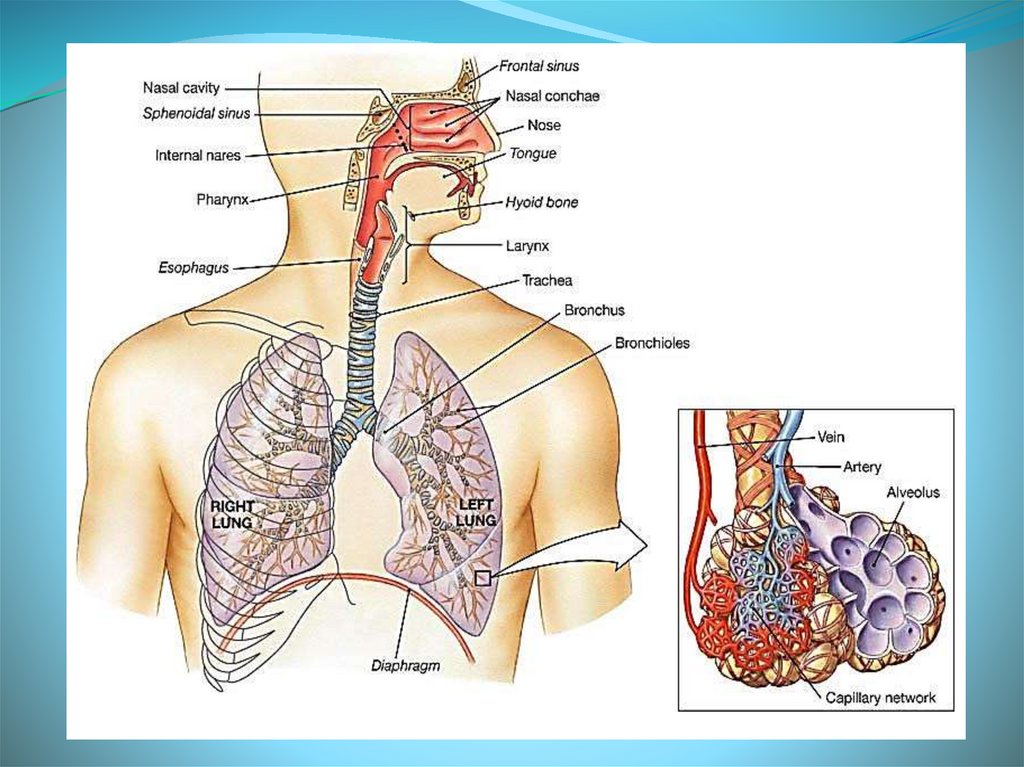
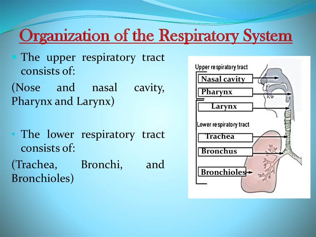
Organization of the Respiratory SystemThe upper respiratory tract
consists of:
(Nose and nasal
Pharynx and Larynx)
cavity,
Pharynx
Larynx
• The lower respiratory tract
consists of:
(Trachea,
Bronchi,
Bronchioles)
Nasal cavity
Trachea
Bronchus
and
Bronchioles
6.
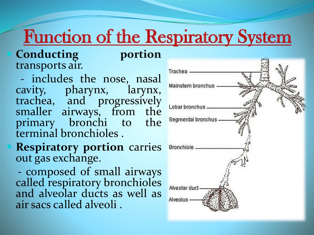
Function of the Respiratory SystemConducting
portion
transports air.
- includes the nose, nasal
cavity,
pharynx,
larynx,
trachea, and progressively
smaller airways, from the
primary bronchi to the
terminal bronchioles .
Respiratory portion carries
out gas exchange.
- composed of small airways
called respiratory bronchioles
and alveolar ducts as well as
air sacs called alveoli .
7.
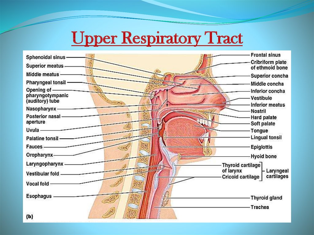
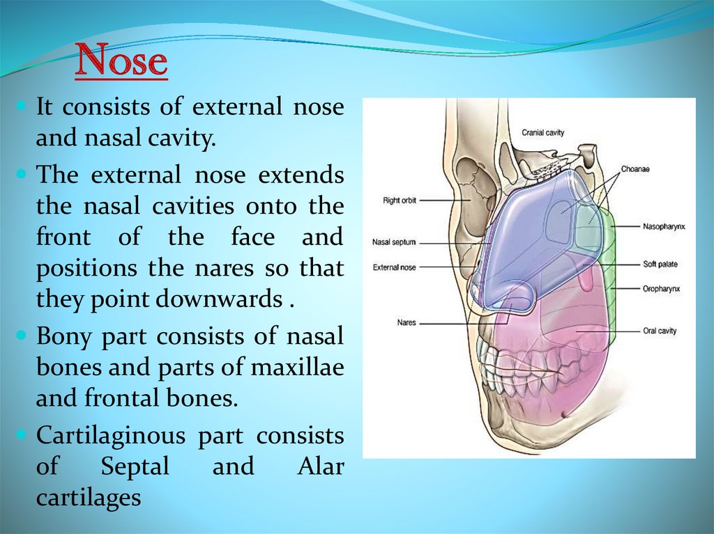
Upper Respiratory Tract8.
NoseIt consists of external nose
and nasal cavity.
The external nose extends
the nasal cavities onto the
front of the face and
positions the nares so that
they point downwards .
Bony part consists of nasal
bones and parts of maxillae
and frontal bones.
Cartilaginous part consists
of
Septal
and
Alar
cartilages
9.
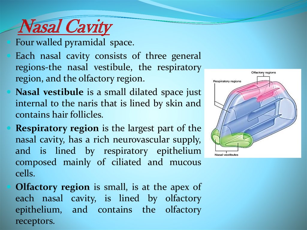
Nasal CavityFour walled pyramidal space.
Each nasal cavity consists of three general
regions-the nasal vestibule, the respiratory
region, and the olfactory region.
Nasal vestibule is a small dilated space just
internal to the naris that is lined by skin and
contains hair follicles.
Respiratory region is the largest part of the
nasal cavity, has a rich neurovascular supply,
and is lined by respiratory epithelium
composed mainly of ciliated and mucous
cells.
Olfactory region is small, is at the apex of
each nasal cavity, is lined by olfactory
epithelium, and contains the olfactory
receptors.
10.
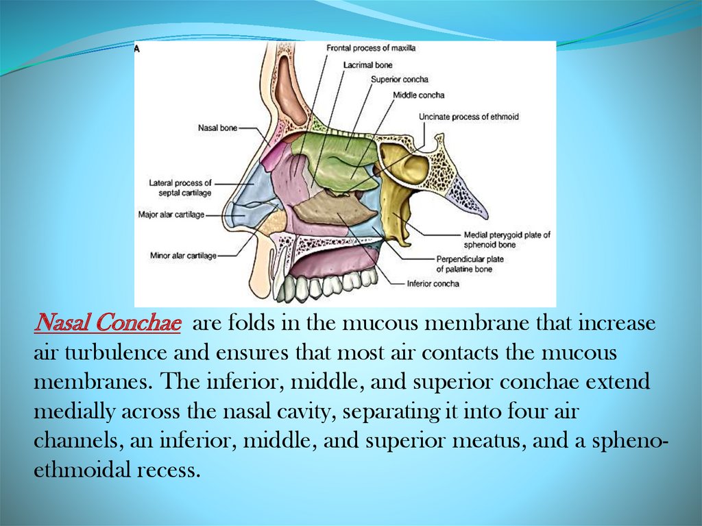
Nasal Conchae are folds in the mucous membrane that increaseair turbulence and ensures that most air contacts the mucous
membranes. The inferior, middle, and superior conchae extend
medially across the nasal cavity, separating it into four air
channels, an inferior, middle, and superior meatus, and a sphenoethmoidal recess.
11.
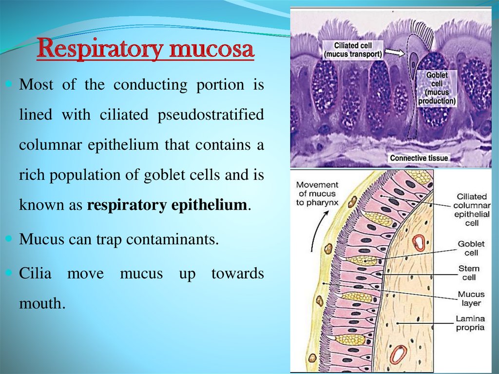
Respiratory mucosaMost of the conducting portion is
lined with ciliated pseudostratified
columnar epithelium that contains a
rich population of goblet cells and is
known as respiratory epithelium.
Mucus can trap contaminants.
Cilia move mucus up towards
mouth.
12.
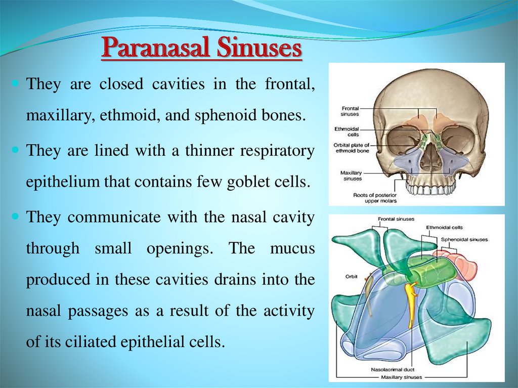
Paranasal SinusesThey are closed cavities in the frontal,
maxillary, ethmoid, and sphenoid bones.
They are lined with a thinner respiratory
epithelium that contains few goblet cells.
They communicate with the nasal cavity
through small openings. The mucus
produced in these cavities drains into the
nasal passages as a result of the activity
of its ciliated epithelial cells.
13.
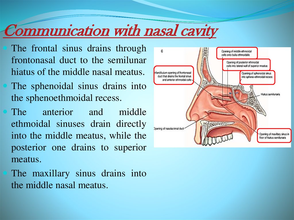
Communication with nasal cavityThe frontal sinus drains through
frontonasal duct to the semilunar
hiatus of the middle nasal meatus.
The sphenoidal sinus drains into
the sphenoethmoidal recess.
The
anterior
and
middle
ethmoidal sinuses drain directly
into the middle meatus, while the
posterior one drains to superior
meatus.
The maxillary sinus drains into
the middle nasal meatus.
14.
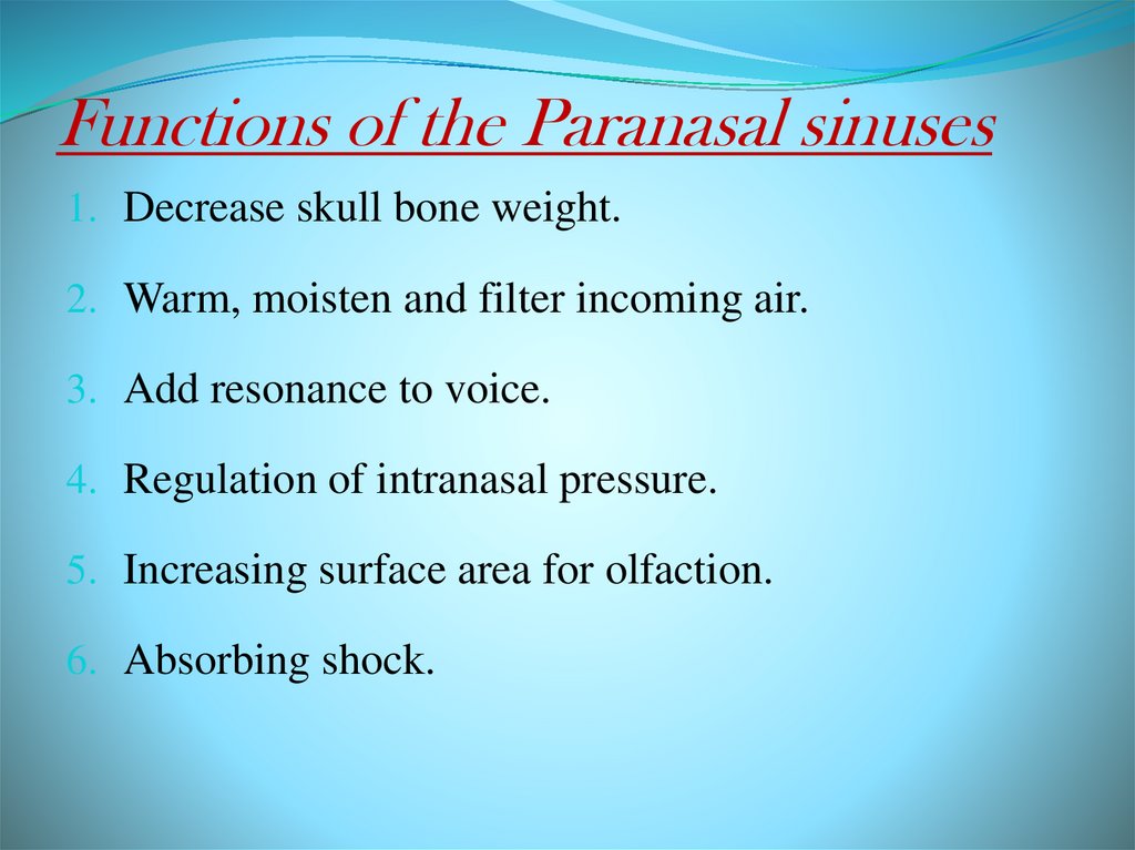
Functions of the Paranasal sinuses1. Decrease skull bone weight.
2. Warm, moisten and filter incoming air.
3. Add resonance to voice.
4. Regulation of intranasal pressure.
5. Increasing surface area for olfaction.
6. Absorbing shock.
15.
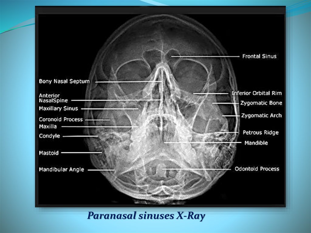
Paranasal sinuses X-Ray16.
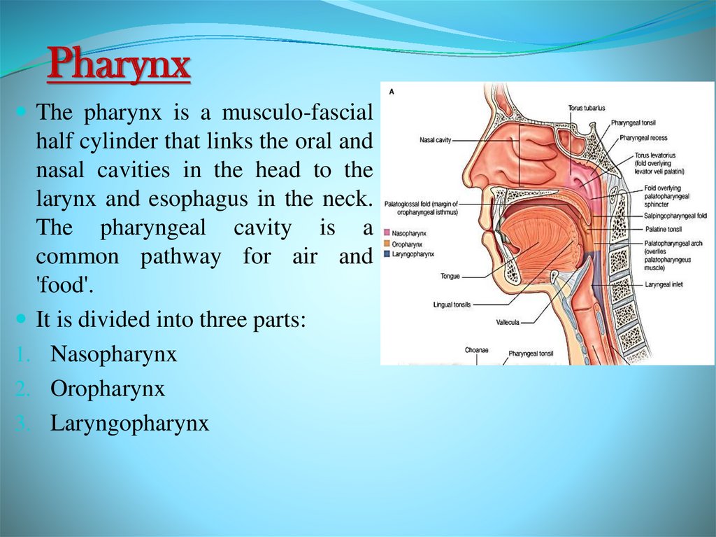
PharynxThe pharynx is a musculo-fascial
half cylinder that links the oral and
nasal cavities in the head to the
larynx and esophagus in the neck.
The pharyngeal cavity is a
common pathway for air and
'food'.
It is divided into three parts:
1. Nasopharynx
2. Oropharynx
3. Laryngopharynx
17.
Pharyngeal mucosaSuperior-most region of the nasopharynx is covered
with pseudostratified ciliated columnar epithelium.
Posterior nasopharynx wall also houses a single
pharyngeal tonsil (commonly called the adenoids).
The oropharynx contains non-keratinized stratified
squamous epithelium.
Palatine tonsils are on the lateral wall between the
arches, and the lingual tonsils are at the base of the
tongue.
Laryngopharynx lined with a nonkeratinized
stratified squamous epithelium.
18.
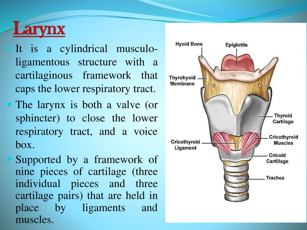
LarynxIt is a cylindrical musculo-
ligamentous structure with a
cartilaginous framework that
caps the lower respiratory tract.
The larynx is both a valve (or
sphincter) to close the lower
respiratory tract, and a voice
box.
Supported by a framework of
nine pieces of cartilage (three
individual pieces and three
cartilage pairs) that are held in
place by ligaments and
muscles.
19.
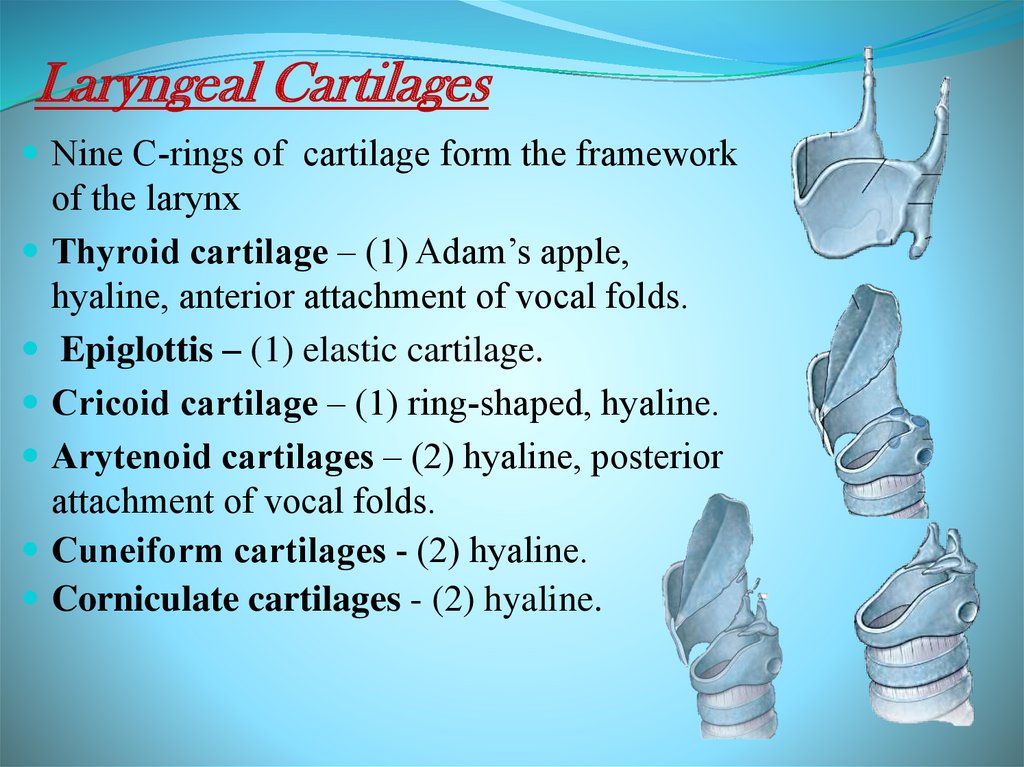
Laryngeal CartilagesNine C-rings of cartilage form the framework
of the larynx
Thyroid cartilage – (1) Adam’s apple,
hyaline, anterior attachment of vocal folds.
Epiglottis – (1) elastic cartilage.
Cricoid cartilage – (1) ring-shaped, hyaline.
Arytenoid cartilages – (2) hyaline, posterior
attachment of vocal folds.
Cuneiform cartilages - (2) hyaline.
Corniculate cartilages - (2) hyaline.
20.
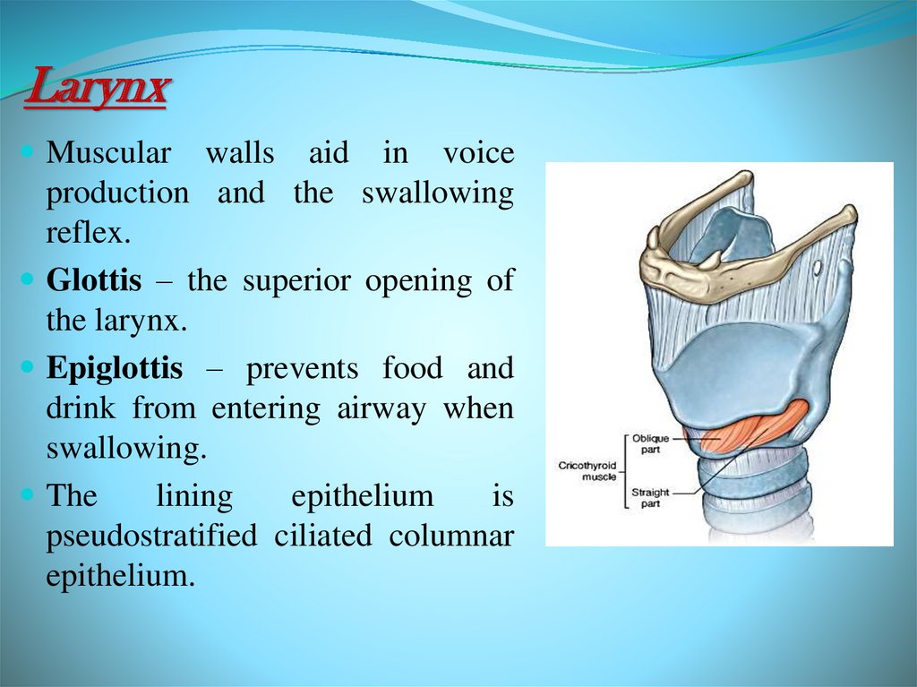
LarynxMuscular
walls aid in voice
production and the swallowing
reflex.
Glottis – the superior opening of
the larynx.
Epiglottis – prevents food and
drink from entering airway when
swallowing.
The
lining
epithelium
is
pseudostratified ciliated columnar
epithelium.
21.
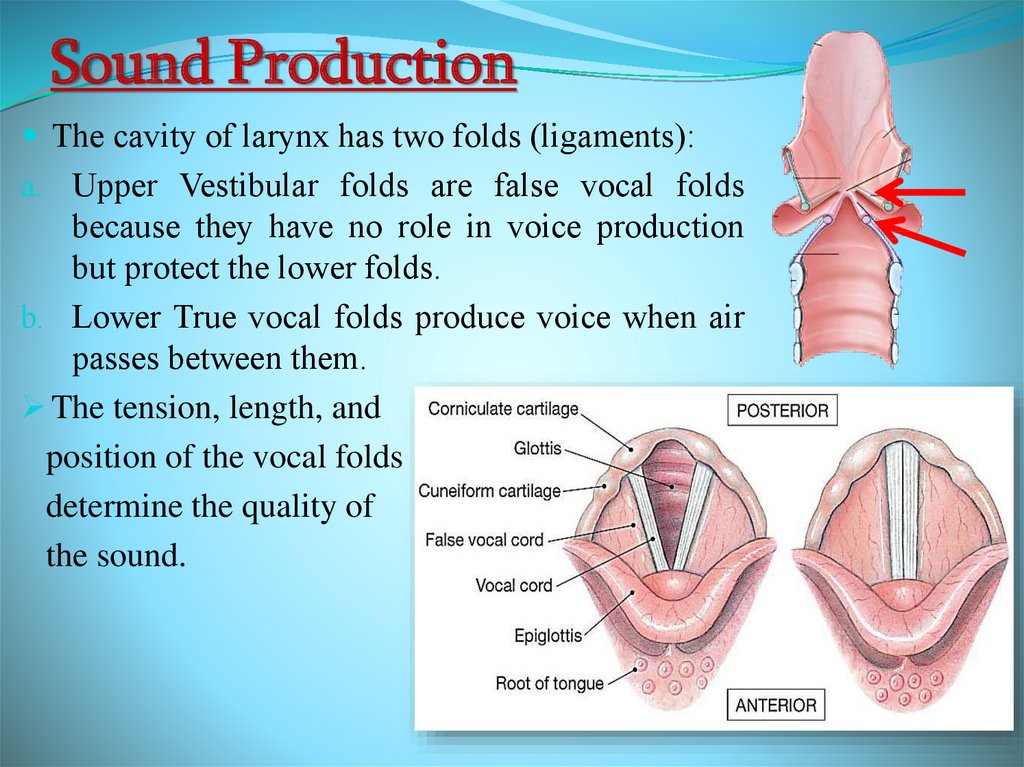
Sound ProductionThe cavity of larynx has two folds (ligaments):
a. Upper Vestibular folds are false vocal folds
because they have no role in voice production
but protect the lower folds.
b. Lower True vocal folds produce voice when air
passes between them.
The tension, length, and
position of the vocal folds
determine the quality of
the sound.
22.
Sound ProductionIntermittent release of exhaled air through the vocal folds
Loudness – depends on the force with which air is exhaled
through the cords
Pharynx, oral cavity, nasal cavity, paranasal sinuses act as
resonating chambers that add quality to the sound
Muscles of the face, tongue, and lips help with expression of
words.
23.
Conducting zone of lower respiratorytract
24.
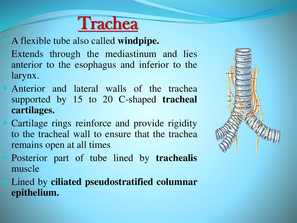
TracheaA flexible tube also called windpipe.
Extends through the mediastinum and lies
anterior to the esophagus and inferior to the
larynx.
Anterior and lateral walls of the trachea
supported by 15 to 20 C-shaped tracheal
cartilages.
Cartilage rings reinforce and provide rigidity
to the tracheal wall to ensure that the trachea
remains open at all times
Posterior part of tube lined by trachealis
muscle
Lined by ciliated pseudostratified columnar
epithelium.
25.
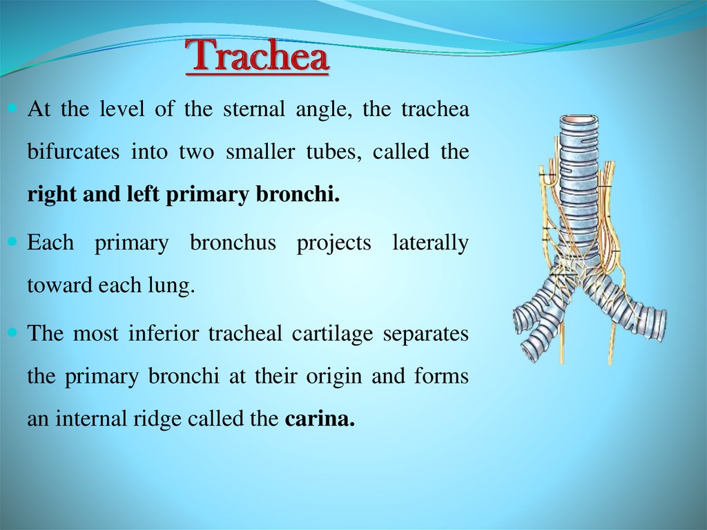
TracheaAt the level of the sternal angle, the trachea
bifurcates into two smaller tubes, called the
right and left primary bronchi.
Each primary bronchus projects laterally
toward each lung.
The most inferior tracheal cartilage separates
the primary bronchi at their origin and forms
an internal ridge called the carina.
26.
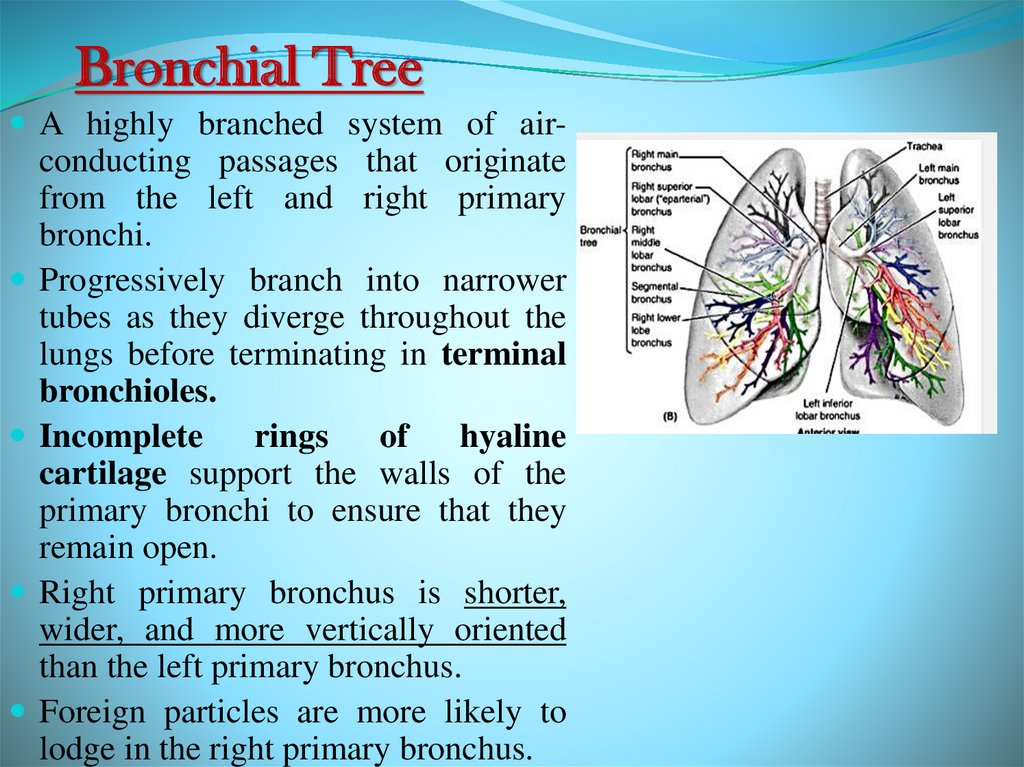
Bronchial TreeA highly branched system of air-
conducting passages that originate
from the left and right primary
bronchi.
Progressively branch into narrower
tubes as they diverge throughout the
lungs before terminating in terminal
bronchioles.
Incomplete rings of
hyaline
cartilage support the walls of the
primary bronchi to ensure that they
remain open.
Right primary bronchus is shorter,
wider, and more vertically oriented
than the left primary bronchus.
Foreign particles are more likely to
lodge in the right primary bronchus.
27.
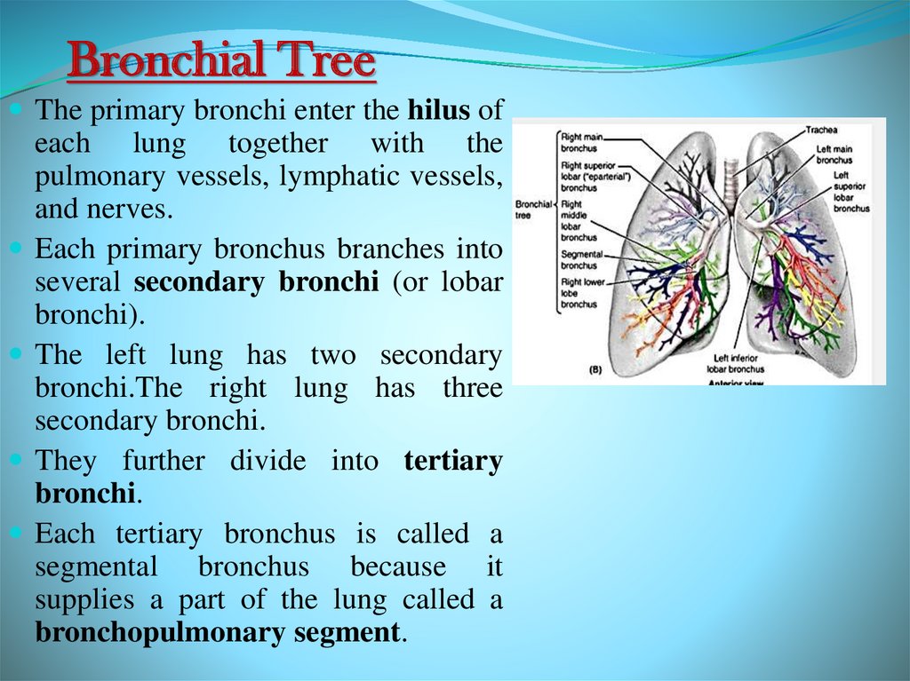
Bronchial TreeThe primary bronchi enter the hilus of
each lung together with the
pulmonary vessels, lymphatic vessels,
and nerves.
Each primary bronchus branches into
several secondary bronchi (or lobar
bronchi).
The left lung has two secondary
bronchi.The right lung has three
secondary bronchi.
They further divide into tertiary
bronchi.
Each tertiary bronchus is called a
segmental bronchus because it
supplies a part of the lung called a
bronchopulmonary segment.
28.
Bronchial TreeSecondary bronchi Tertiary bronchi Bronchioles
Terminal bronchioles.
With successive branching amount of cartilage decreases and
amount of smooth muscle increases, this allows for variation
in airway diameter.
During exertion and when sympathetic division active
bronchodilation.
Mediators of allergic reactions like histamine
bronchoconstriction.
Epithelium gradually changes from ciliated pseudostratified
columnar epithelium to simple cuboidal epithelium in
terminal bronchioles.
29.
Respiratory Zone of Lower RespiratoryTract
30.
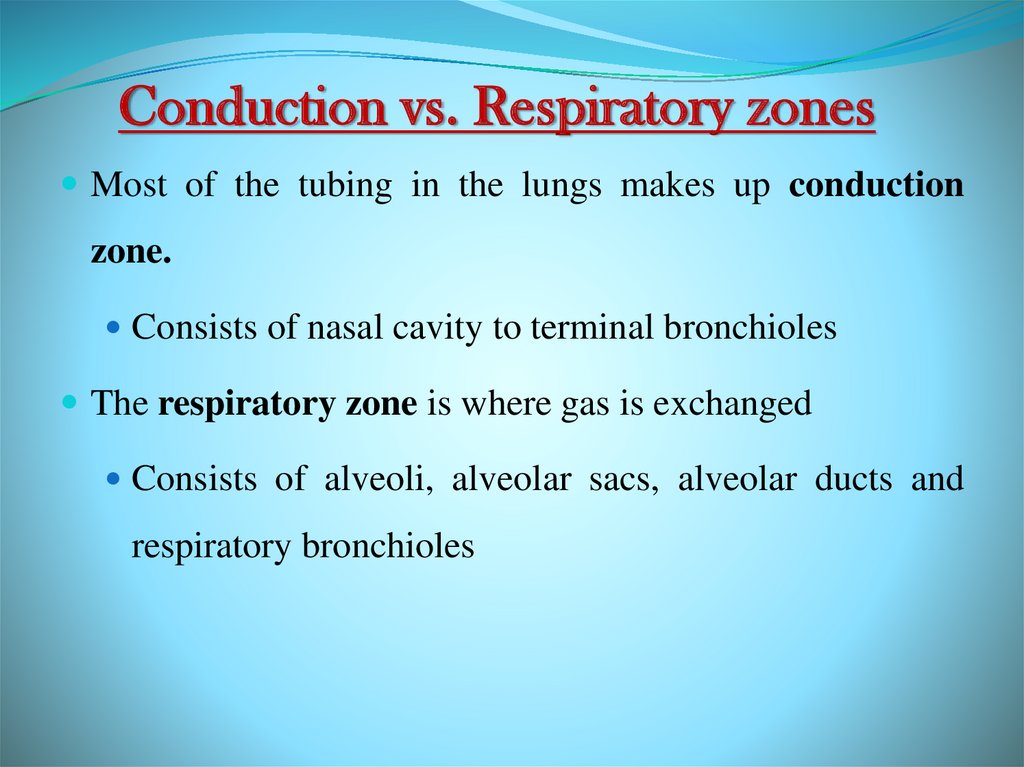
Conduction vs. Respiratory zonesMost of the tubing in the lungs makes up conduction
zone.
Consists of nasal cavity to terminal bronchioles
The respiratory zone is where gas is exchanged
Consists of alveoli, alveolar sacs, alveolar ducts and
respiratory bronchioles
31.
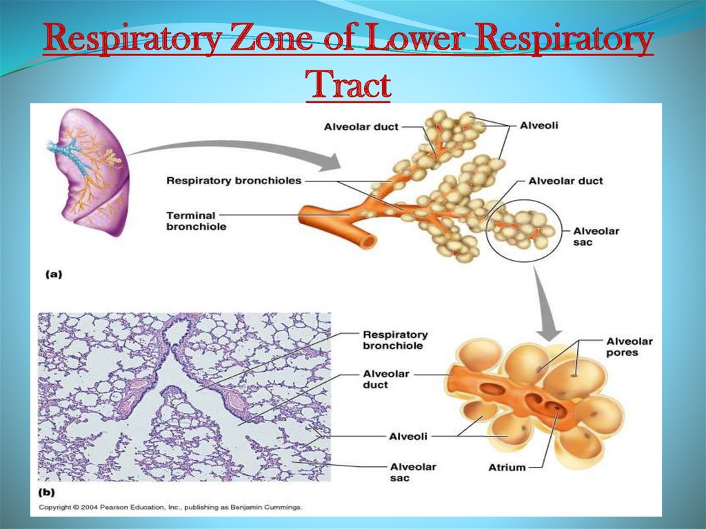
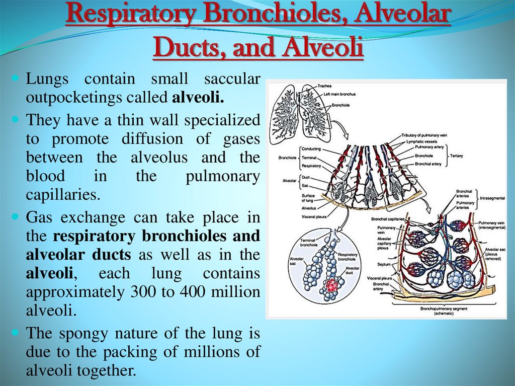
Respiratory Bronchioles, AlveolarDucts, and Alveoli
Lungs contain small saccular
outpocketings called alveoli.
They have a thin wall specialized
to promote diffusion of gases
between the alveolus and the
blood
in
the
pulmonary
capillaries.
Gas exchange can take place in
the respiratory bronchioles and
alveolar ducts as well as in the
alveoli, each lung contains
approximately 300 to 400 million
alveoli.
The spongy nature of the lung is
due to the packing of millions of
alveoli together.
32.
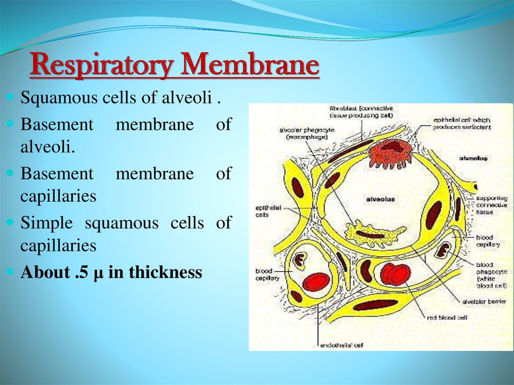
Respiratory MembraneSquamous cells of alveoli .
Basement
membrane
of
alveoli.
Basement membrane of
capillaries
Simple squamous cells of
capillaries
About .5 μ in thickness
33.
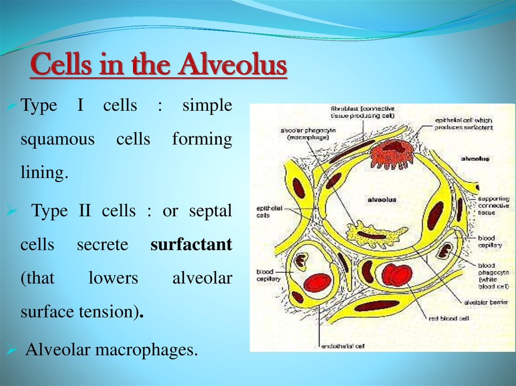
Cells in the AlveolusType
I
cells
squamous
:
cells
simple
forming
lining.
Type II cells : or septal
cells
(that
secrete
lowers
surfactant
alveolar
surface tension).
Alveolar macrophages.
34.
Gross Anatomy of the LungsEach lung has a conical shape.
Its superior region called the apex
projects superiorly to a point that is
slightly superior and posterior to the
clavicle.
Both lungs are bordered by the
thoracic wall anteriorly, laterally, and
posteriorly, and supported by the rib
cage.
Toward the midline, the lungs are
separated from each other by the
mediastinum.
The relatively broad, rounded
surface in contact with the thoracic
wall is called the costal surface of
the lung.
35.
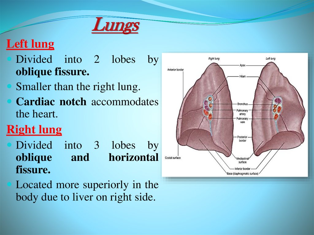
LungsLeft lung
Divided
into 2 lobes by
oblique fissure.
Smaller than the right lung.
Cardiac notch accommodates
the heart.
Right lung
Divided
into 3 lobes by
and
horizontal
oblique
fissure.
Located more superiorly in the
body due to liver on right side.
36.
Further Reading1) Clinically Oriented Anatomy (Moore). 5th Edition, 2006.
Chapters (1,7,8).
2) Gray’s Anatomy for Students (Elsevier 2007). Chapters
(3,8).
3) Basic Histology. Text and Atlas. 11th Edition, 2007.
Chapter 17.