Similar presentations:
Abdominal Cavity (1)
1.
2.
3.
4.
5.
6.
7.
8.
9.
10.
11.
12.
13.
14.
15.
16.
17.
18.
19.
20.
21.
22.
23.
24.
25.
26.
27.
28.
29.
30.
31.
32.
33.
34.
35.
36.
37.
38.
39.
40.
41.
42.
43.
44.
45.
46.
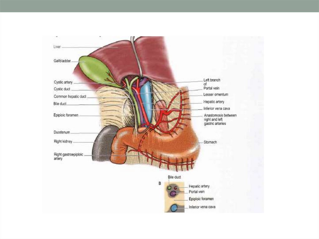
47. Epiploic foramen
48.
49.
50.
51.
52. Meso…. attachment
53.
54.
55.
56.
57.
58.
59.
Abdominal recess60.
61.
62.
63.
64.
65.
66.
67.
68.
69.
70. Stomach Anatomy
OpeningsGastroesophageal:
To esophagus
Pyloric: To
duodenum
Regions
Cardiac
Fundus
Body
Pyloric
71.
3 muscle layersOblique
Circular
Longitudinal
Regions
Cardiac sphincter
Fundus
BODY
Pyloric sphincter
Inner surface thrown into folds –
Rugae
Contains enzymes that work best at
pH 1-2
72.
73.
74.
75.
76.
• Divisions– Duodenum (5%)
– Jejunum (<40%)
– Ileum (<60%)
• Runs from pyloric
sphincter to large
intestine
• About 3-6 hours to
move food through
77.
Longest part of alimentary canalDuodenum
Jejunum
Ileocecal valve
Ileum
Duodenum (“twelve finger widths”) : 25 cm
5%
Jejunum (“empty”):
2.5 m
40 %
Ileum (“twisted”):
3.5 m
55%
78.
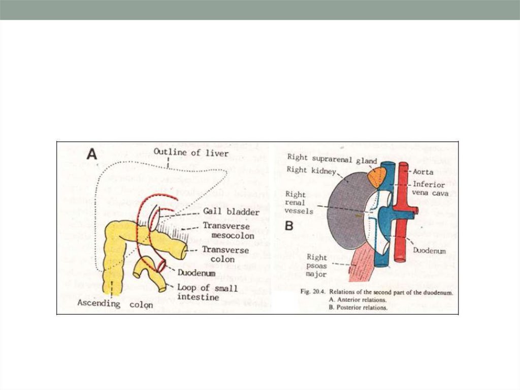
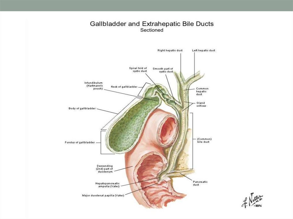
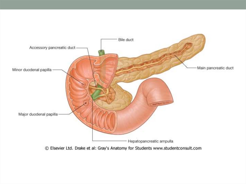
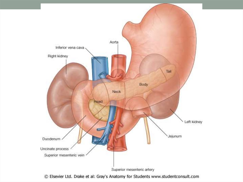
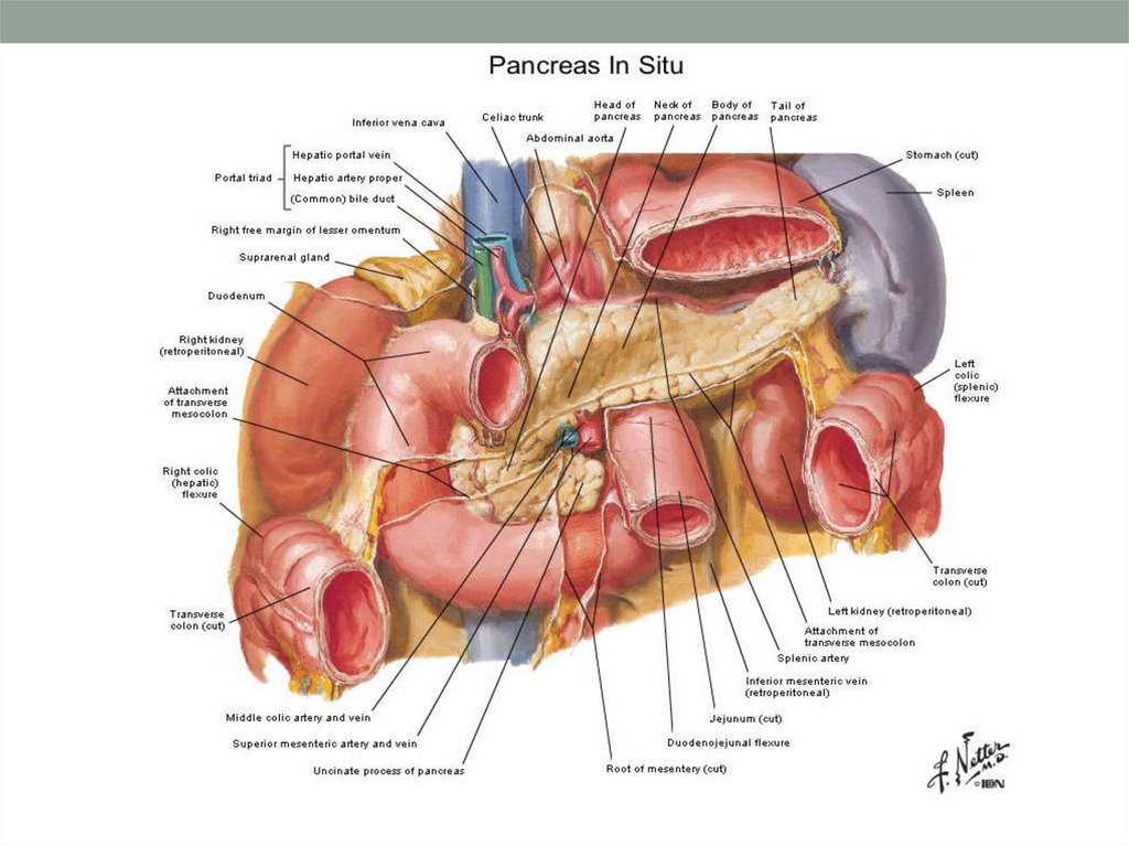
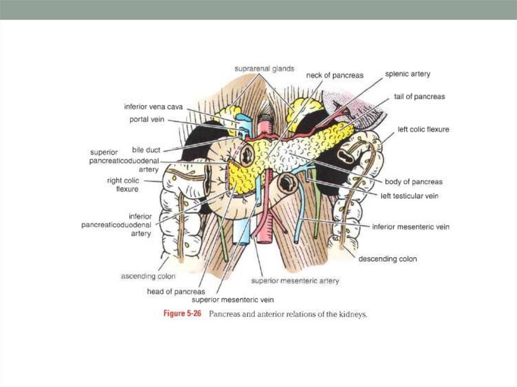
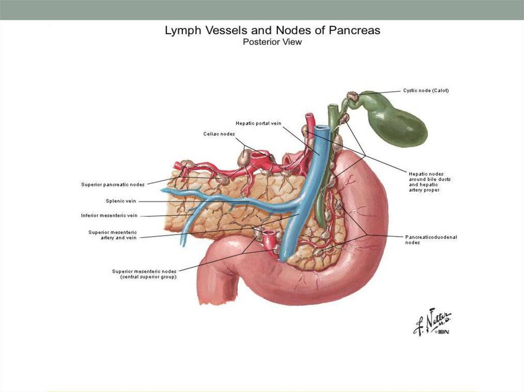
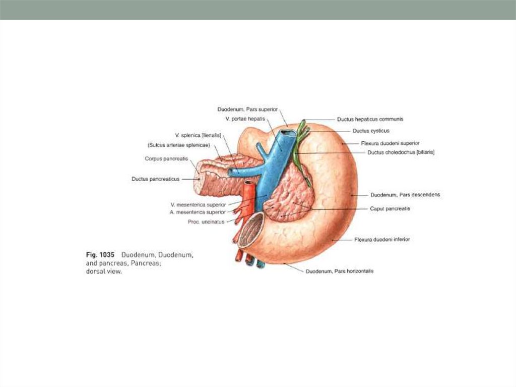
79. Duodenum and Pancreas
80.
81.
82.
83.
PANCREATIC DUCTSBile Duct
1st
Duodenal
Papillae:
B
Minor
2nd
Major
4th
A
PLICAE
CIRCULARIS
3rd
A – Main Pancreatic Duct
B – Accessory Pancreatic Duct
84.
85.
86.
87.
88.
89.
90.
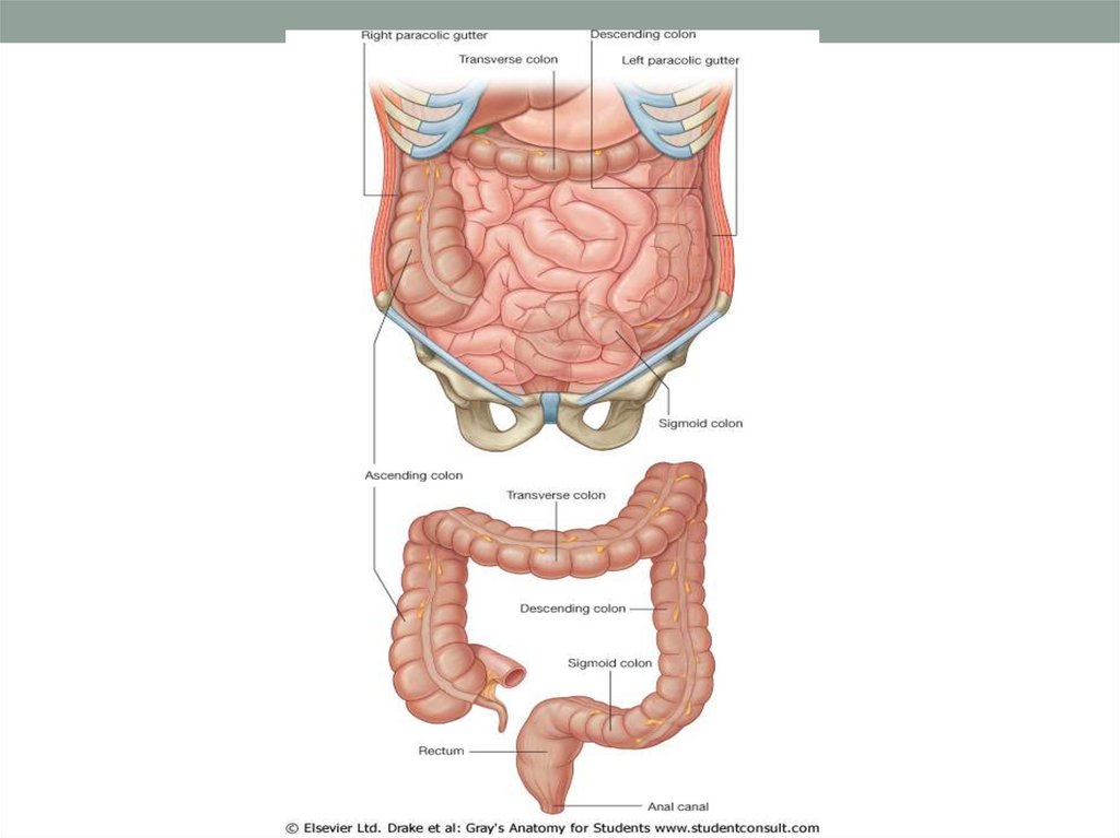
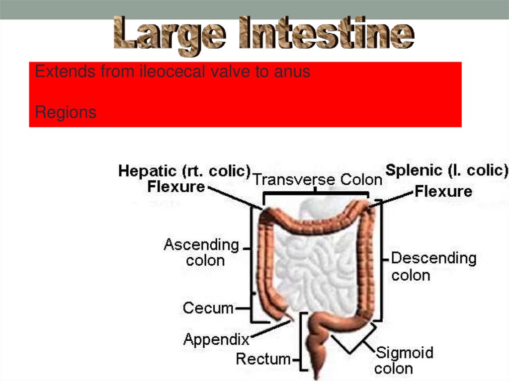
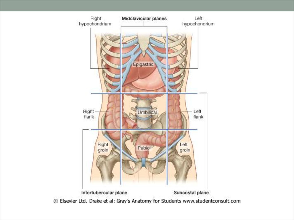
Extends from ileocecal valve to anusRegions
91.
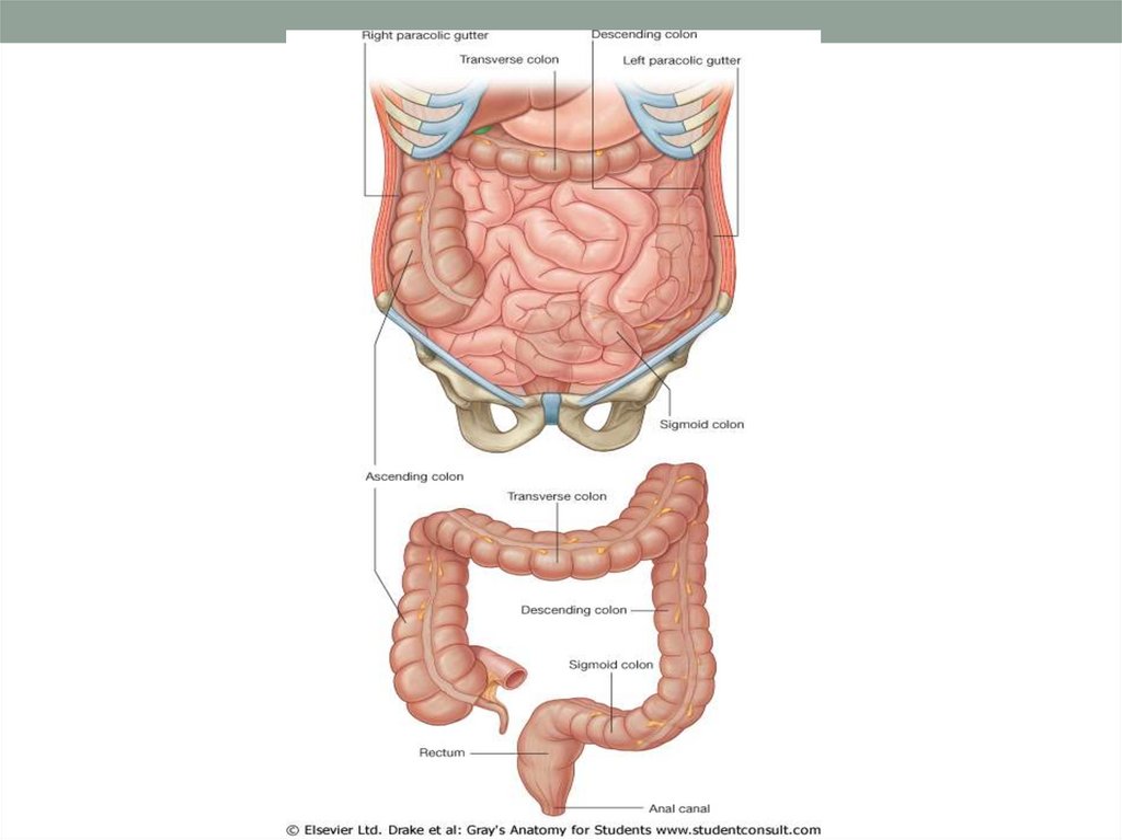
Extends from ileocecal valve to anus6 cm in diameter
1.5 to 1.8 m in length
Consists of
Cecum
Colon
Ascending
Transverse
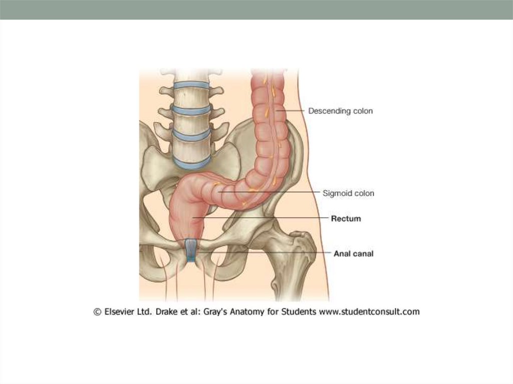
Descending
sigmoid
Rectum
Anus
.1
.2
.3
.4
hepatic and splenic flexures
Movements sluggish (18-24 hours)
92.
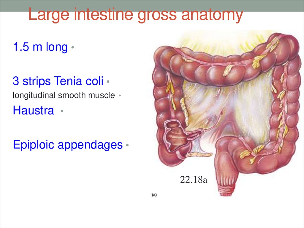
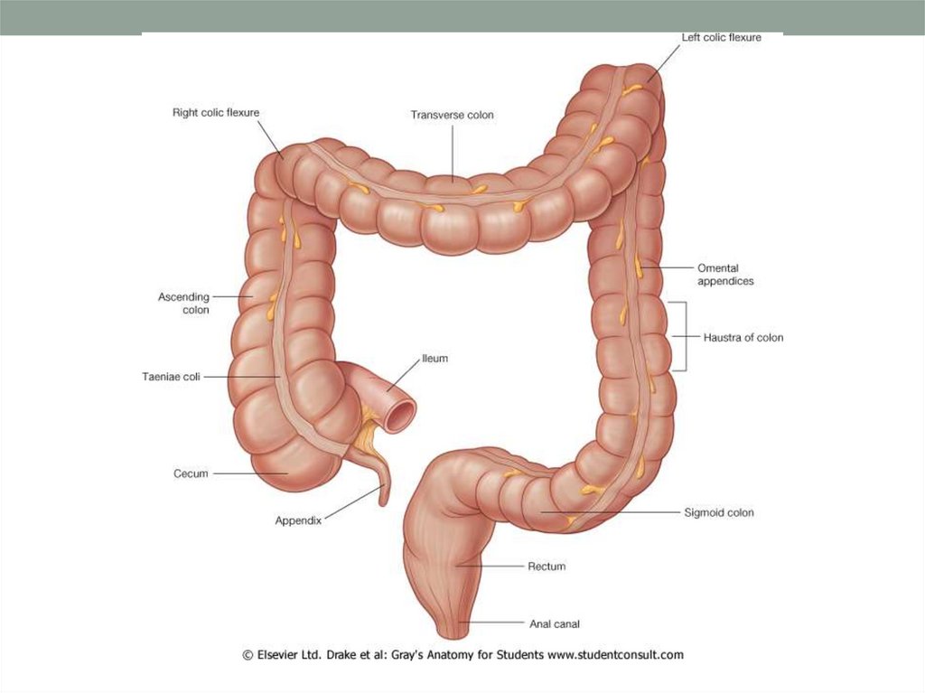
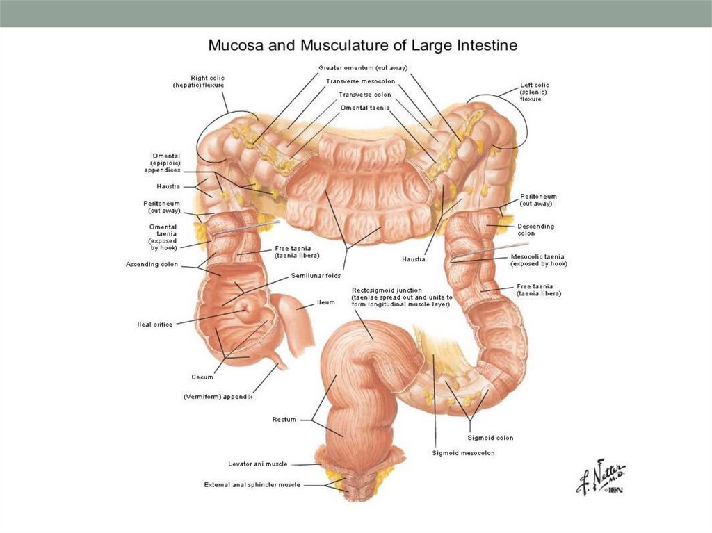
I.The large intestine exhibits 3 features not seen in other
area of the digestive system.
the teniae coli
II. Haustra
III. epiploic appendages.
I.
93. Large intestine gross anatomy
1.5 m long3 strips Tenia coli
longitudinal smooth muscle
Haustra
Epiploic appendages
22.18a
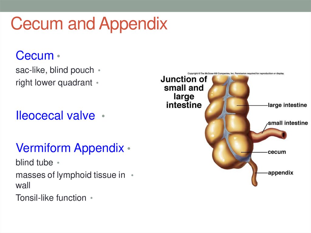
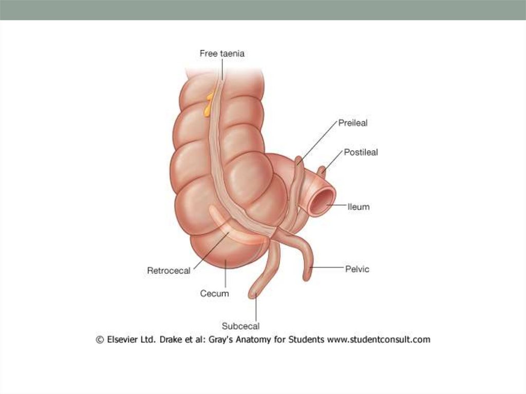
94. Cecum and Appendix
Cecumsac-like, blind pouch
right lower quadrant
Ileocecal valve
Vermiform Appendix
blind tube
masses of lymphoid tissue in
wall
Tonsil-like function





















 medicine


