


 biology
Similar presentations:


Цианобактерии
1. Лекция 3
2.
3.
4.
5.
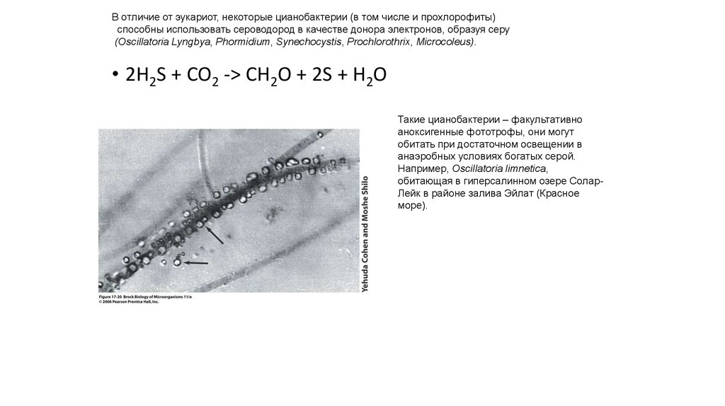
В отличие от эукариот, некоторые цианобактерии (в том числе и прохлорофиты)способны использовать сероводород в качестве донора электронов, образуя серу
(Oscillatoria Lyngbya, Phormidium, Synechocystis, Prochlorothrix, Microcoleus).
• 2H2S + CO2 -> CH2O + 2S + H2O
Такие цианобактерии – факультативно
аноксигенные фототрофы, они могут
обитать при достаточном освещении в
анаэробных условиях богатых серой.
Например, Oscillatoria limnetica,
обитающая в гиперсалинном озере СоларЛейк в районе залива Эйлат (Красное
море).
6.
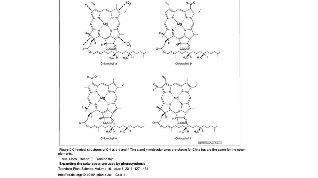
Figure 2 Chemical structures of Chl a, b d and f. The x and y molecular axes are shown for Chl a but are the same for the otherpigments.
Min Chen , Robert E. Blankenship
Expanding the solar spectrum used by photosynthesis
Trends in Plant Science, Volume 16, Issue 8, 2011, 427 - 431
http://dx.doi.org/10.1016/j.tplants.2011.03.011
7.
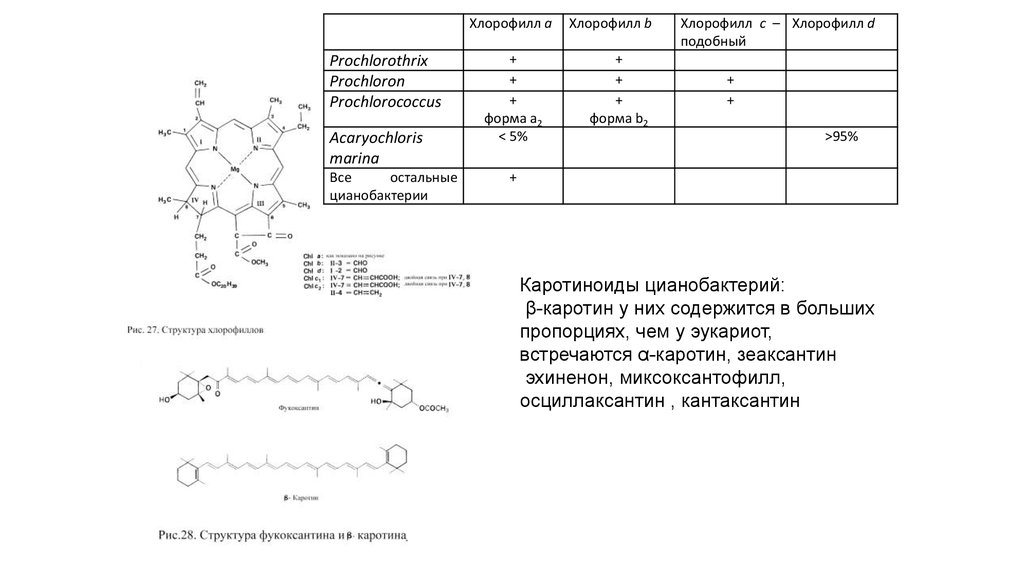
Хлорофилл aProchlorothrix
Prochloron
Prochlorococcus
Acaryochloris
marina
Все
остальные
цианобактерии
+
+
+
форма а2
< 5%
Хлорофилл b
+
+
+
форма b2
Хлорофилл c – Хлорофилл d
подобный
+
+
>95%
+
Каротиноиды цианобактерий:
β-каротин у них содержится в больших
пропорциях, чем у эукариот,
встречаются α-каротин, зеаксантин
эхиненон, миксоксантофилл,
осциллаксантин , кантаксантин
8.
9.
10.
11.
12.
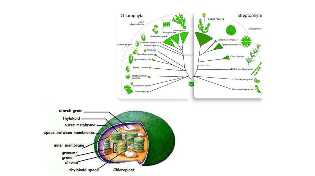
SARH13.
The distribution of photosynthesis across the eukaryotes.Dorrell R G , Howe C J J Cell Sci 2012;125:1865-1875
©2012 by The Company of Biologists Ltd
14.
15.
16. Отдел Glaucocystophyta
17. Отдел Rhodophyta
18.
19.
20.
21.
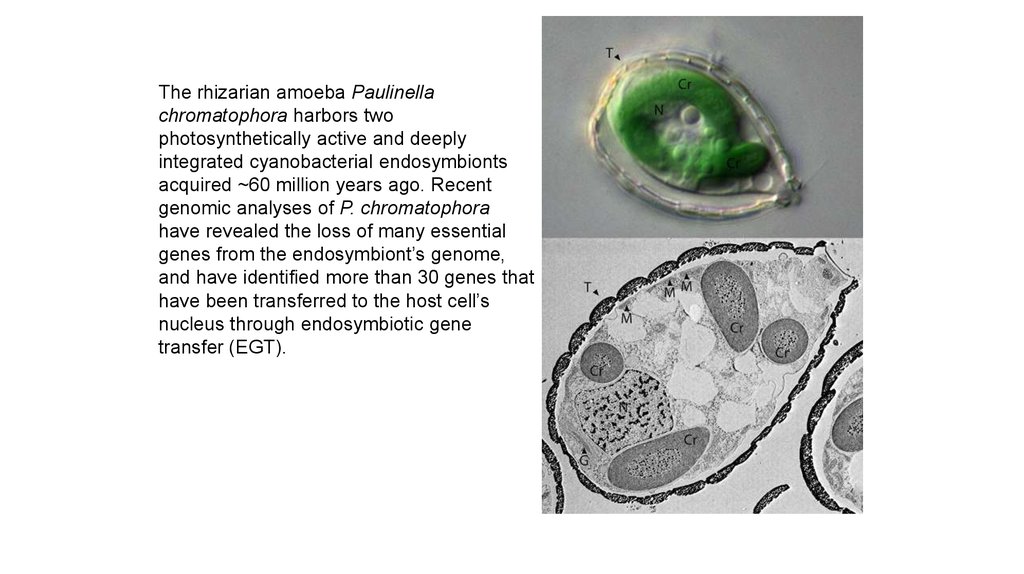
The rhizarian amoeba Paulinellachromatophora harbors two
photosynthetically active and deeply
integrated cyanobacterial endosymbionts
acquired ~60 million years ago. Recent
genomic analyses of P. chromatophora
have revealed the loss of many essential
genes from the endosymbiont’s genome,
and have identified more than 30 genes that
have been transferred to the host cell’s
nucleus through endosymbiotic gene
transfer (EGT).
22.
The endosymbiont genome has alreadybeen reduced compared to free-living
cyanobacteria, but not as much as the
primary plastids of the Archaeplastida
Figure 2. Evolution of
photosynthetic Paulinella species.
A, Schematic maximum likelihood
(RAxML) phylogenetic tree of
plastid-derived 16S rDNA from P.
chromatophora, P. microporus,
algal and plant (Plantae) plastids,
and the cyanobacterial donors of
these organelles. Note the clear
independent origins of Plantae and
photosynthetic Paulinella plastids.
See Yoon et al. (2009) for details.
B, Light micrograph images of P.
chromatophora (top two images)
and P. microporus (bottom two
images). The scale bar indicates 5
μm.
23.
24.
25.
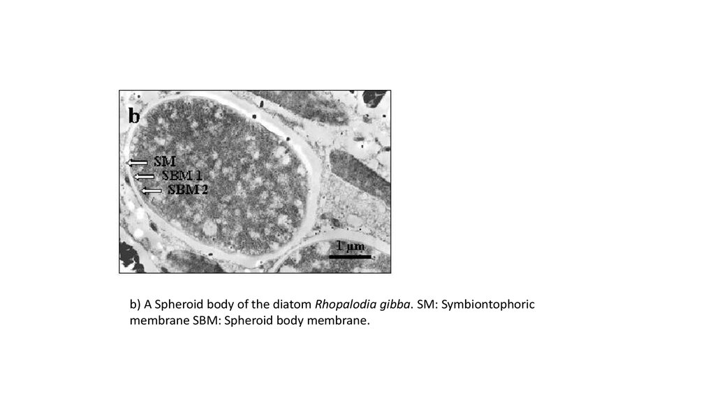
b) A Spheroid body of the diatom Rhopalodia gibba. SM: Symbiontophoricmembrane SBM: Spheroid body membrane.
26.
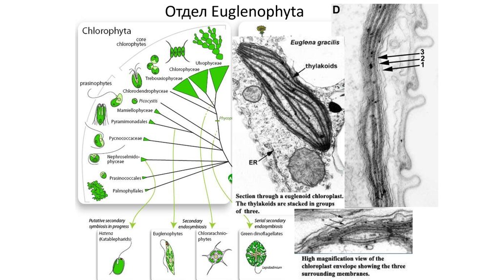
ОтделEuglenophyta
Отдел
Euglenophyta
27. Отдел Chlorarachniophyta
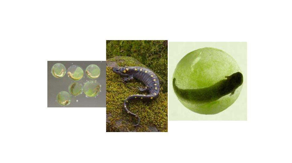
28. Hatena
29.
30.
31.
Fig. 1ERAD, SELMA, and protein import into complex plastids ofdiatoms. Entry into the (chloroplast) ER is host-derived (h): nucleusencoded preproteins are equipped with an N-terminal signal peptide
(SP) and enter the (c)ER cotranslationally through the Sec61 complex.
During translocation, the SP of ER-resident or secretory proteins and,
most likely, plastidal preproteins is cleaved off by the signal peptidase
complex (SPC). The majority of ER or secretory proteins are Nglycosylated in the (c)ER lumen by an oligosaccharyl transferase (OST),
folded into their native conformation via chaperones (hBiP) and are
further transported through vesicles (e.g., COPII) towards the
endomembrane system. The host machinery of the ERAD-L pathway
recognizes misfolded or defective proteins (asterisks) in the (c)ER
lumen, translocates them into the cytosol and polyubiquitinates the
substrates for proteasomal degradation. For detailed information on the
hostERAD-L system, see text. Passage of the second outermost
membrane (PPM) and subsequent transport steps are symbiont derived
(s): In addition to a SP, plastidal preproteins possess a transit peptidelike sequence (TPL) at their N-terminus, which is recognized in the cER
lumen by an as yet unknown factor. Preproteins cross the PPM through
the symbiont-specific ERAD-like machinery (SELMA), representing a
recycled symbiontic ERAD system installed for translocation of plastidal
preproteins. The ubiquitin-dependent translocation is supposed to be
mediated by the AAA-ATPase Cdc48 with its cofactors (see text). PPCresident proteins (+X) are processed by an as yet unidentified transit
peptide peptidase and folded in the PPC. In addition to the SELMA
system, an incomplete proteasomal s20S complex is present in the PPC
of diatoms, which is believed to be uncoupled from translocation.
Passage of the two inner membranes: stromal preproteins (+F) are
further transported across the two inner membranes of the complex
plastid via TOC- (ptOmp85) and TIC-related systems prior processing
and folding in the stroma. Note that vesicular traffic (not shown) may be
an alternative model for transport of stromal preproteins between PPM
and OEM. Question mark unknown function; cER chloroplast
endoplasmic reticulum;OEM/IEM outer/inner chloroplast envelope
membrane; IMS intermembrane space; TOC/TIC translocon at the
outer/inner chloroplast membrane
32. Отдел Cryptophyta
33.
Genome and proteome mosaicism in complex algae.John M. Archibald PNAS 2015;112:10147-10153
©2015 by National Academy of Sciences
34. Summary of known or estimated nucleomorph genome sizes in cryptophyte and chlorarachniophyte algae
Guilardia theta CCMP327551
Hanusia phi CCMP325
Proteomonas sulcata
~550
~570
Chroomonas sp. CCMP270
Chroomonas sp. SAG980
~815
~785
Ch. mesostigmatica CCMP1168
~805
Hemiselmis tepida CCMP443
~640
H.rufescens RCC659
~640
H.andersenii CCMP644
572
Cryptomonas erosa CCAP979/67
C.pyrenoidifera CCAP979/61
~655
~640
C.tetrapyrenoidosa CCAP979/63
~635
C.lundii CCAP979/69
~600
C.lucens CCAP979/52
~650
Cryptomonas sp. CCAP979/52
~640
C.ovata CCAC0064
~640
C.paramecium 9772a
~450
Rhodomonas salina X55032
R. salina CCMP 1170
~660
~715
R.salina CCMP1319
Rhodomonas sp. CCMP768
~755
~650
R.lens
Unindentified CCMP1178
~730
~845
Archibald, J. M. et al. J Hered 2009 100:582-590
35.
36.
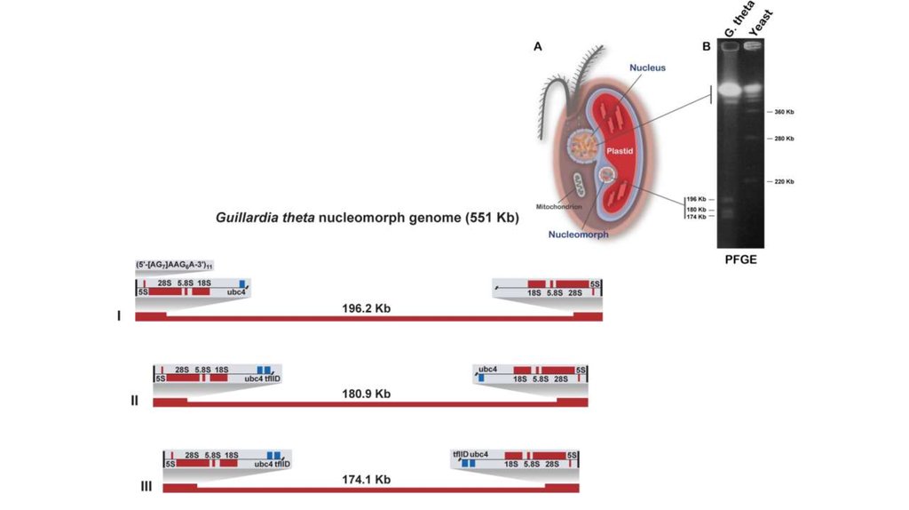
37. Характеристики геномов нуклеоморф
Genome characteristicsGuillardia theta
Hemiselmis
andersenii
Bigelowiella natans
Evolutionary origin
Genome size (bp)
Chromosome number/size
Chromosome structure
Red algae
551 264
3 (196.2, 180.9, 174.1 kbp)
Subtelomeric inverted
repeats including rDNA
genes
Red algae
571 872
3 (207.5, 184.7, 179.6 kbp)
Subtelomeric inverted
repeats, only 3 with
complete rDNAs
Green algae
372 870
3 (140.6, 134.1, 98.1 kbp)
Subtelomeric inverted repeats
including rDNA genes
Telomeric sequence/length
([AG]7AAG6A)11
(G[A]17)4–7
(TCTAGGG)25–45
Genomic A + T content
Inverted repeats
(including rDNA) (%)
Single-copy DNA (%)
~55
~60
~50
65–77
~ 75
>65
465
67
472
53
293
42
1
513
1.07 kb/gene
17 (42–52 bp)
30
1
525
1.09 kb/gene
None
30
5
340
1.10 kb/gene
852 (18–21 bp)
17
Number of genes
Protein genes
Non-mRNA (rRNA, tRNA,
snRNA, and snoRNA)
Pseudogenes
Total
Gene density
Introns and size range
Plastid genes
38.
39.
40.
41.
42.
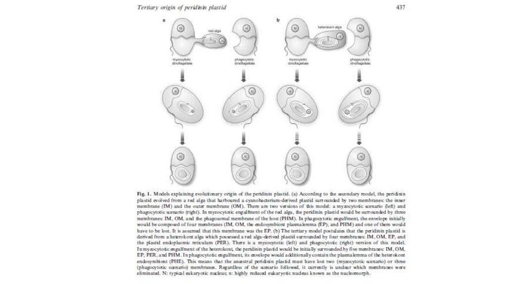
Наиболее часто встречающийся типпластид у динофитов – перединин
содержащие, окруженные тремя
мембранами. В этих пластидах
обнаружена форма II РуБисКо,
известная также для некоторых бактерий,
состоящая только из 2-х больших
субъединиц и кодирующаяся ядерным
геномом. У ряда динофитовых с такими
пластидами хлоропластный геном
сильно редуцирован (осталось менее 20
действующих генов) и пластидная ДНК
фрагментирована на отдельные
кольцевые фрагменты 2-3 кб, содержащие
по одному гену. Такая форма
хлоропластной ДНК и максимальная
передача хлоропластных генов в ядро уникальна для водорослей. В
хлоропластах встречаются пиреноиды
различной формы.
4. Пресноводные формы запасают
преимущественно крахмал,
откладываемый в цитоплазме, а морские
– липиды и стеролы.
43.
44.
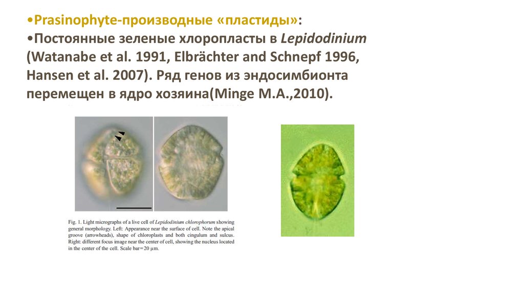
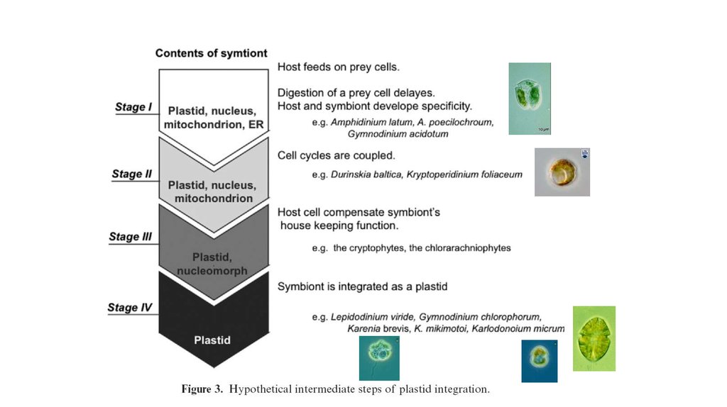
•Prasinophyte-производные «пластиды»:•Постоянные зеленые хлоропласты в Lepidodinium
(Watanabe et al. 1991, Elbrächter and Schnepf 1996,
Hansen et al. 2007). Ряд генов из эндосимбионта
перемещен в ядро хозяина(Minge M.A.,2010).
45.
•Haptophyte-произошедшие пластиды:•В этой ветви (Karenia, Karlodinium, Takayama) фотосинтезирующие
органеллы представляют настоящие пластиды – гены для многих белков,
участвующие в фотосинтезе, находятся в ядре (Ishida and Green 2002,
Patron et al. 2006, Nosenko et al. 2006). При таком типе возникновения
пластид, эукариота съела другую эукариоту, у которой пластида
произошла в результате вторичного эндосимбиоза. Такой тип
эндосимбиоза называется третичным. У Dinophysis mitra гаптофит
существует как клептопластида (Koike et al. 2005).
Karenia brevis
Karlodinium micrum
46.
•Dictyophyte-производные пластиды:Один вид динофлагеллят (Podolampas bipes) содержит постоянный
диктиохофициевый эндосимбионт с хлоропластами и ядром
(Schnepf and Elbrächter 1999, Schweikert and Elbrächter 2004).
47.
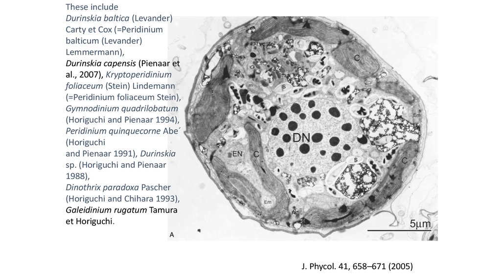
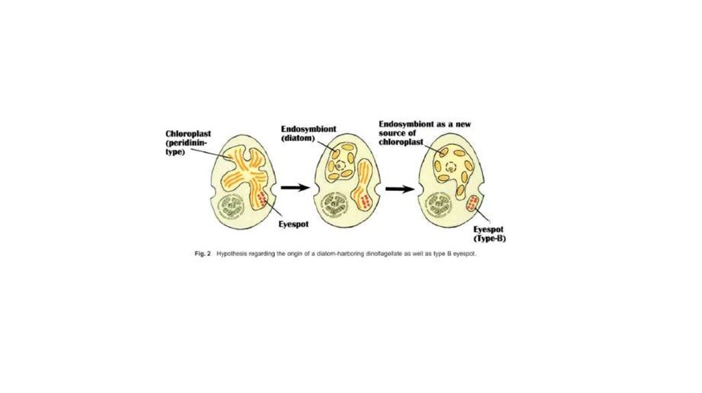
•Diatom-производные фотосинтезирующие эндосимбионты:
некоторые динофдагелляты (e.g., Durinskia baltica, Kryptoperidinium
foliaceum, Peridinium quinquecorne) постоянно содержат диатомовые
эндосимбионты с хлоропластами и ядром (e.g., Dodge 1971, Horiguchi
and Pienaar 1991, 1994, Schnepf and Elbrächter 1999). Такие водоросли
содержат 2 ядра. Диатомоморфные динофлагелляты культивируются,
более года поддерживаются в культуре. Одноядерные
нефотосинтезирующие представители некоторых из этих видов
встречаются в природе, указывая, что эндосимбиоз произошел
недавно.
Durinskia baltica
Peridinium quinquecorne
48.
49.
These includeDurinskia baltica (Levander)
Carty et Cox (=Peridinium
balticum (Levander)
Lemmermann),
Durinskia capensis (Pienaar et
al., 2007), Kryptoperidinium
foliaceum (Stein) Lindemann
(=Peridinium foliaceum Stein),
Gymnodinium quadrilobatum
(Horiguchi and Pienaar 1994),
Peridinium quinquecorne Abe´
(Horiguchi
and Pienaar 1991), Durinskia
sp. (Horiguchi and Pienaar
1988),
Dinothrix paradoxa Pascher
(Horiguchi and Chihara 1993),
Galeidinium rugatum Tamura
et Horiguchi.
J. Phycol. 41, 658–671 (2005)
50.
51.
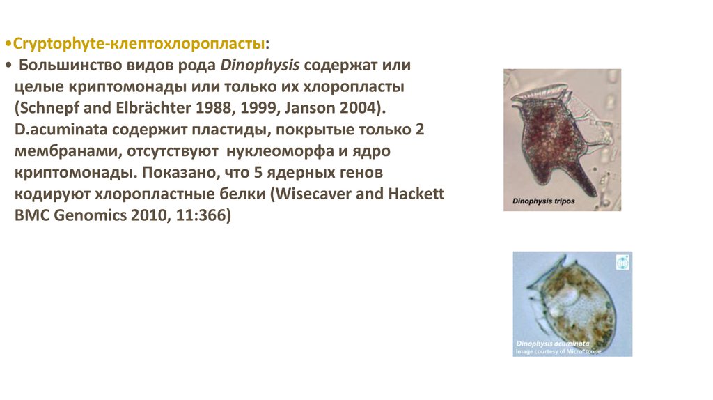
•Cryptophyte-клептохлоропласты:• Большинство видов рода Dinophysis содержат или
целые криптомонады или только их хлоропласты
(Schnepf and Elbrächter 1988, 1999, Janson 2004).
D.acuminata содержит пластиды, покрытые только 2
мембранами, отсутствуют нуклеоморфа и ядро
криптомонады. Показано, что 5 ядерных генов
кодируют хлоропластные белки (Wisecaver and Hackett
BMC Genomics 2010, 11:366)
52. Myrionecta rubra
53.
54.
55.
The relationship between Convoluta roscoffensisand Tetraselmis convolutae
Tetraselmis spp. in Symsagittifera (=Convoluta) spp.),
diatoms (Licmophora spp. in Convoluta convoluta
(Abildgaard 1806))
and dinoflagellates (Amphidinium spp. in Amphiscolops
spp.;e.g. Douglas, 1992; McCoy & Balzer, 2002; Venn
et al., 2008).
Symsagittifera roscoffensis
2–7.104 endosymbionts per animal
56.
57.
• Функционирующие клептопласты широко распространены средимоллюсков рода Elysia (менее 10 дней у E. hedgpethi (Greene, 1970), до 6
недель для E. (=Tridachia) crispata, E. (=Tridachiella) diomedea и Placobranchus
ianthobapsus) (R.Trench, 1969; Greene,1970), до нескольких месяцев (три у E.
viridis) (Hinde and Smith, 1972), 9 и более у E.chlorotica) (Pierce et al., 1996;
Rumpho et al., 2001;Mondy and Pierce, 2003).
• E.viridis - Codium fragile;
• Elysia timida - Acetabularia acetabulum;
• E. furvacauda - Codium и Microdictyon , красные водоросли и бурая
Sargassum
• E. crispata (R. Trench et al., 1969) и Е.diomedea (R. Trench,1975) - Caulerpa.
58.
59.
60.
Species of Costasiella investigated.Figure 1. Species of Costasiella investigated. A. C.
ocellifera (Florida Keys). B. C. nonatoi (Florida Keys). C.
C. kuroshimae (Guam). D. C. sp. 2 (Guam).
E. C. sp. 1 (Guam). F. Phylogenetic relationship of
Costasiella based on partial sequences of 16S, 1st and
2nd positions of COI, H3 and 28S. Shown is
a 50% majority-rule tree based on a Bayesian analysis.
Numbers at nodes represent posterior probabilities
(Bayesian analysis) and bootstrap values
(maximum-likelihood analysis). Siphonaria pectinata was
chosen as outgroup. Stars indicate food sources of
Costasiella species identified by barcoding
using rbcL: brown, Avrainvillea; red, Tydemania; orange,
Rhipilia; green, Pseudochlorodesmis; beige, Bryopsis.
Blue clade shows Costasiella with no functional
retention of kleptoplasts, green clade indicates species
with functional retention.
Christa G et al. J. Mollus. Stud. 2014;mollus.eyu026
© The Author 2014. Published by Oxford University Press on behalf of The Malacological Society
of London, all rights reserved
61.
62.
63.
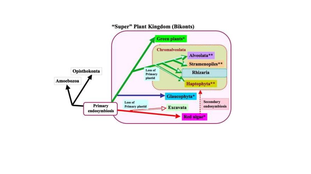
(a) Origin of complex algae with red plastids via a single secondary endosymbiosis with a redalga and successive tertiary and quaternary endosymbioses.
N: nucleus; M: mitochondrion; P: plastid. (b) Scenario of
plastid evolution among CASH lineages according to the
rhodoplex hypothesis. X-ray images of the Russian
Matryoshka dolls indicate independent events of plastid
endosymbioses. All CASH plastids originate from an
initial engulfment of a rhodophyte (see [a]), but the
genuine secondary endosymbiont and the order of
subsequent endosymbioses remains to be determined
(indicated by 2nd/3rd and 3rd/4th). The typical plastid of
PCD may represent a reduced apicomplexan alga (see
current study). The gain of rhodophycean plastids as well
as the loss of photosynthesis/plastids is indicated by the
red horizontal lines. With respect to stramenopiles, only a
subset of separate lineages is shown. Micrograph
courtesy of Peter Vontobel, Sven Gould, Woody
Hastings, and Manfred Rohde.
Petersen J et al. Genome Biol Evol 2014;6:666-684
© The Author(s) 2014. Published by Oxford University Press on behalf of the Society for
Molecular Biology and Evolution.