


 english
Similar presentations:


Initiation of translation in prokaryotes: initiation factors, initiator codons, 3'end of RNA small ribosomal subunit
1. Initiation of translation in prokaryotes: initiation factors, initiator codons, 3'end of RNA small ribosomal subunit and the
Initiation of translation inprokaryotes: initiation factors,
initiator codons, 3'end of RNA
small ribosomal subunit and the
Shine-Dalgarno sequence in mRNA»
Done by: Maulenova R.
Moldakozhayev A.
Naizabayeva D.
2.
In molecularbiology and genetic,
translation is the
process in
which ribosomes in
a cell's
cytoplasm create
proteins,
following transcription of DNA to
RNA in the
cell's nucleus. The
entire process is a
part of gene
expression.
3.
Translation proceeds in three phases:Initiation: The ribosome assembles around the target mRNA. The
first tRNA is attached at the start codon.
Elongation: The tRNA transfers an amino acid to the tRNA
corresponding to the next codon. The ribosome then moves
(translocates) to the next mRNA codon to continue the process, creating
an amino acid chain.
Termination: When a stop codon is reached, the ribosome releases
the polypeptide.
4.
In bacteria, translation occurs in the cytoplasm,where the large and small subunits of the ribosome bind to
the mRNA. In eukaryotes, translation occurs in
the cytosol or across the membrane of the endoplasmic
reticulum in a process called vectorial synthesis. In many
instances, the entire ribosome/mRNA complex binds to the
outer membrane of the rough endoplasmic reticulum (ER);
the newly created polypeptide is stored inside the ER for
later vesicle transport and secretion outside of the cell.
Many types of transcribed RNA, such as transfer
RNA, ribosomal RNA, and small nuclear RNA, do not
undergo translation into proteins.
5.
A number of antibiotics act by inhibitingtranslation. These include anisomycin, cycloheximide,
chloramphenicol, tetracycline, streptomycin,
erythromycin, and puromycin. Prokaryotic ribosomes
have a different structure from that of eukaryotic
ribosomes, and thus antibiotics can specifically
target bacterial infections without any harm to a
eukaryotic host's cells.
6.
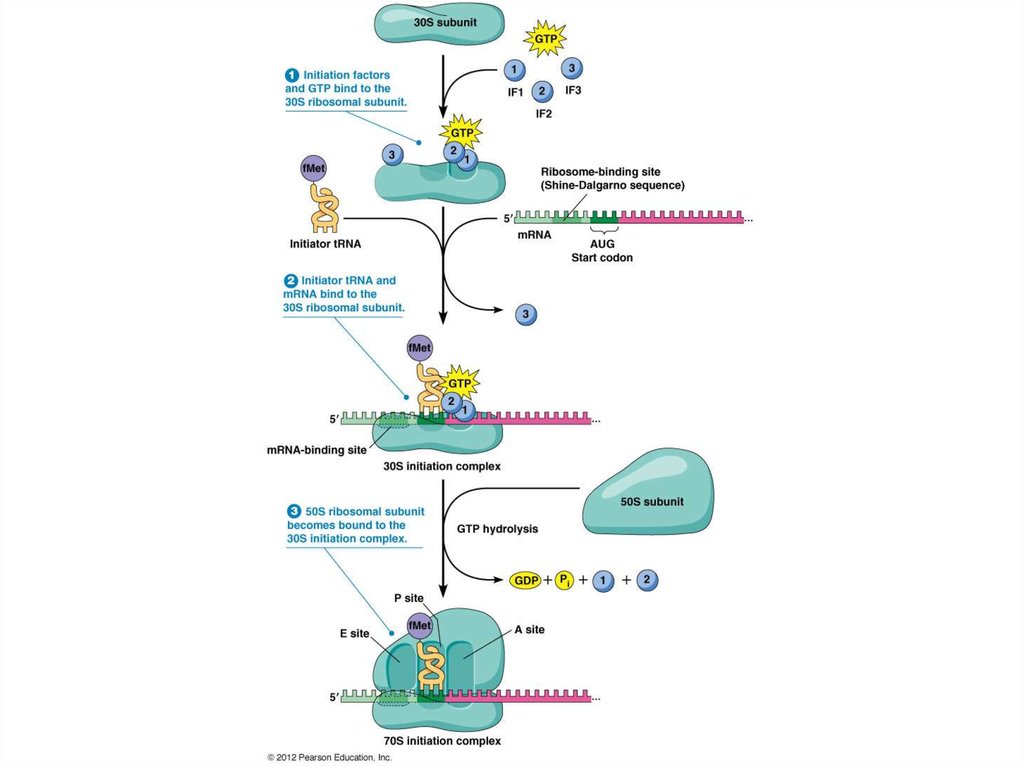
Translation initiation: Initiation factorsProkaryotes require the use of three initiation
factors: IF1, IF2, and IF3, for translation.
IF1 associates with the 30S ribosomal subunit in the
A site and prevents an aminoacyl-tRNA from entering. It
modulates IF2 binding to the ribosome by increasing its
affinity. It may also prevent the 50S subunit from binding,
stopping the formation of the 70S subunit. It also contains
a β-domain fold common for nucleic acid binding proteins.
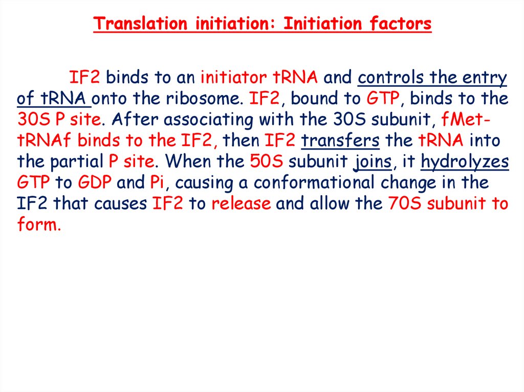
7.
Translation initiation: Initiation factorsIF2 binds to an initiator tRNA and controls the entry
of tRNA onto the ribosome. IF2, bound to GTP, binds to the
30S P site. After associating with the 30S subunit, fMettRNAf binds to the IF2, then IF2 transfers the tRNA into
the partial P site. When the 50S subunit joins, it hydrolyzes
GTP to GDP and Pi, causing a conformational change in the
IF2 that causes IF2 to release and allow the 70S subunit to
form.
8.
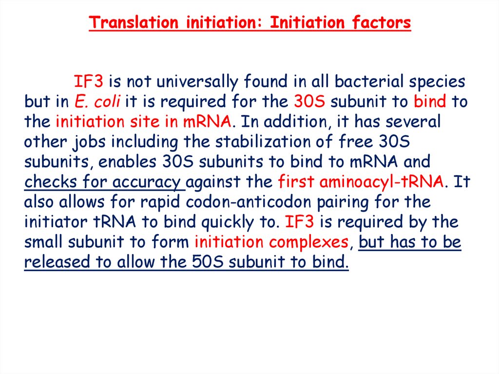
Translation initiation: Initiation factorsIF3 is not universally found in all bacterial species
but in E. coli it is required for the 30S subunit to bind to
the initiation site in mRNA. In addition, it has several
other jobs including the stabilization of free 30S
subunits, enables 30S subunits to bind to mRNA and
checks for accuracy against the first aminoacyl-tRNA. It
also allows for rapid codon-anticodon pairing for the
initiator tRNA to bind quickly to. IF3 is required by the
small subunit to form initiation complexes, but has to be
released to allow the 50S subunit to bind.
9.
10.
RibosomeThe fact that cells typically contain many ribosomes
reflects the central importance of protein synthesis in cell
metabolism. E. coli, for example, contain about 20,000
ribosomes, which account for approximately 25% of the dry
weight of the cell, and rapidly growing mammalian cells
contain about 10 million ribosomes.
11.
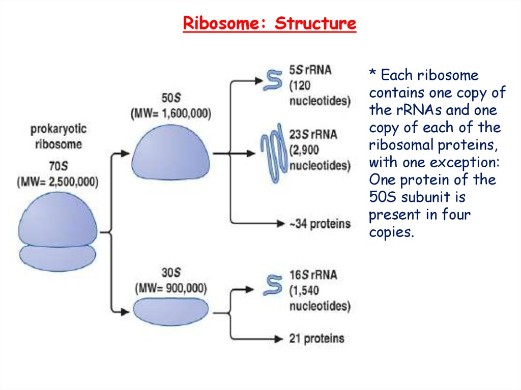
Ribosome: Structure* Each ribosome
contains one copy of
the rRNAs and one
copy of each of the
ribosomal proteins,
with one exception:
One protein of the
50S subunit is
present in four
copies.
12.
Ribosome: rRNAA noteworthy feature of ribosomes is that they can be
formed in vitro by self-assembly of their RNA and protein constituents.
As first described in 1968 by Masayasu Nomura, purified
ribosomal proteins and rRNAs can be mixed together and, under
appropriate conditions, will reform a functional ribosome.
Initially, rRNAs were thought to play a structural role, providing
a scaffold upon which ribosomal proteins assemble. However, with the
discovery of the catalytic activity of other RNA molecules, the possible
catalytic role of rRNA became widely considered. Consistent with this
hypothesis, rRNAs were found to be absolutely required for the in
vitro assembly of functional ribosomes.
13.
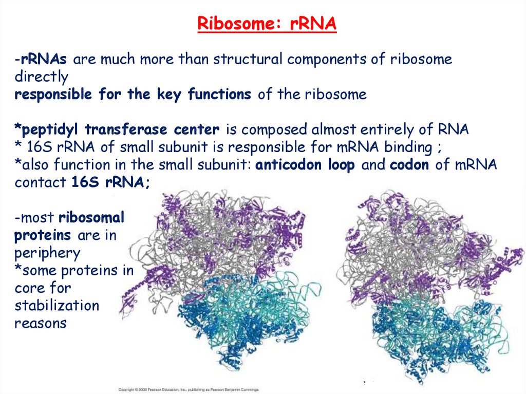
Ribosome: rRNA-rRNAs are much more than structural components of ribosome
directly
responsible for the key functions of the ribosome
*peptidyl transferase center is composed almost entirely of RNA
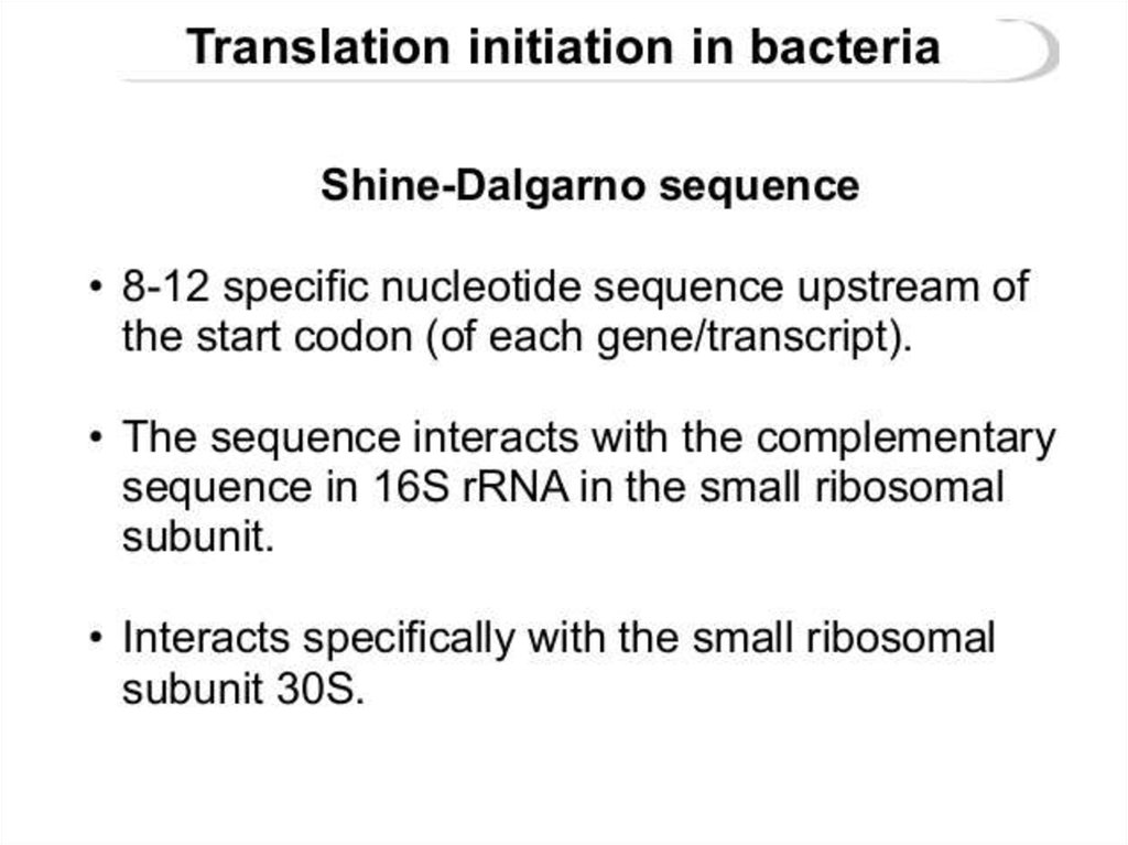
* 16S rRNA of small subunit is responsible for mRNA binding ;
*also function in the small subunit: anticodon loop and codon of mRNA
contact 16S rRNA;
-most ribosomal
proteins are in
periphery
*some proteins in
core for
stabilization
reasons
14.
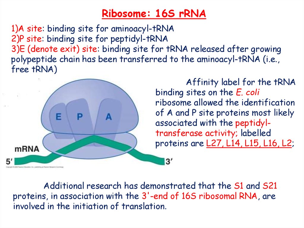
Ribosome: 16S rRNA1)A site: binding site for aminoacyl-tRNA
2)P site: binding site for peptidyl-tRNA
3)E (denote exit) site: binding site for tRNA released after growing
polypeptide chain has been transferred to the aminoacyl-tRNA (i.e.,
free tRNA)
Affinity label for the tRNA
binding sites on the E. coli
ribosome allowed the identification
of A and P site proteins most likely
associated with the peptidyltransferase activity; labelled
proteins are L27, L14, L15, L16, L2;
Additional research has demonstrated that the S1 and S21
proteins, in association with the 3'-end of 16S ribosomal RNA, are
involved in the initiation of translation.
15.
Ribosome: 16S rRNA(Zwieb & Brimacombe I979)
16.
Ribosome: 16S rRNAThe arrangement of the 16S rRNA creates a 5' domain, central
domain, 3' major domain, and a 3' minor domain.
The 5' domain consists of 19
double helices that makes up
the bulk of the body.
The central domain of the rRNA generates
the platform and is an elongated, curved
structure of nine helices, with the junction
of helices 20, 21, and 22 being at the
heart of it.
17.
Ribosome: 16S rRNAThe 3' major domain contains 15 helical
elements and composes the head.
The 3' minor domain contains 2
helices and projects from the subunit
to interact with the 50S subunit.
18.
19.
Ribosome: 3’ end of 16S rRNAFirst, the small subunit
associates with the mRNAby
base-pairing interaction
between RBS and 16S
rRNA
-the small subunit is
positioned on
mRNA such that the start
codon will be in the P site
when the large subunit
joins the complex
20.
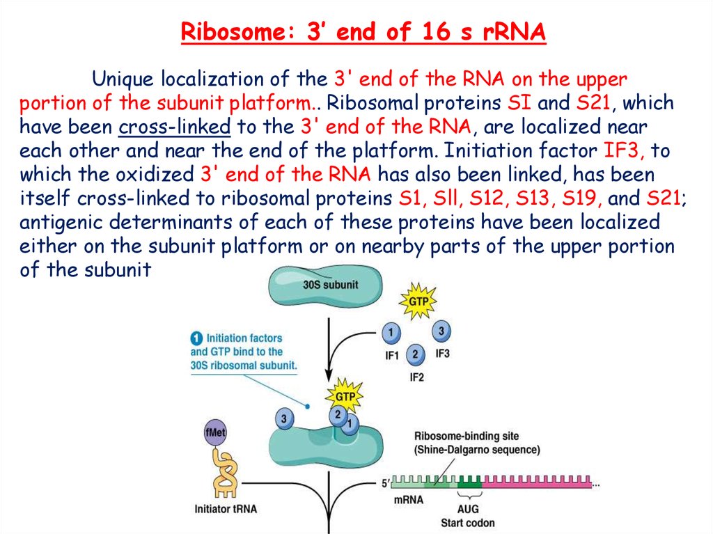
Ribosome: 3’ end of 16 s rRNAUnique localization of the 3' end of the RNA on the upper
portion of the subunit platform.. Ribosomal proteins SI and S21, which
have been cross-linked to the 3' end of the RNA, are localized near
each other and near the end of the platform. Initiation factor IF3, to
which the oxidized 3' end of the RNA has also been linked, has been
itself cross-linked to ribosomal proteins S1, Sll, S12, S13, S19, and S21;
antigenic determinants of each of these proteins have been localized
either on the subunit platform or on nearby parts of the upper portion
of the subunit
21.
Ribosome: 3’ end of 16 s rRNACross-linking studies have
shown that the nucleic acid-binding
domain of S1 is aligned with a region
of the mRNA upstream of the SD,
suggesting that S1 may interact with
5' parts of the Translation initiation
region. Consistent with this
observation, A/U-rich sequences in
front of the SD or downstream of
the initiator codon enhance protein
synthesis. Disruption of the E. coli
gene coding for S1 has been reported
to be lethal.
22.
Antibiotics affecting 16S rRNAColicin E3 (protein antibiotic from E.coli) makes a
single cut in the 16S rRNA of 70S ribosomes, these include the
loss of activity.
Pactamycin (Pct) was isolated from Streptomyces pactum
as a potential new human antitumor drug, but in fact a potent
inhibitor of translation in all three kingdoms, eukarya, bacteria,
and archaea (Bhuyan et al., 1961; Mankin, 1997). For this reason,
the drug is expected to interact with highly conserved regions of
16S RNA.
Streptomycin and spectinomycin are typical examples
which function by binding to specific sites on prokaryotic rRNA
and affecting the fidelity of protein synthesis. Binding of drug to
the 16S subunit near the A-site of the 30S subunit leads to a
decrease in translational accuracy and inhibition of the
translocation of the ribosome.
23. Shine-Dalgarno sequence
24.
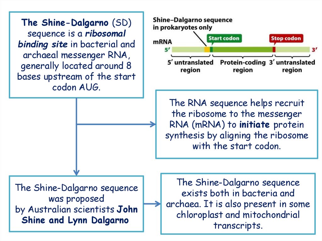
The Shine-Dalgarno (SD)sequence is a ribosomal
binding site in bacterial and
archaeal messenger RNA,
generally located around 8
bases upstream of the start
codon AUG.
The RNA sequence helps recruit
the ribosome to the messenger
RNA (mRNA) to initiate protein
synthesis by aligning the ribosome
with the start codon.
The Shine-Dalgarno sequence
was proposed
by Australian scientists John
Shine and Lynn Dalgarno
The Shine-Dalgarno sequence
exists both in bacteria and
archaea. It is also present in some
chloroplast and mitochondrial
transcripts.
25.
26.
27.
• several examples of the Shine-Dalgarno sequence28.
REFERENCE1. The Cell: A Molecular Approach. 2nd edition.Cooper GM. Sunderland
(MA): Sinauer Associates; 2000.
2. Watson J D, Baker T A , Bell S P, Gann A, Levine M, Losick R. Molecular Biology
of the Gene. 5th edition. Pearson education 2004.
3. J. E. KREBS, E.S. GOLDSTEIN, S.T. KILPATRICK. Lewin’s genes XI. Copyright©
2 0 1 4 by Jones & B artlett Learning, LLC, an Ascend Learning Company
4. Daniel H. Lackner et al., Translational Control of Gene Expression: From
Transcripts to Transcriptomes, International Review of Cell and Molecular
Biology, 2008, 271, 200-238.
5. The Cell: A Molecular Approach. 2nd edition.Cooper GM. Sunderland
(MA): Sinauer Associates; 2000.
6. Czernilofsky, A; Kurland, C.G.; Stöffler, G. (1975). "30S RIBOSOMALPROTEINS ASSOCIATED WITH 3'-TERMINUS OF 16S RNA". FEBS Letters.
ELSEVIER SCIENCE BV. 58 (1): 281–284.
7. H. G. WITTMANN . Structure and evolution of ribosomes . Proc. R. Soc. Lond. B
216, 117-135 (1982)
8. Zwieb, C. & Brimacombe, R. I979 Nucl. Acids Res. 6, 1775-1790.
9. HELEN MCKUSKIE OLSON* AND DOHN G. GLITZ. Ribosome structure:
Localization of 3' end of RNA in small subunit by immunoelectronmicroscopy.
Proc. Natl. Acad. Sci. USA. Vol. 76, No. 8, pp. 3769-3773, August 1979
10. Heinz-Giinter WITTMANN. Structure, Function and Evolution of Ribosomes.
Eur. J. Biochem. 61, 1 - 13 (1976)
29.
REFERENCE11. A. P. CZERNILOFSKY, C. G. KURLAND and G. STOFFLER. 30s RIBOSOMAL
PROTEINS ASSOCIATED WITH THE 3’-TERMINUS OF 16s RNA . FEBS
LETTERS Volume 58, number 1, October 1975 .
12. Vladimir Vimberg1, Age Tats2, Maido Remm2 and Tanel Tenson. Translation
initiation region sequence preferences in Escherichia coli. BMC Molecular
Biology 2007, 8:100
13. Weiling Hong, Jie Zeng, and Jianping Xie. Antibiotic drugs targeting
bacterial RNAs. Acta Pharm Sin B. 2014 Aug; 4(4): 258–265.
14. Ditlev E. Brodersen,* William M. Clemons, Jr.,*† Andrew P. Carter,* Robert J.
Morgan-Warren,* Brian T. Wimberly,* and V. Ramakrishnan*. The Structural
Basis for the Action of the Antibiotics Tetracycline, Pactamycin, and
Hygromycin B on the 30S Ribosomal Subunit. Cell, Vol. 103, 1143–1154,
December 22, 2000, Copyright 2000 by Cell Press.
15. Hale WG, Margham JP, Saunders VA, Collins Dictionary of Biology, (2nd ed)
Shine-Dalgarno (SD) sequence
Links:
1. http://www.biochem.umd.edu/biochem/kahn/bchm46501/ribosome/16SrRNA.
html
2. http://rna.ucsc.edu/rnacenter/ribosome_images.html
3. https://en.wikipedia.org/wiki/Shine-Dalgarno_sequence
4. thermofisher.com/Ribosomal Binding Site Sequence Requirements