


 medicine
Similar presentations:


")


Traumatic injuries of kidneys, ureter, bladder
1. SEMEY STATE MEDICAL UNIVERSITY
Department of visual diagnosticsSIW
Discipline: Visual diagnostics
Theme: «Traumatic injuries of kidneys, ureter, bladder»
Prepared: Amir D.N. 343 GM
Checked: Madieva M.R.
Semey
2018
2.
Closed kidney damage- Damage to the fat and fibrous capsules
with the formation of a hematoma in
perirenal cellulose
- Breaks of the parenchyma of the kidney,
not penetrating into the renal cups and the
pelvis
- Tears of the parenchyma of the kidney,
penetrating into the renal cups and the
pelvis
- Crushing a kidney
- The separation of the kidney from the
vessels and ureter
3.
Mechanism of closed kidney damageCauses:
Blunt blunt objects
Shaking
Pressure
The degree of damage depends on:
Forces and directions of impact, places of its application
Anatomical location of the kidney
Topographic relation to XI and XII edges, spine
Development of musculature, fat and perirenal fiber
The degree of filling the intestines
Values of intra-abdominal and retroperitoneal pressure
Hydrodynamic pressure inside the kidney (urine, blood)
If there are pathological changes in the kidney that precede the trauma
(hydronephrosis, pyonephrosis, kidney abnormalities, chronic pyelonephritis),
kidney damage occurs with minor strokes - the so-called spontaneous rupture
of the kidney.
4.
Open kidney damageBy the type of the hurting projectile:
•firearms (bullet, shrapnel, explosive);
•non-fireable
In the course of the wound channel:
•the blind
•through;
•tangents.
By the nature of the damage:
•injury;
•wound;
•crush kidney;
•injury to the vascular pedicle.
Is accompanied by shock, bleeding, phlegmon, peritonitis
5.
Iatrogenic exposureRetrograde pyelography
Puncture
Shockwave remote lithotripsy
6.
Clinical manifestationsLumbar pain
Hematuria
Dysuria
Symptoms of peritoneal irritation
Nausea
Vomiting
Fever
Gastrointestinal dysfunction
Swelling
7.
Three degrees of severity1. Mild kidney injury - the general condition of the victim is poorly impaired, there are
moderate pains in the lumbar region, short-term minor micro- or gross hematuria,
pararenal hematoma is absent, no signs of peritoneal irritation. This type of damage is
referred to as kidney contusion.
2. Medium-grade kidney injury - the general condition from a satisfactory quickly
becomes a moderate severity state (pulse quickens, blood pressure decreases),
hematuria is pronounced and can continue to increase. The accumulation of blood in
the bladder can cause dysuria (urinary disturbance), up to a complete retention of urine.
Under the skin in the area of injury, in some patients, a hematoma is clearly visible. The
pain is insignificant and often radiates to the lower abdomen, groin and genitals.
Obstruction of the ureter by blood clots can lead to the development of renal colic. The
urogematoma may lead to the development of symptoms of peritoneal irritation.
3. Severe kidney injury - collapse and shock come to the fore, severe pain in the lumbar
region on the affected side, profuse and prolonged gross hematuria. Urogematoma and
signs of internal bleeding tend to increase
8.
DiagnosticsOn examination:
Hematoma, swelling in the lumbar region
Local muscle tension
Rib fractures
Paleness of the skin
Rachiocampsis
AS / BH (hematocrit, hemoglobin)
OAM (hematuria)
CT scan with contrast enhancement (mandatory in the presence of
hematuria)
MRI
Ultrasound (fluid in the abdominal cavity)
Excretory urography
Renal Angiography
Survey urography
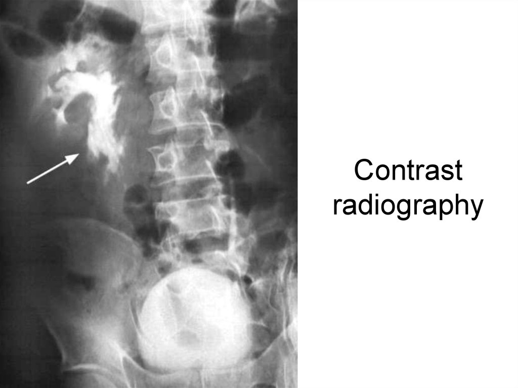
9.
Contrastradiography
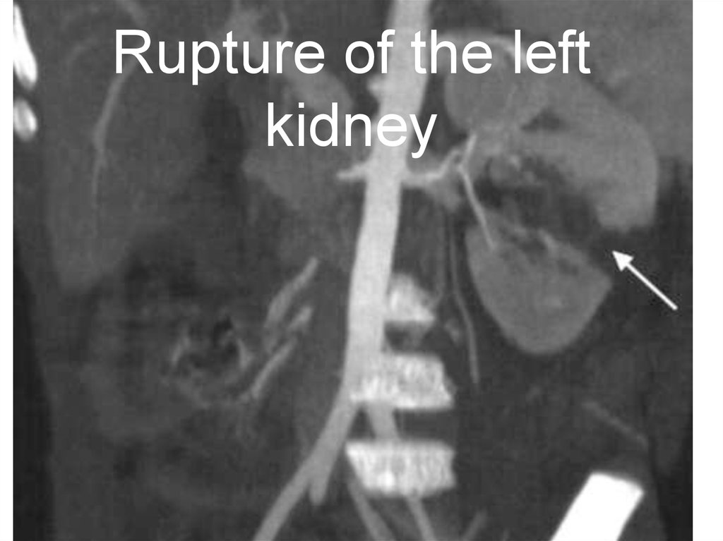
10.
Rupture of the leftkidney
11.
TreatmentStopping bleeding
Bed rest 10-15 days
Control of hemodynamics and hematocrit
Preventive parenteral administration of antibiotics and uroantiseptics
Analgesics
Surgical treatment:
An organ-preserving operation (nephro / pyelostomy) is performed
with removal of the urohematoma, perirenal hematoma of the prva, resection of a part of the kidney with impaired blood circulation,
closure of gaps, drainage of the retroperitoneal space.
Nephrectomy is performed at breaks, tears of the kidney, provided
that the second kidney is functionally active.
12.
Damage of the uretersUreters are rarely damaged due to elasticity,
displaceability and location.
Iatrogenic damage
More often closed damage
Ureteroscopy
Cystoscopy
Ureteral stent
Bladder catheterization
During operations on the pelvic organs, large intestine,
external ileal vessels, lymphadenectomy and suturing of the
posterior leaflet of the parietal peritoneum, in gynecology.
13.
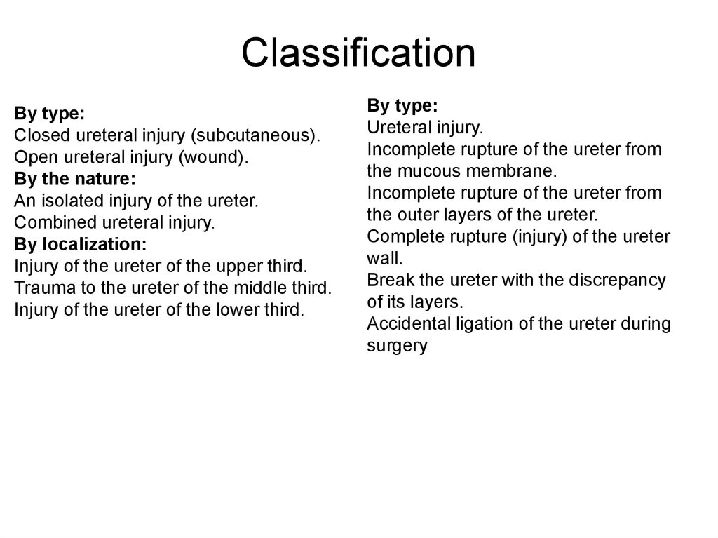
ClassificationBy type:
Closed ureteral injury (subcutaneous).
Open ureteral injury (wound).
By the nature:
An isolated injury of the ureter.
Combined ureteral injury.
By localization:
Injury of the ureter of the upper third.
Trauma to the ureter of the middle third.
Injury of the ureter of the lower third.
By type:
Ureteral injury.
Incomplete rupture of the ureter from
the mucous membrane.
Incomplete rupture of the ureter from
the outer layers of the ureter.
Complete rupture (injury) of the ureter
wall.
Break the ureter with the discrepancy
of its layers.
Accidental ligation of the ureter during
surgery
14.
DiagnosticsDiagnosis is based on an analysis of the circumstances and mechanism of
injury, clinical manifestations and data of special research methods. Diagnostics
includes 3 stages:
Clinical: localization of the wound, direction of the wound channel, evaluation of
urine and wound discharge, clinical manifestations - should suggest the
possibility of ureteral injury.
Instrumental: ultrasound of the abdominal and retroperitoneal space; general
radiography; excretory urography; infusion urography with the implementation of
deferred urogramm (if indicated); retrograde pyeloureterography, computed
tomography. The severity of the patient's condition may contraindicate to some
instrumental method of examination.
Operative - the most accurate method for diagnosing damage to the ureter.
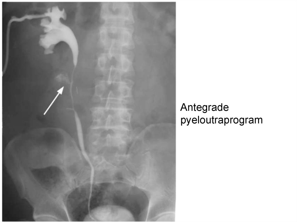
15.
Antegradepyeloutraprogram
16.
Differential diagnosticsTo distinguish between injuries of the ureter and bladder,
use the method of filling the bladder with a colored fluid
(methylene blue, indigo carmine). If the bladder is
damaged, the colored fluid is released from the urinary
fistula; in case of damage to the ureter, unpainted urine is
still excreted from the fistula.
Treatment
Nephrostomy or ureteral stenting with mandatory bladder
keterization
17.
Bladder damageCauses: blunt or penetrating injury leading to
rupture
Mechanism
•Blunt blow to full bladder;
•Iatrogenic damage (cystoscopy, endoscopy,
catheterization)
•Catatrauma
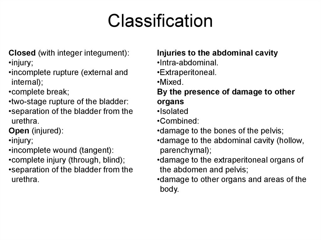
18.
ClassificationClosed (with integer integument):
•injury;
•incomplete rupture (external and
internal);
•complete break;
•two-stage rupture of the bladder:
•separation of the bladder from the
urethra.
Open (injured):
•injury;
•incomplete wound (tangent):
•complete injury (through, blind);
•separation of the bladder from the
urethra.
Injuries to the abdominal cavity
•Intra-abdominal.
•Extraperitoneal.
•Mixed.
By the presence of damage to other
organs
•Isolated
•Combined:
•damage to the bones of the pelvis;
•damage to the abdominal cavity (hollow,
parenchymal);
•damage to the extraperitoneal organs of
the abdomen and pelvis;
•damage to other organs and areas of the
body.
19.
Clinical manifestationsIntraperitoneal
Pain over pubis
Anuria
Signs of peritonitis
Bloating
Symptom "Vanka-Vstanki"
Extraperitoneal
Pain over the bosom and pelvis
Hematuria
State of shock
Frequent false and painful urge to
urinate
The appearance of swelling of the
skin in the suprapubic area
Increasing intoxication
20.
DiagnosticsCatheterization
Zeldovich positive symptom (inconsistency between the injected and exiting
fluid from the catheter)
AS / OAM
Overview of the pelvic region
Retrograde cystography with the introduction of at least 250 ml of contrast
media
Ultrasound
CT
MRI
21.
Retrograde cystogram.Extraperitoneal bladder rupture
22.
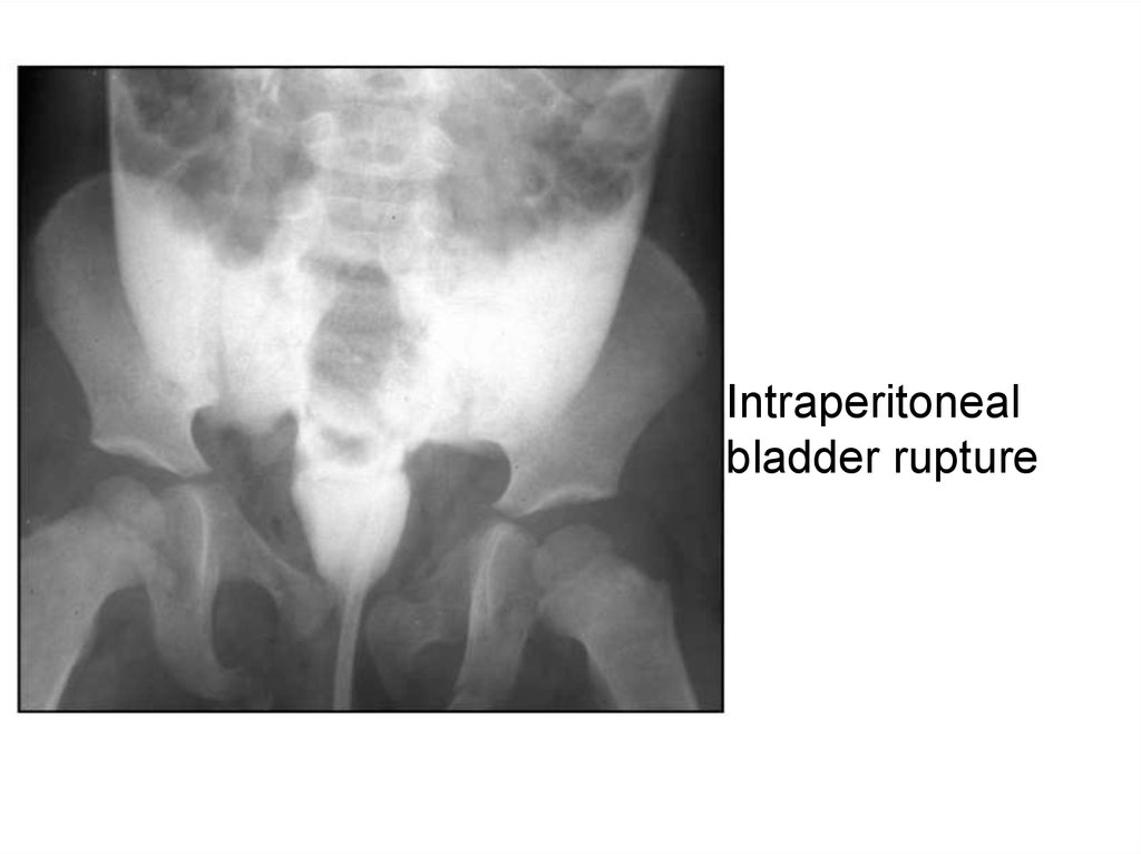
Intraperitonealbladder rupture
23.
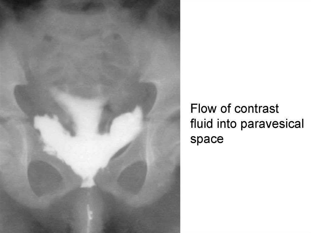
Flow of contrastfluid into paravesical
space
24.
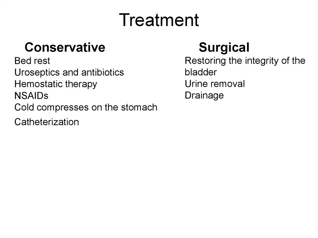
TreatmentConservative
Bed rest
Uroseptics and antibiotics
Hemostatic therapy
NSAIDs
Cold compresses on the stomach
Catheterization
Surgical
Restoring the integrity of the
bladder
Urine removal
Drainage
25.
Drainage by Buyalsky-McWorthier26.
LiteratureGrattan-Smith JD. MR urography: anatomy and
physiology. Pediatr Radiol. 2008;
McAninch J. Renal injuries. In: Gillenwater J,
Grayhack J, Howards S, Duckett J, editors. Adult and
pediatric urologyMosby. Mo: St Louis; 1996. pp. 539–
553.
Cerwinka WH, Damien Grattan-Smith J, Kirsch AJ
(2008) Magnetic resonance urography in pediatric
urology. J Pediatr Urol 4:74–82, quiz 82-83
Wikipedia
https://images.google.com/