


")


 integrates multiple cell-signaling pathways")


 internet
Similar presentations:


")

Cell Communication
1. Chapter 11
Cell CommunicationPowerPoint® Lecture Presentations for
Biology
Eighth Edition
Neil Campbell and Jane Reece
Lectures by Chris Romero, updated by Erin Barley with contributions from Joan Sharp
Copyright © 2008 Pearson Education, Inc., publishing as Pearson Benjamin Cummings
2. Overview: The Cellular Internet
• Cell-to-cell communication is essential formulticellular organisms
• Biologists have discovered some universal
mechanisms of cellular regulation
• The combined effects of multiple signals
determine cell response
• For example, the dilation of blood vessels is
controlled by multiple molecules
Copyright © 2008 Pearson Education, Inc., publishing as Pearson Benjamin Cummings
3.
Fig. 11-14. Concept 11.1: External signals are converted to responses within the cell
• Microbes are a window on the role of cellsignaling in the evolution of life
Copyright © 2008 Pearson Education, Inc., publishing as Pearson Benjamin Cummings
5. Evolution of Cell Signaling
• A signal transduction pathway is a seriesof steps by which a signal on a cell’s surface
is converted into a specific cellular response
• Signal transduction pathways convert signals
on a cell’s surface into cellular responses
Copyright © 2008 Pearson Education, Inc., publishing as Pearson Benjamin Cummings
6.
Fig. 11-2factor
Receptor
1
Exchange
of mating
factors
a
a factor
Yeast cell,
mating type a
2
Mating
3
New a/
cell
Yeast cell,
mating type
a
a/
7.
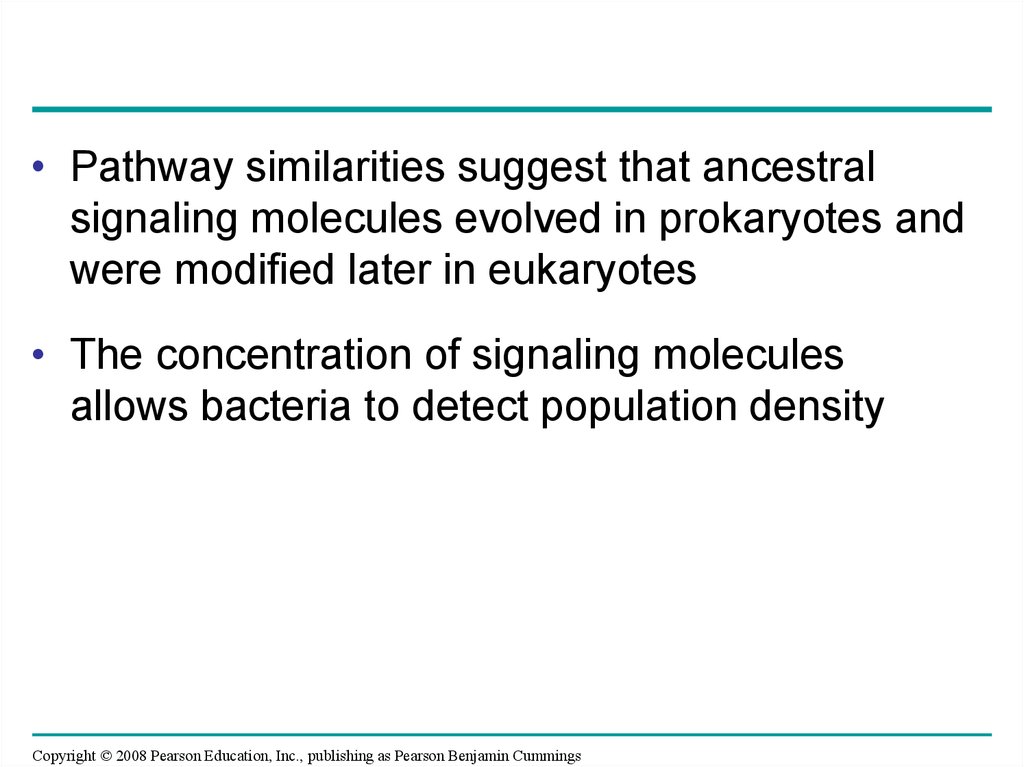
• Pathway similarities suggest that ancestralsignaling molecules evolved in prokaryotes and
were modified later in eukaryotes
• The concentration of signaling molecules
allows bacteria to detect population density
Copyright © 2008 Pearson Education, Inc., publishing as Pearson Benjamin Cummings
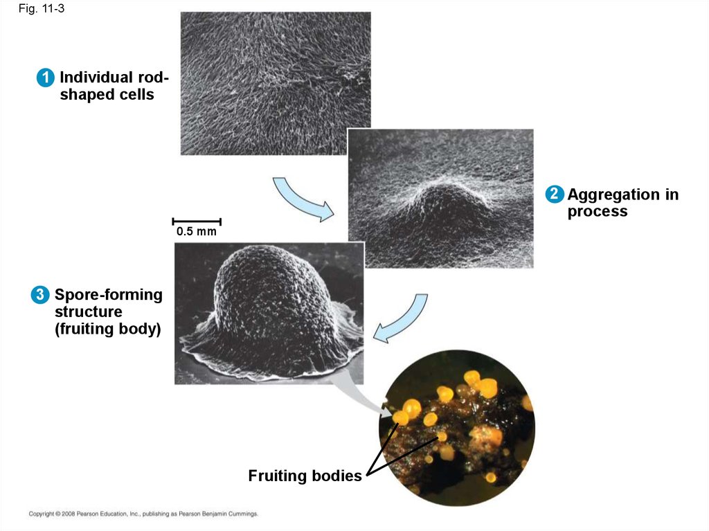
8.
Fig. 11-31 Individual rodshaped cells
2 Aggregation in
process
0.5 mm
3 Spore-forming
structure
(fruiting body)
Fruiting bodies
9. Local and Long-Distance Signaling
• Cells in a multicellular organism communicateby chemical messengers
• Animal and plant cells have cell junctions that
directly connect the cytoplasm of adjacent cells
• In local signaling, animal cells may
communicate by direct contact, or cell-cell
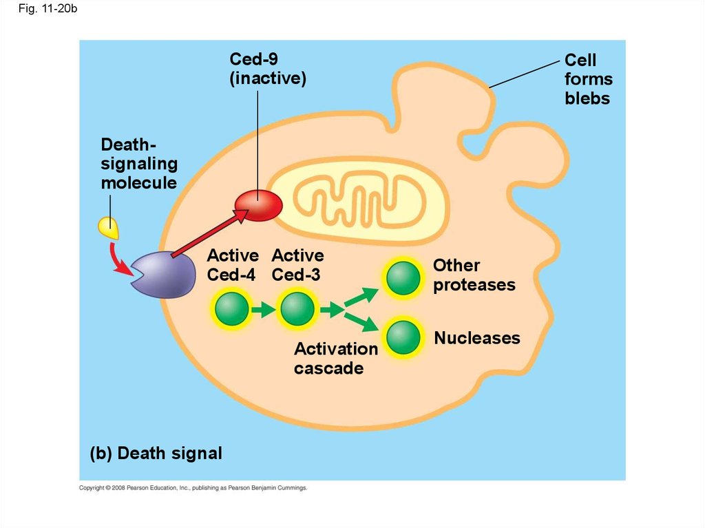
recognition
Copyright © 2008 Pearson Education, Inc., publishing as Pearson Benjamin Cummings
10.
Fig. 11-4Plasma membranes
Gap junctions
between animal cells
(a) Cell junctions
(b) Cell-cell recognition
Plasmodesmata
between plant cells
11.
• In many other cases, animal cells communicateusing local regulators, messenger molecules
that travel only short distances
• In long-distance signaling, plants and animals
use chemicals called hormones
Copyright © 2008 Pearson Education, Inc., publishing as Pearson Benjamin Cummings
12.
Fig. 11-5Long-distance signaling
Local signaling
Electrical signal
along nerve cell
triggers release of
neurotransmitter
Target cell
Secreting
cell
Local regulator
diffuses through
extracellular fluid
(a) Paracrine signaling
Endocrine cell
Neurotransmitter
diffuses across
synapse
Secretory
vesicle
Target cell
is stimulated
Blood
vessel
Hormone travels
in bloodstream
to target cells
Target
cell
(b) Synaptic signaling
(c) Hormonal signaling
13.
Fig. 11-5abLocal signaling
Electrical signal
along nerve cell
triggers release of
neurotransmitter
Target cell
Secreting
cell
Local regulator
diffuses through
extracellular fluid
(a) Paracrine signaling
Neurotransmitter
diffuses across
synapse
Secretory
vesicle
Target cell
is stimulated
(b) Synaptic signaling
14.
Fig. 11-5cLong-distance signaling
Endocrine cell
Blood
vessel
Hormone travels
in bloodstream
to target cells
Target
cell
(c) Hormonal signaling
15. The Three Stages of Cell Signaling: A Preview
• Earl W. Sutherland discovered how thehormone epinephrine acts on cells
• Sutherland suggested that cells receiving
signals went through three processes:
– Reception
– Transduction
– Response
Animation: Overview of Cell Signaling
Copyright © 2008 Pearson Education, Inc., publishing as Pearson Benjamin Cummings
16.
Fig. 11-6-1EXTRACELLULAR
FLUID
1 Reception
Receptor
Signaling
molecule
CYTOPLASM
Plasma membrane
17.
Fig. 11-6-2CYTOPLASM
EXTRACELLULAR
FLUID
Plasma membrane
1 Reception
2 Transduction
Receptor
Relay molecules in a signal transduction pathway
Signaling
molecule
18.
Fig. 11-6-3CYTOPLASM
EXTRACELLULAR
FLUID
Plasma membrane
1 Reception
2 Transduction
3 Response
Receptor
Activation
of cellular
response
Relay molecules in a signal transduction pathway
Signaling
molecule
19. Concept 11.2: Reception: A signal molecule binds to a receptor protein, causing it to change shape
• The binding between a signal molecule(ligand) and receptor is highly specific
• A shape change in a receptor is often the initial
transduction of the signal
• Most signal receptors are plasma membrane
proteins
Copyright © 2008 Pearson Education, Inc., publishing as Pearson Benjamin Cummings
20. Receptors in the Plasma Membrane
• Most water-soluble signal molecules bind tospecific sites on receptor proteins in the
plasma membrane
• There are three main types of membrane
receptors:
– G protein-coupled receptors
– Receptor tyrosine kinases
– Ion channel receptors
Copyright © 2008 Pearson Education, Inc., publishing as Pearson Benjamin Cummings
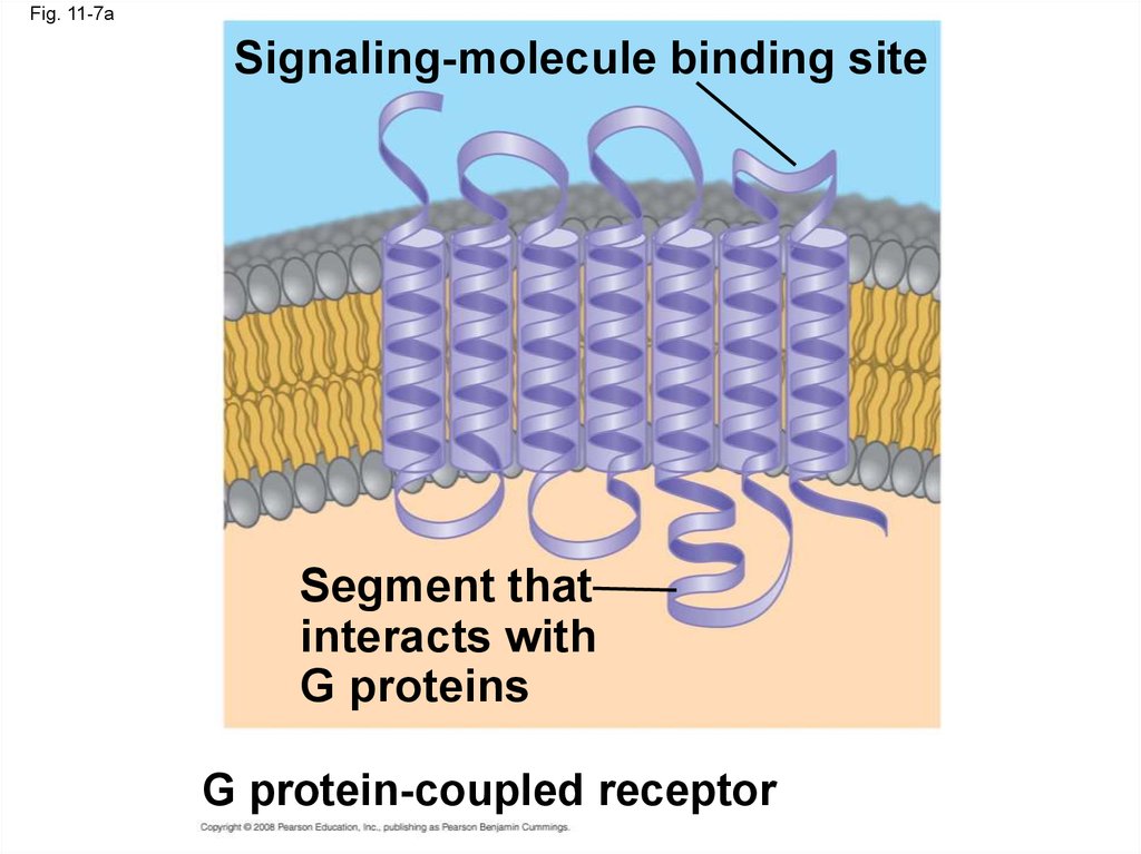
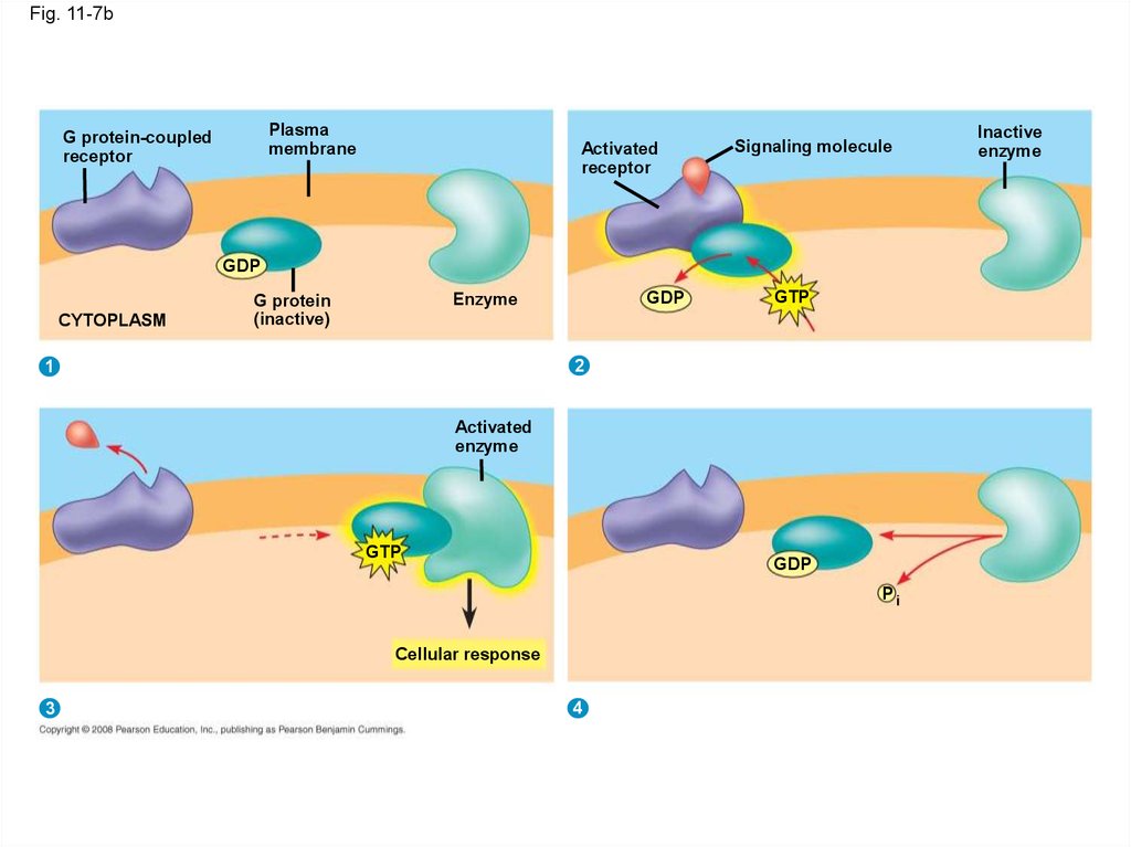
21.
• A G protein-coupled receptor is a plasmamembrane receptor that works with the help of
a G protein
• The G protein acts as an on/off switch: If GDP
is bound to the G protein, the G protein is
inactive
Copyright © 2008 Pearson Education, Inc., publishing as Pearson Benjamin Cummings
22.
Fig. 11-7aSignaling-molecule binding site
Segment that
interacts with
G proteins
G protein-coupled receptor
23.
Fig. 11-7bPlasma
membrane
G protein-coupled
receptor
Activated
receptor
Signaling molecule
GDP
CYTOPLASM
GDP
Enzyme
G protein
(inactive)
GTP
2
1
Activated
enzyme
GTP
GDP
Pi
Cellular response
3
4
Inactive
enzyme
24.
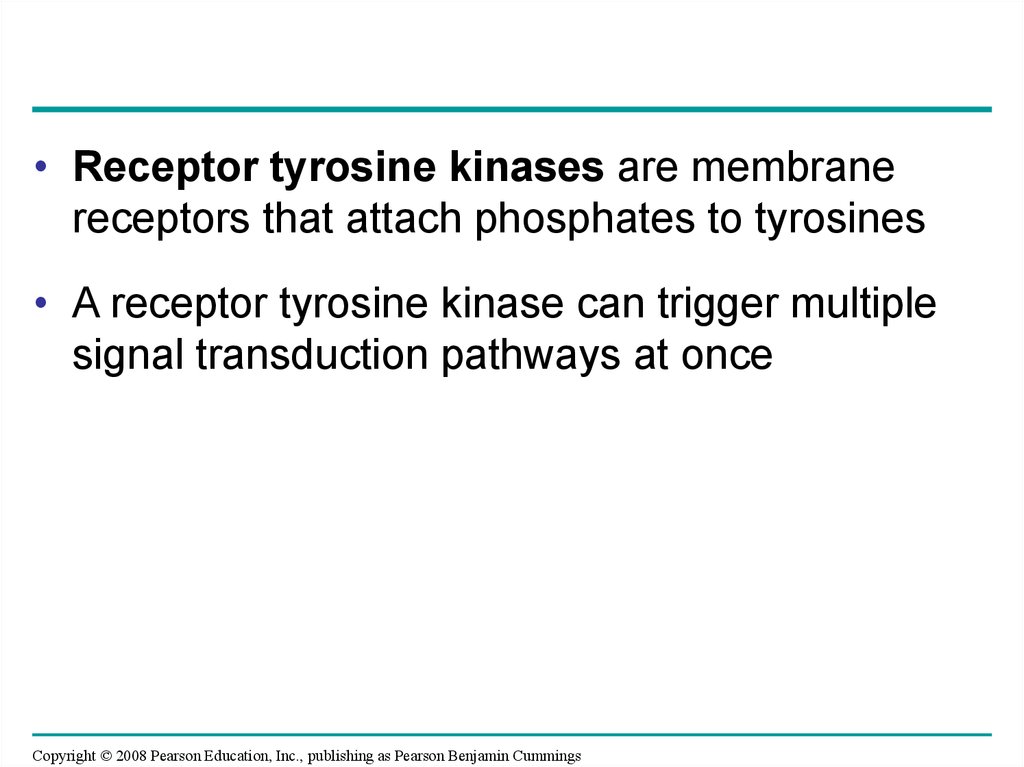
• Receptor tyrosine kinases are membranereceptors that attach phosphates to tyrosines
• A receptor tyrosine kinase can trigger multiple
signal transduction pathways at once
Copyright © 2008 Pearson Education, Inc., publishing as Pearson Benjamin Cummings
25.
Fig. 11-7cLigand-binding site
Signaling
molecule (ligand)
Signaling
molecule
Helix
Tyrosines
Tyr
Tyr
Tyr
Tyr
Tyr
Tyr
Tyr
Tyr
Tyr
Tyr
Tyr
Tyr
Tyr
Tyr
Tyr
Tyr
Tyr
Tyr
Receptor tyrosine
kinase proteins
CYTOPLASM
Dimer
1
2
Activated relay
proteins
Tyr
Tyr
Tyr
Tyr
P Tyr
P Tyr
Tyr
Tyr
P
6 ATP
Activated tyrosine
kinase regions
6 ADP
Tyr
Tyr
P Tyr
Tyr
P
Tyr
P Tyr
P Tyr
Tyr
P
P
P
P
Tyr P
Tyr
Fully activated receptor
tyrosine kinase
Inactive
relay proteins
3
4
Cellular
response 1
Cellular
response 2
26.
• A ligand-gated ion channel receptor acts as agate when the receptor changes shape
• When a signal molecule binds as a ligand to
the receptor, the gate allows specific ions, such
as Na+ or Ca2+, through a channel in the
receptor
Copyright © 2008 Pearson Education, Inc., publishing as Pearson Benjamin Cummings
27.
Fig. 11-7d1 Signaling
molecule
(ligand)
Gate
closed
Ligand-gated
ion channel receptor
2
Ions
Plasma
membrane
Gate open
Cellular
response
3
Gate closed
28. Intracellular Receptors
• Some receptor proteins are intracellular, foundin the cytosol or nucleus of target cells
• Small or hydrophobic chemical messengers
can readily cross the membrane and activate
receptors
• Examples of hydrophobic messengers are the
steroid and thyroid hormones of animals
• An activated hormone-receptor complex can
act as a transcription factor, turning on specific
genes
Copyright © 2008 Pearson Education, Inc., publishing as Pearson Benjamin Cummings
29.
Fig. 11-8-1Hormone
(testosterone)
EXTRACELLULAR
FLUID
Plasma
membrane
Receptor
protein
DNA
NUCLEUS
CYTOPLASM
30.
Fig. 11-8-2Hormone
(testosterone)
EXTRACELLULAR
FLUID
Plasma
membrane
Receptor
protein
Hormonereceptor
complex
DNA
NUCLEUS
CYTOPLASM
31.
Fig. 11-8-3Hormone
(testosterone)
EXTRACELLULAR
FLUID
Plasma
membrane
Receptor
protein
Hormonereceptor
complex
DNA
NUCLEUS
CYTOPLASM
32.
Fig. 11-8-4Hormone
(testosterone)
EXTRACELLULAR
FLUID
Plasma
membrane
Receptor
protein
Hormonereceptor
complex
DNA
mRNA
NUCLEUS
CYTOPLASM
33.
Fig. 11-8-5Hormone
(testosterone)
EXTRACELLULAR
FLUID
Plasma
membrane
Receptor
protein
Hormonereceptor
complex
DNA
mRNA
NUCLEUS
CYTOPLASM
New protein
34. Concept 11.3: Transduction: Cascades of molecular interactions relay signals from receptors to target molecules in the cell
• Signal transduction usually involves multiplesteps
• Multistep pathways can amplify a signal: A few
molecules can produce a large cellular
response
• Multistep pathways provide more opportunities
for coordination and regulation of the cellular
response
Copyright © 2008 Pearson Education, Inc., publishing as Pearson Benjamin Cummings
35. Signal Transduction Pathways
• The molecules that relay a signal from receptorto response are mostly proteins
• Like falling dominoes, the receptor activates
another protein, which activates another, and
so on, until the protein producing the response
is activated
• At each step, the signal is transduced into a
different form, usually a shape change in a
protein
Copyright © 2008 Pearson Education, Inc., publishing as Pearson Benjamin Cummings
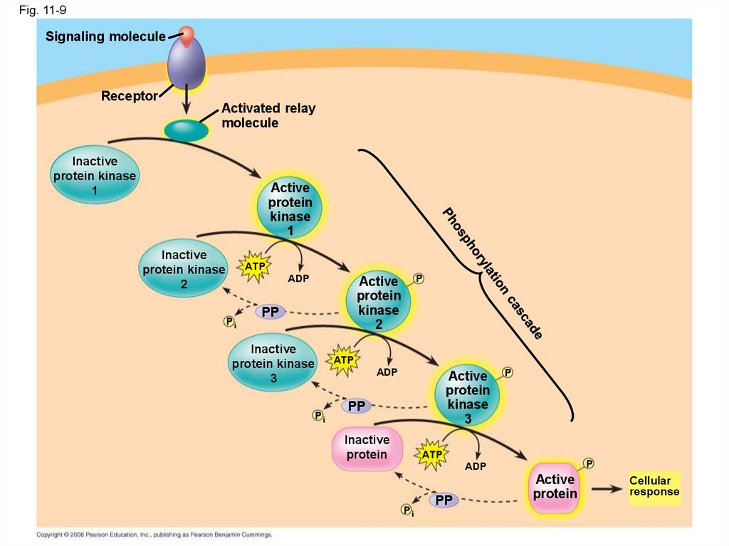
36. Protein Phosphorylation and Dephosphorylation
• In many pathways, the signal is transmitted bya cascade of protein phosphorylations
• Protein kinases transfer phosphates from ATP
to protein, a process called phosphorylation
Copyright © 2008 Pearson Education, Inc., publishing as Pearson Benjamin Cummings
37.
• Protein phosphatases remove thephosphates from proteins, a process called
dephosphorylation
• This phosphorylation and dephosphorylation
system acts as a molecular switch, turning
activities on and off
Copyright © 2008 Pearson Education, Inc., publishing as Pearson Benjamin Cummings
38.
Fig. 11-9Signaling molecule
Receptor
Activated relay
molecule
Inactive
protein kinase
1
Active
protein
kinase
1
Inactive
protein kinase
2
ATP
ADP
Pi
P
Active
protein
kinase
2
PP
Inactive
protein kinase
3
Pi
ATP
ADP
Active
protein
kinase
3
PP
Inactive
protein
P
ATP
P
ADP
Pi
PP
Active
protein
Cellular
response
39. Small Molecules and Ions as Second Messengers
• The extracellular signal molecule that binds tothe receptor is a pathway’s “first messenger”
• Second messengers are small, nonprotein,
water-soluble molecules or ions that spread
throughout a cell by diffusion
• Second messengers participate in pathways
initiated by G protein-coupled receptors and
receptor tyrosine kinases
• Cyclic AMP and calcium ions are common
second messengers
Copyright © 2008 Pearson Education, Inc., publishing as Pearson Benjamin Cummings
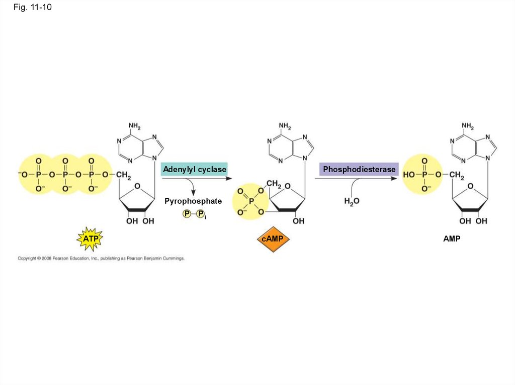
40. Cyclic AMP
• Cyclic AMP (cAMP) is one of the most widelyused second messengers
• Adenylyl cyclase, an enzyme in the plasma
membrane, converts ATP to cAMP in response
to an extracellular signal
Copyright © 2008 Pearson Education, Inc., publishing as Pearson Benjamin Cummings
41.
Fig. 11-10Adenylyl cyclase
Phosphodiesterase
Pyrophosphate
P
ATP
Pi
cAMP
AMP
42.
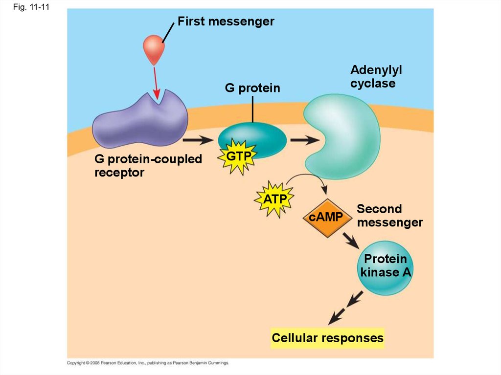
• Many signal molecules trigger formation ofcAMP
• Other components of cAMP pathways are G
proteins, G protein-coupled receptors, and
protein kinases
• cAMP usually activates protein kinase A, which
phosphorylates various other proteins
• Further regulation of cell metabolism is
provided by G-protein systems that inhibit
adenylyl cyclase
Copyright © 2008 Pearson Education, Inc., publishing as Pearson Benjamin Cummings
43.
Fig. 11-11First messenger
Adenylyl
cyclase
G protein
G protein-coupled
receptor
GTP
ATP
cAMP
Second
messenger
Protein
kinase A
Cellular responses
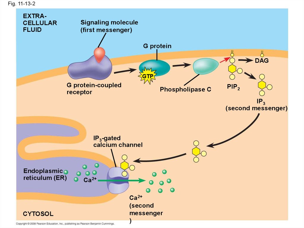
44. Calcium Ions and Inositol Triphosphate (IP3)
• Calcium ions (Ca2+) act as a secondmessenger in many pathways
• Calcium is an important second messenger
because cells can regulate its concentration
Copyright © 2008 Pearson Education, Inc., publishing as Pearson Benjamin Cummings
45.
Fig. 11-12EXTRACELLULAR
FLUID
Plasma
membrane
Ca2+ pump
ATP
Mitochondrion
Nucleus
CYTOSOL
Ca2+
pump
Endoplasmic
reticulum (ER)
ATP
Key
High [Ca2+]
Low [Ca2+]
Ca2+
pump
46.
• A signal relayed by a signal transductionpathway may trigger an increase in calcium in
the cytosol
• Pathways leading to the release of calcium
involve inositol triphosphate (IP3) and
diacylglycerol (DAG) as additional second
messengers
Animation: Signal Transduction Pathways
Copyright © 2008 Pearson Education, Inc., publishing as Pearson Benjamin Cummings
47.
Fig. 11-13-1EXTRACELLULAR
FLUID
Signaling molecule
(first messenger)
G protein
DAG
GTP
G protein-coupled
receptor
Phospholipase C
PIP2
IP3
(second messenger)
IP3-gated
calcium channel
Endoplasmic
reticulum (ER)
CYTOSOL
Ca2+
48.
Fig. 11-13-2EXTRACELLULAR
FLUID
Signaling molecule
(first messenger)
G protein
DAG
GTP
G protein-coupled
receptor
Phospholipase C
PIP2
IP3
(second messenger)
IP3-gated
calcium channel
Endoplasmic
reticulum (ER)
CYTOSOL
Ca2+
Ca2+
(second
messenger
)
49.
Fig. 11-13-3EXTRACELLULAR
FLUID
Signaling molecule
(first messenger)
G protein
DAG
GTP
G protein-coupled
receptor
PIP2
Phospholipase C
IP3
(second messenger)
IP3-gated
calcium channel
Endoplasmic
reticulum (ER)
CYTOSOL
Various
proteins
activated
Ca2+
Ca2+
(second
messenger
)
Cellular
responses
50. Concept 11.4: Response: Cell signaling leads to regulation of transcription or cytoplasmic activities
• The cell’s response to an extracellular signal issometimes called the “output response”
Copyright © 2008 Pearson Education, Inc., publishing as Pearson Benjamin Cummings
51. Nuclear and Cytoplasmic Responses
• Ultimately, a signal transduction pathway leadsto regulation of one or more cellular activities
• The response may occur in the cytoplasm or
may involve action in the nucleus
• Many signaling pathways regulate the
synthesis of enzymes or other proteins, usually
by turning genes on or off in the nucleus
• The final activated molecule may function as a
transcription factor
Copyright © 2008 Pearson Education, Inc., publishing as Pearson Benjamin Cummings
52.
Fig. 11-14Growth factor
Reception
Receptor
Phosphorylatio
n
cascade
Transduction
CYTOPLASM
Inactive
transcription
factor
Active
transcription
factor
P
Response
DNA
Gene
NUCLEUS
mRNA
53.
• Other pathways regulate the activity ofenzymes
Copyright © 2008 Pearson Education, Inc., publishing as Pearson Benjamin Cummings
54.
Fig. 11-15Reception
Binding of epinephrine to G protein-coupled receptor (1 molecule)
Transduction
Inactive G protein
Active G protein (102 molecules)
Inactive adenylyl cyclase
Active adenylyl cyclase (102)
ATP
Cyclic AMP (104)
Inactive protein kinase A
Active protein kinase A (104)
Inactive phosphorylase kinase
Active phosphorylase kinase (105)
Inactive glycogen phosphorylase
Active glycogen phosphorylase (106)
Response
Glycogen
Glucose-1-phosphate
(108 molecules)
55.
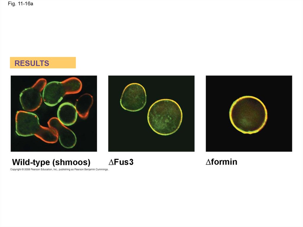
• Signaling pathways can also affect the physicalcharacteristics of a cell, for example, cell shape
Copyright © 2008 Pearson Education, Inc., publishing as Pearson Benjamin Cummings
56.
Fig. 11-16RESULTS
∆Fus3
Wild-type (shmoos)
∆formin
CONCLUSION
1
Mating
factor G protein-coupled
receptor
Shmoo projection
forming
Formin
P
Fus3
GTP
GDP
Phosphorylation
cascade
2
Actin
subunit
P
Formin
Formin
P
4
Fus3
Fus3
P
Microfilament
5
3
57.
Fig. 11-16aRESULTS
Wild-type (shmoos)
∆Fus3
∆formin
58.
Fig. 11-16bCONCLUSION
1
Mating
factor G protein-coupled
receptor
Shmoo projection
forming
Formin
P
Fus3
GTP
GDP
Phosphorylation
cascade
2
Actin
subunit
P
Formin
Formin
P
4
Fus3
Fus3
P
Microfilament
5
3
59. Fine-Tuning of the Response
• Multistep pathways have two importantbenefits:
– Amplifying the signal (and thus the response)
– Contributing to the specificity of the response
Copyright © 2008 Pearson Education, Inc., publishing as Pearson Benjamin Cummings
60. Signal Amplification
• Enzyme cascades amplify the cell’s response• At each step, the number of activated products
is much greater than in the preceding step
Copyright © 2008 Pearson Education, Inc., publishing as Pearson Benjamin Cummings
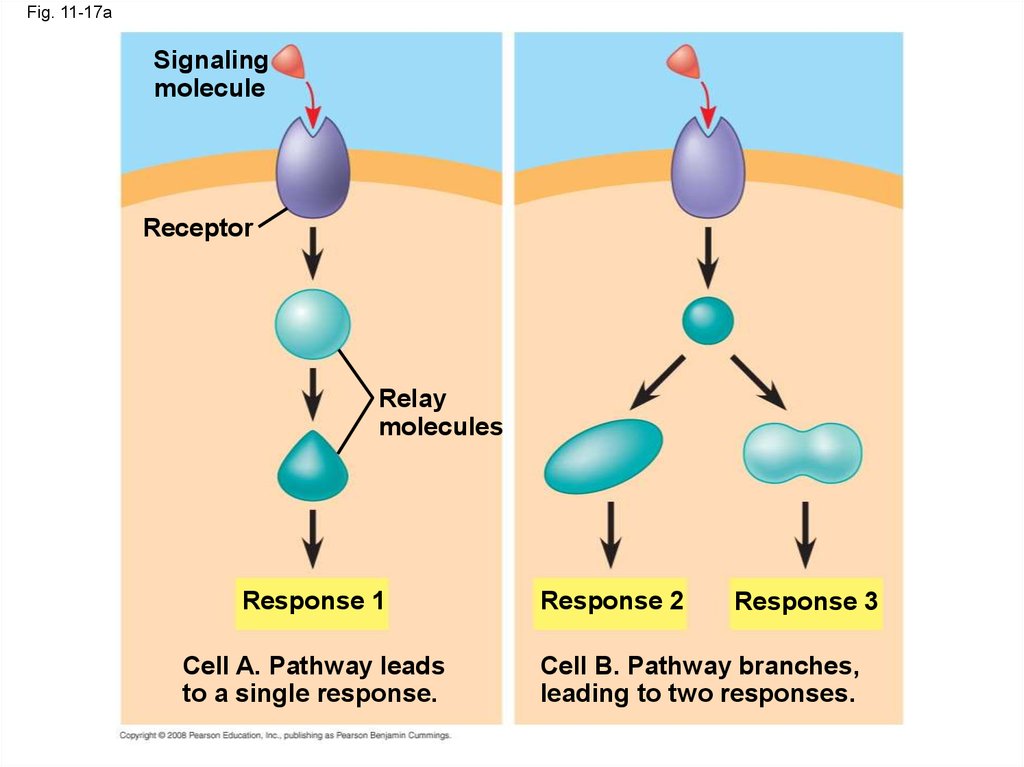
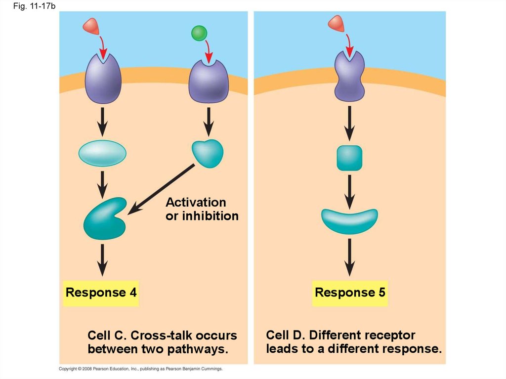
61. The Specificity of Cell Signaling and Coordination of the Response
• Different kinds of cells have differentcollections of proteins
• These different proteins allow cells to detect
and respond to different signals
• Even the same signal can have different effects
in cells with different proteins and pathways
• Pathway branching and “cross-talk” further help
the cell coordinate incoming signals
Copyright © 2008 Pearson Education, Inc., publishing as Pearson Benjamin Cummings
62.
Fig. 11-17Signaling
molecule
Receptor
Relay
molecules
Response 1
Cell A. Pathway leads
to a single response.
Response 2
Response 3
Cell B. Pathway branches,
leading to two responses.
Activation
or inhibition
Response 4
Cell C. Cross-talk occurs
between two pathways.
Response 5
Cell D. Different receptor
leads to a different response.
63.
Fig. 11-17aSignaling
molecule
Receptor
Relay
molecules
Response 1
Cell A. Pathway leads
to a single response.
Response 2
Response 3
Cell B. Pathway branches,
leading to two responses.
64.
Fig. 11-17bActivation
or inhibition
Response 4
Cell C. Cross-talk occurs
between two pathways.
Response 5
Cell D. Different receptor
leads to a different response.
65. Signaling Efficiency: Scaffolding Proteins and Signaling Complexes
• Scaffolding proteins are large relay proteinsto which other relay proteins are attached
• Scaffolding proteins can increase the signal
transduction efficiency by grouping together
different proteins involved in the same pathway
Copyright © 2008 Pearson Education, Inc., publishing as Pearson Benjamin Cummings
66.
Fig. 11-18Signaling
molecule
Plasma
membrane
Receptor
Three
different
protein
kinases
Scaffolding
protein
67. Termination of the Signal
• Inactivation mechanisms are an essentialaspect of cell signaling
• When signal molecules leave the receptor, the
receptor reverts to its inactive state
Copyright © 2008 Pearson Education, Inc., publishing as Pearson Benjamin Cummings
68. Concept 11.5: Apoptosis (programmed cell death) integrates multiple cell-signaling pathways
• Apoptosis is programmed or controlled cellsuicide
• A cell is chopped and packaged into vesicles
that are digested by scavenger cells
• Apoptosis prevents enzymes from leaking out
of a dying cell and damaging neighboring cells
Copyright © 2008 Pearson Education, Inc., publishing as Pearson Benjamin Cummings
69.
Fig. 11-192 µm
70. Apoptosis in the Soil Worm Caenorhabditis elegans
• Apoptosis is important in shaping an organismduring embryonic development
• The role of apoptosis in embryonic
development was first studied in
Caenorhabditis elegans
• In C. elegans, apoptosis results when specific
proteins that “accelerate” apoptosis override
those that “put the brakes” on apoptosis
Copyright © 2008 Pearson Education, Inc., publishing as Pearson Benjamin Cummings
71.
Fig. 11-20Ced-9
protein (active)
inhibits Ced-4
activity
Mitochondrion
Ced-4 Ced-3
Receptor
for deathsignaling
molecule
Inactive proteins
(a) No death signal
Ced-9
(inactive)
Cell
forms
blebs
Deathsignaling
molecule
Active Active
Ced-4 Ced-3
Activation
cascade
(b) Death signal
Other
proteases
Nucleases
72.
Fig. 11-20aCed-9
protein (active)
inhibits Ced-4
activity
Mitochondrion
Receptor
for deathsignaling
molecule
Ced-4 Ced-3
Inactive proteins
(a) No death signal
73.
Fig. 11-20bCed-9
(inactive)
Cell
forms
blebs
Deathsignaling
molecule
Active Active
Ced-4 Ced-3
Activation
cascade
(b) Death signal
Other
proteases
Nucleases
74. Apoptotic Pathways and the Signals That Trigger Them
• Caspases are the main proteases (enzymesthat cut up proteins) that carry out apoptosis
• Apoptosis can be triggered by:
– An extracellular death-signaling ligand
– DNA damage in the nucleus
– Protein misfolding in the endoplasmic
reticulum
Copyright © 2008 Pearson Education, Inc., publishing as Pearson Benjamin Cummings
75.
• Apoptosis evolved early in animal evolutionand is essential for the development and
maintenance of all animals
• Apoptosis may be involved in some diseases
(for example, Parkinson’s and Alzheimer’s);
interference with apoptosis may contribute to
some cancers
Copyright © 2008 Pearson Education, Inc., publishing as Pearson Benjamin Cummings
76.
Fig. 11-21Interdigital tissue
1 mm
77.
Fig. 11-UN11
Reception
2
Transduction
3 Response
Receptor
Relay molecules
Signaling
molecule
Activation
of cellular
response
78.
Fig. 11-UN279. You should now be able to:
1. Describe the nature of a ligand-receptorinteraction and state how such interactions
initiate a signal-transduction system
2. Compare and contrast G protein-coupled
receptors, tyrosine kinase receptors, and ligandgated ion channels
3. List two advantages of a multistep pathway in the
transduction stage of cell signaling
4. Explain how an original signal molecule can
produce a cellular response when it may not
even enter the target cell
Copyright © 2008 Pearson Education, Inc., publishing as Pearson Benjamin Cummings
80.
5. Define the term second messenger; brieflydescribe the role of these molecules in
signaling pathways
6. Explain why different types of cells may
respond differently to the same signal
molecule
7. Describe the role of apoptosis in normal
development and degenerative disease in
vertebrates
Copyright © 2008 Pearson Education, Inc., publishing as Pearson Benjamin Cummings