


:")
:")


")


 medicine
Similar presentations:


")

Генетика рака
1. ГЕНЕТИКА РАКА Understanding Cancer Genomics
2.
Mutations: Somatic and GermlineSomatic mutations
Germline mutations
Occur in nongermline tissues
Present in egg or sperm
Are nonheritable
Are heritable
Cause cancer family syndrome
Nonheritable
Somatic mutation
(e.g., breast)
Mutation in
egg or
sperm
All cells
affected in
offspring
3.
Tumors Are ClonalNormal
cell
First
mutation
Second
mutation
Third
mutation
Malignant cells
Fourth or
later
mutation
4.
Somatic MutationsNormal lung cell
Normal islet cell
Many years later
Lung cancer cell
Diabetic islet cell
5.
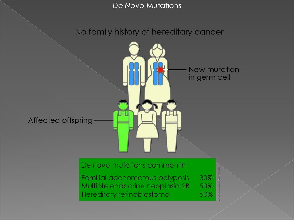
De Novo MutationsNo family history of hereditary cancer
New mutation
in germ cell
Affected offspring
De novo mutations common in:
Familial adenomatous polyposis
Multiple endocrine neoplasia 2B
Hereditary retinoblastoma
30%
50%
50%
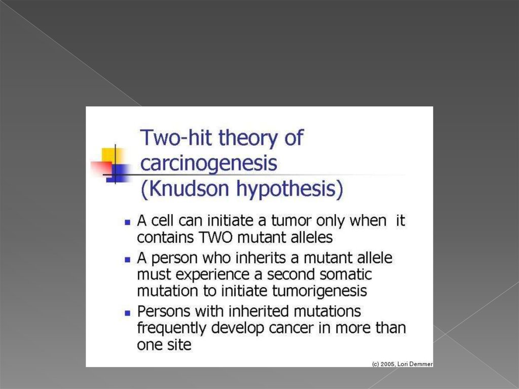
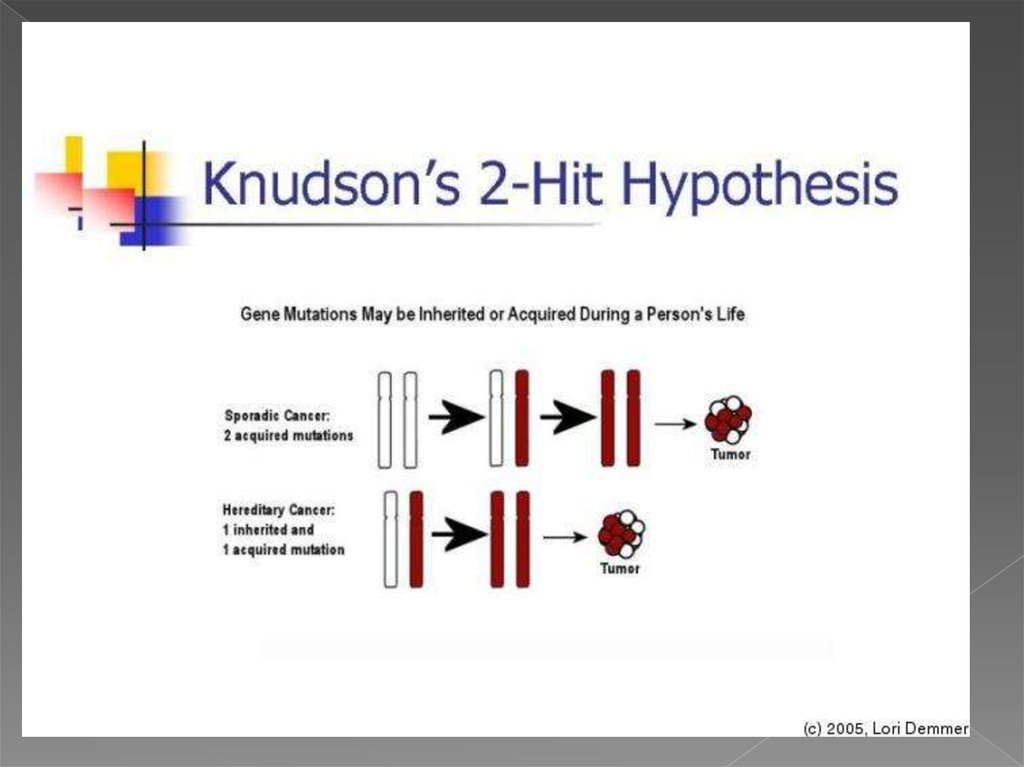
6. теория двойного удара или двойной мутации
В 1971 году Альфред Кнудсон предложил гипотезу, известнуюсейчас как теория двойного удара или двойной мутации,
объясняющую механизм возникновения наследственной и
спорадической форм ретинобластомы — злокачественной
опухоли сетчатки глаза.
для возникновения опухоли в клетке должны произойти две
последовательные мутации. В случае наследственной
ретинобластомы первая мутация происходит в клетках
зародышевой линии (наследственная мутация), а вторая
мутация (второй удар) — в соматических. Спорадическая
ретинобластома встречается реже и является результатом
двух мутаций в соматической клетке. Вероятность того, что в
одной клетке произойдёт две последовательные мутации,
невелика, поэтому спорадическая ретинобластома
встречается реже, чем наследственная, опухоли при этом
формируются позже и в меньшем количестве
7.
8.
9.
ОНКОГЕН — это ген, продукт которогоможет стимулировать образование
злокачественной опухоли. Мутации,
вызывающие активацию онкогенов,
повышают шанс того, что клетка
превратится в раковую клетку.
гены-супрессоры опухолей (ГСО)
предохраняют клетки от ракового
перерождения
рак возникает либо в случае нарушения
работы генов-супрессоров опухолей,
либо при появлении онкогенов
10.
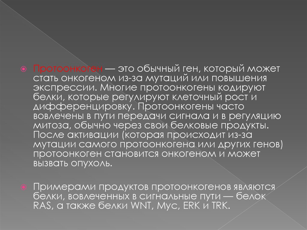
Протоонкоген — это обычный ген, который можетстать онкогеном из-за мутаций или повышения
экспрессии. Многие протоонкогены кодируют
белки, которые регулируют клеточный рост и
дифференцировку. Протоонкогены часто
вовлечены в пути передачи сигнала и в регуляцию
митоза, обычно через свои белковые продукты.
После активации (которая происходит из-за
мутации самого протоонкогена или других генов)
протоонкоген становится онкогеном и может
вызвать опухоль.
Примерами продуктов протоонкогенов являются
белки, вовлеченных в сигнальные пути — белок
RAS, а также белки WNT, Myc, ERK и TRK.
11.
Протоонкоген может стать онкогеном путем относительнонезначительной модификации его естественной функции.
три основных пути активации:
1. Мутация внутри протоонкогена, которая меняет структуру белка и
повышает активность белка (фермента)
при этом утрачивается регуляция экспрессии соответствующего
гена
2. Повышение концентрации белка путем
повышения экспрессии гена (нарушение регуляции экспрессии)
повышение стабильности белка, увеличение периода полужизни и,
соответственно, активности в клетке
дупликация гена (хромосомная перестройка), в результате чего
повышается концентрация белка в клетке
3.Транслокация (хромосомная перестройка), которая вызывает
повышение экспрессии гена в нетипичных клетках или в нетипичное
время
экспрессия постоянно активного гибридного белка. Такой тип
перестройки в делящихся стволовых клетках костного мозга
приводит к лейкемии у взрослых.
Мутации в микроРНК могут также приводить к активации онкогенов
Исследования показали, что малые молекулы РНК длиной 21-25
нуклеотидов, называемые микроРНК, контролируют экспрессию
генов путем понижения их активности. Антисмысловые мРНК могут
теоретически быть использованы для блокировки действия онкогенов.
12.
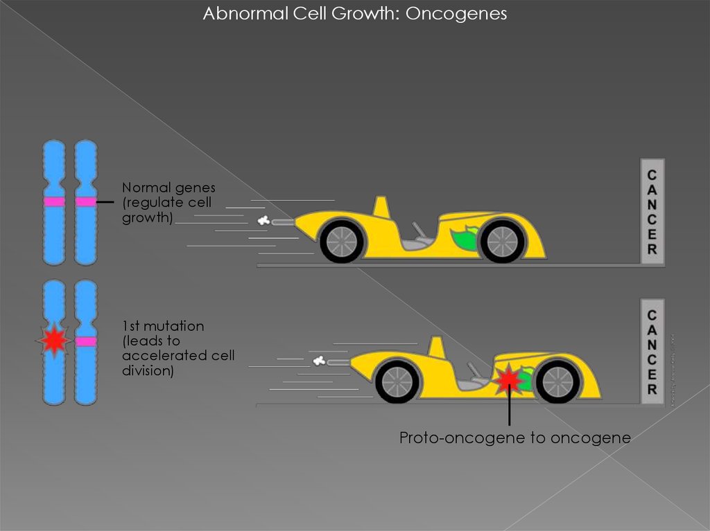
Abnormal Cell Growth: OncogenesNormal genes
(regulate cell
growth)
1st mutation
(leads to
accelerated cell
division)
Proto-oncogene to oncogene
13.
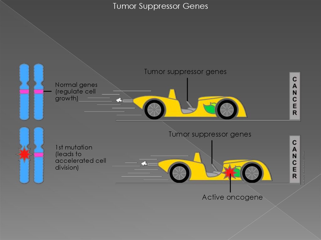
Tumor Suppressor GenesTumor suppressor genes
Normal genes
(regulate cell
growth)
Tumor suppressor genes
1st mutation
(leads to
accelerated cell
division)
Active oncogene
14.
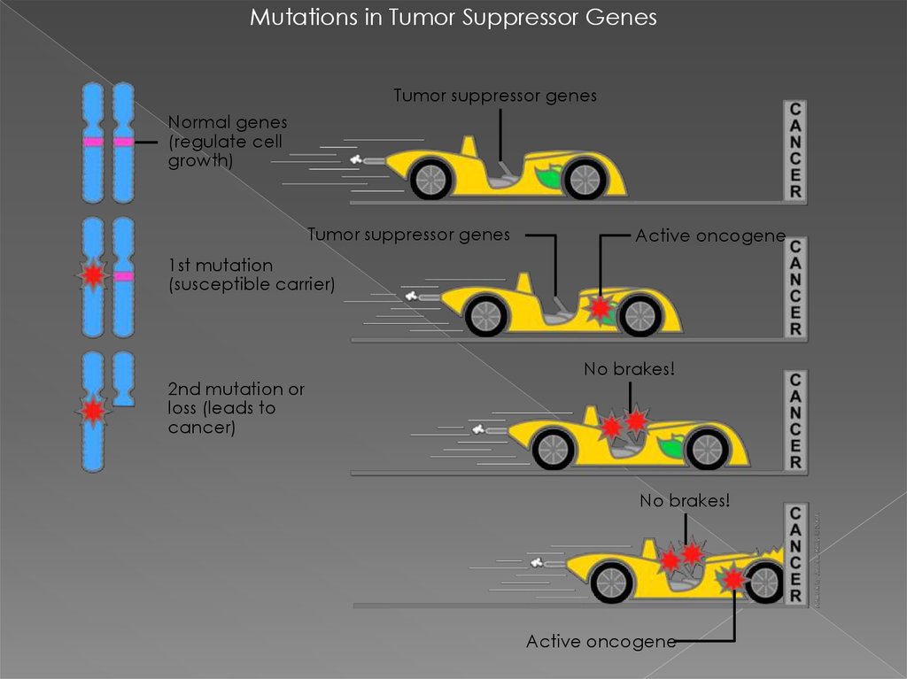
Mutations in Tumor Suppressor GenesTumor suppressor genes
Normal genes
(regulate cell
growth)
Tumor suppressor genes
Active oncogene
1st mutation
(susceptible carrier)
2nd mutation or
loss (leads to
cancer)
No brakes!
No brakes!
Active oncogene
15.
Two-Hit HypothesisNo cancer
Germline mutation
Somatic mutation
Cancer
If first hit is a germline
mutation, second
somatic mutation more
likely to enable cancer
16.
Regulatory MutationsNormal expression
Overexpression
Her2 protein
Her2 protein
Messenger
RNA
Chromosome 17
Her2 gene
Her2 gene amplification
17.
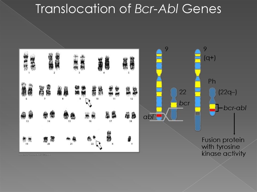
Translocation of Bcr-Abl Genes9
9
(q+)
Ph
22
bcr
(22q–)
bcr-abl
abl
Fusion protein
with tyrosine
kinase activity
18.
Different Locus, Different Allele,Same Phenotype
Chromosome 17
Chromosome 13
Allele
(gene)
Locus
(spot on
gene)
Allele
(gene)
Locus
(spot on
gene)
BRCA1
BRCA2
Hereditary breast and ovarian cancer
19.
Founder Effect inAshkenazi Jewish Population
An estimated 1 in 40 Ashkenazi Jews
carries a BRCA1 or BRCA2 mutation
BRCA1
185delAG
Prevalence = ~1%
5382insC
Prevalence = ~0.15%
BRCA2
6174delT
Prevalence = ~1.5%
20.
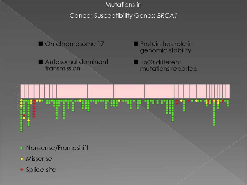
Mutations inCancer Susceptibility Genes: BRCA1
On chromosome 17
Protein has role in
genomic stability
Autosomal dominant
transmission
~500 different
mutations reported
Nonsense/Frameshift
Missense
Splice-site
21.
Mutations inCancer Susceptibility Genes: BRCA2
On chromosome 13
Protein has role in
genomic stability
Autosomal dominant
transmission
~300 different
mutations reported
Nonsense/Frameshift
Missense
22.
Autosomal Dominant InheritanceEqually transmitted
by men and women
No skipped generations
Each child has a 50%
chance of inheriting
the mutation
Normal
Affected
23.
Examples ofDominantly Inherited Cancer Syndromes
24.
Autosomal Recessive InheritanceTwo germline mutations
(one from each parent)
to develop disease
Equally transmitted by
men and women
Nonaffected carrier
Noncarrier
Affected carrier
25.
Some Recessively InheritedCancer Syndromes
26.
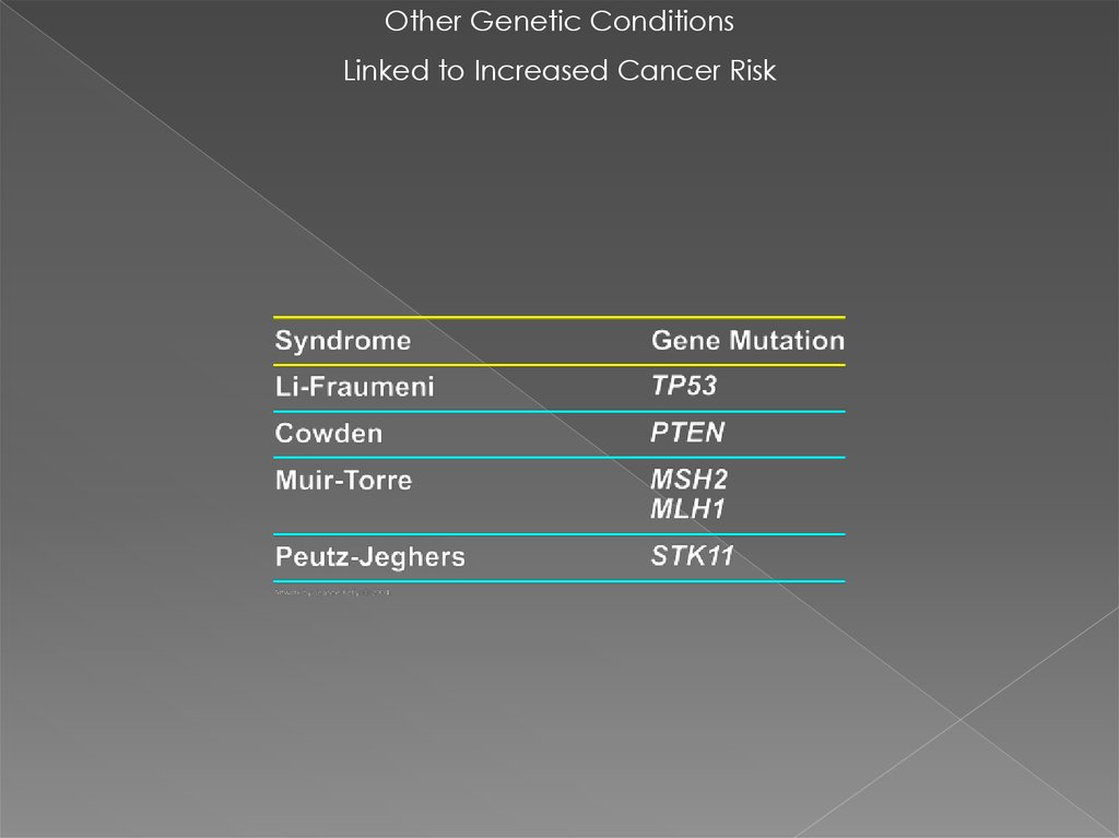
Other Genetic ConditionsLinked to Increased Cancer Risk
27.
Repair FailureDamaging Agent
X-rays
Oxygen
radicals
Alkylating
agents
Spontaneous
reactions
UV light
X-rays
Polycyclic
Anti-tumor
aromatic
agents
hydrocarbons (cis-Pt, MMC)
G
G
(6-4)PP
Bulky
adduct
CPD
G
G
A
Interstrand
cross-link
Double-strand
break
Single-strand
break
Baseexcision
repair (BER)
Replication
errors
T
C
T
T
Uracil
Abasic
site
B-oxoguanine
Consequences
G
A-G
mismatch
T-C
mismatch
Insertion
(Transient)
cell cycle
arrest
C
T
Inhibition of:
–Transcription
–Replication
–Chromosome
replication
Apoptosis
(cell death)
Mutations
Chromosome
aberrations
Cancer
Aging
Inborn
disease
Deletion
Nucleotide- Recombinational Mismatch
excision repair
repair
Repair
(NER)
(HR, EJ)
Repair Process
28.
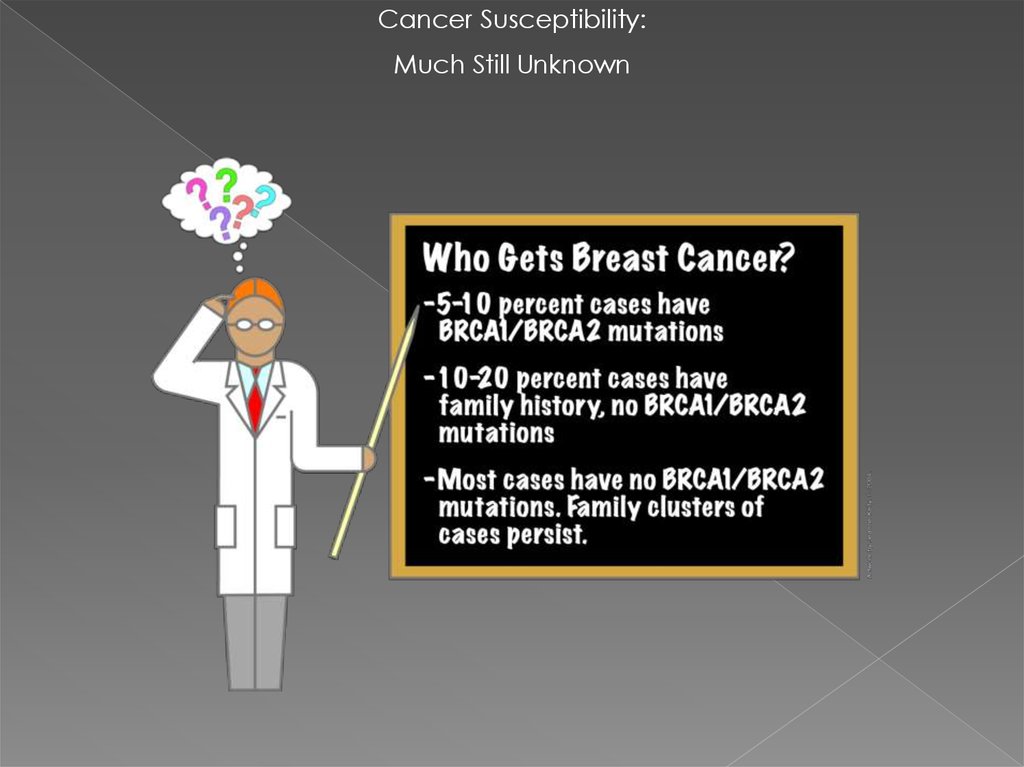
Cancer Susceptibility:Much Still Unknown
29. How do people know if they should consider genetic testing for BRCA1 and BRCA2 mutations?
For women who are not of Ashkenazi Jewish descent:two first-degree relatives (mother, daughter, or sister) diagnosed with breast cancer,
one of whom was diagnosed at age 50 or younger;
three or more first-degree or second-degree (grandmother or aunt) relatives diagnosed
with breast cancer regardless of their age at diagnosis;
a combination of first- and second-degree relatives diagnosed with breast cancer and
ovarian cancer (one cancer type per person);
a first-degree relative with cancer diagnosed in both breasts (bilateral breast cancer);
a combination of two or more first- or second-degree relatives diagnosed with ovarian
cancer regardless of age at diagnosis;
a first- or second-degree relative diagnosed with both breast and ovarian cancer
regardless of age at diagnosis; and
breast cancer diagnosed in a male relative.
For women of Ashkenazi Jewish descent:
any first-degree relative diagnosed with breast or ovarian cancer; and
two second-degree relatives on the same side of the family diagnosed with breast or
ovarian cancer.
These family history patterns apply to about 2 percent of adult women in the general
population. Women who have none of these family history patterns have a low
probability of having a harmful BRCA1 or BRCA2 mutation.
30. Li-Fraumeni Syndrome
(LFS) was first described in 1969 by Drs.Frederick Li and Joseph F. Fraumeni, Jr., who were working at
the NCI. Their study identified four families with sarcomas,
breast cancer, brain tumors, and leukemia, many of which
were diagnosed at much younger-than-usual ages.
Additional studies showed that other tumors, including
cancers of the adrenal cortex, gastrointestinal tract, lung,
and non-Hodgkin lymphoma, also occurred more often than
expected in these families.
31. Classic Li-Fraumeni Syndrome (LFS):
Three features must be present in a family to fit theclassic LFS criteria. Often more than 3 family members
have had cancers.
A person with a sarcoma diagnosed under the age
of 45; AND
At least one first-degree relative (meaning parents,
brothers, sisters and children) with a cancer of any
kind diagnosed under the age of 45; AND
A third family member who is either a first- or seconddegree relative (such as grandparents, aunts, uncles,
nieces, nephews, and grandchildren) with cancer
diagnosed under the age of 45, or having a sarcoma
at any age
32. Li-Fraumeni-Like Syndrome (LFL):
A person with any childhood cancer orsarcoma, brain tumor, or adrenal cortical
tumor diagnosed under the age of 45 AND
A first- or second-degree relative with a
typical LFS cancer (soft tissue and bone
sarcomas, brain tumors, breast cancer,
adrenocortical carcinomas, leukemia, and
many others) at any age AND
An additional first- or second-degree
relative with any cancer diagnosed under
the age of 60.
33. What Causes LFS?
Changes in a “tumor suppressor” gene called “TP53” were discovered in1990 as the most common cause of LFS. Everyone has two copies of the
TP53 gene – one inherited from the mother, the other from the father – in
every cell of their body. This gene is very important for the normal
growth, function, and division of cells. The gene causes cells that are
damaged beyond repair to die, a process that stops damaged cells
from becoming cancerous. If there is a change (or mutation) in TP53,
the gene fails to work properly and cancer may develop. The kind of
cancer that develops depends on where in the body the abnormal cell
is located. The fact that TP53 is so important to the normal functioning of
most cells in the body may explain why so many different kinds of
cancer occur in LFS.
About 7 out of every 10 patients (or 70%) with classic LFS, and 4 out of
every 10 (40%) of patients with LFL, have a detectable change in the
TP53 gene. We don’t yet fully understand what causes LFS in families that
do not have a TP53 mutation, but there are several ideas. For example
there could be an unusual mutation in TP53 that is not easily found by
the usual testing methods. Or there may be other genes which have not
yet been identified, that can cause LFS.
34. Risk of Cancer in Patients with LFS
The lifetime risk of cancer – all types combined - in aperson who carries a TP53 mutation ranges from 70% to
90% by age 70. Women with LFS have a higher lifetime
cancer risk than men with LFS, most likely due to the high
risk of female breast cancer. The lifetime cancer risk for
women reaches almost 100%. At present, we cannot
predict which individual with a TP53 mutation will
eventually develop cancer and, if they do develop
cancer, which type and when.
If a family member has a known mutation in the TP53
gene, genetic testing can identify other family members
with the same mutation who would also be at high cancer
risk. For those at high risk, early cancer detection and risk
reduction strategies are desirable, but not yet
standardized. Currently, management recommendations
are based on our best clinical judgment.
35.
For now, in persons with a TP53 genemutation, we can try to find cancers as
early as possible (a process called
screening) in the hope that finding
cancer early will lead to more successful
treatment.
36. Cowden syndrome mutations in the PTEN gene
Cowden syndrome is a disordercharacterized by multiple
noncancerous, tumor-like growths called
hamartomas and an increased risk of
developing certain cancers.
37. Cowden syndrome mutations in the PTEN gene (TSG)
Cowden syndrome is associated with an increasedrisk of developing several types of cancer,
particularly cancers of the breast, thyroid, and the
lining of the uterus (the endometrium).
Other cancers that have been identified in people
with Cowden syndrome include colorectal cancer,
kidney cancer, and melanoma. Compared with the
general population, people with Cowden syndrome
develop these cancers at younger ages, often
beginning in their thirties or forties. Other diseases of
the breast, thyroid, and endometrium are also
common in Cowden syndrome. Additional signs and
symptoms can include an enlarged head
(macrocephaly) and a rare, noncancerous brain
tumor called Lhermitte-Duclos disease.
38. Что такое ОНКОГЕН ?
1.ген, стимулирующий образованиеопухоли
2. гены, предохраняющие клетки от
ракового перерождения
3. ген, продукт которого может
стимулировать образование
злокачественной опухоли
39. Рак груди встречается при следующих генетических синдромах, кроме:
BRCA-1/2 мутация2. Cowden syndrom
3. Li-Fraumeni syndrom
4. Все верно
1.