


 medicine
Similar presentations:


Pathogenetic peculiarities and pathoanatomical changes in bronchial pneumonia of calves
1. KAZAKH NATIONAL AGRARIAN UNIVERSITY
FACULTY OF VETERINARYDepartment of Clinical Veterinary Medicine
PATHOGENETIC PECULIARITIES AND
PATHOANATOMICAL CHANGES IN
BRONCHIAL PNEUMONIA OF CALVES
Presented by: Nauatbek Azhar
VM-308
Scientific instructor: Turyspayeva Sholpan Dzhapashevna,
associate professor
Almaty 2017
2. Currently, it revealed a clear tendency to increase the number of patients suffering from diseases of the upper respiratory
INTRODUCTIONCurrently, it revealed a clear tendency to increase the number
of patients suffering from diseases of the upper respiratory
tract, bronchial tubes and lungs bronchial tube.
Bronchial pneumonia is registered in different zones of the
country, and takes the second place in terms of the ratio after
gastrointestinal diseases. As per the data of different authors,
20-30 % of young cattle suffer from bronchial pneumonia
every year.
The occurrence of bronchial pneumonia is caused by low
Natural resistance in young animals, and hence reduced
resistance of hypopneumatic and atelectatic areas of the lungs
due to the small number of ciliary epithelium of the mucous
membrane of the airways, which is a favorable environment
for the development of potentially pathogenic microflora.
Long lying of a weakly developed animal, hyposthenia of
cross-striped muscles and smooth muscles of the bronchi
cause sudden decrease in ventilation of the lungs with
reduction of their respiratory surface and further development
of atelectasis and hypostasis where foci of inflammation occur
3.
THE AIMS AND OBJECTIVES OF RESEARCHThe aim of research was to study the incidence and
prevalence of pneumonia in young cattle Raiymbek district,
Almaty region, its pathogenesis and pathological morphology.
In this connection, the following objectives:
To examine the incidence and distribution of pneumonia in
young animals;
To study the pathogenesis and functional morphology of the
respiratory organs of young animals;
Develop a comprehensive system of protection of young
veterinary;
To study morphological changes in the bronchopneumonia in
young cattle at the organ, tissue, cellular and subcellular levels.
4.
MATERIALS AND RESEARCHMETHODS
The experimental part of the work is carried out at the
Department of “Clinical Veterinary Medicine” of the Kazakh
National Agrarian University and the farms “Aktasty” and
“Sholadyr” Raiymbek District with the total number of cattle-144
head.
In the experimental work were studied the incidence and spread
of pneumonia in young cattle in the area; morphological changes
in the bronchopneumonia in young cattle at the organ, tissue,
cellular and subcellular level. The features of pulmonary
surfactant system and the ultrastructural organization alveolocytes
I and II types for bronchopneumonia. For the first time in calves
described pneumocyte type III. Elucidated pathogenetic
mechanisms of development of pneumonia in young cattle.
The material for histologic and histochemical examination was
fixed in 10-12% solution of neutral formaline, Carnoy's fluid.
Pieces of lung tissue were frozen over liquid nitrogen for enzyme
reactions and research lung surfactant.
5.
Fixation material for electronic microscope studies wereperformed in 2.5%-gluteraldehyde on collodion buffer with post
fixation in 1%- solution of osmium tetroxide, dehydrated in
alcohol, embedded in EPON-812.
Paraffin sections were stained with hematoxylin-eosin and
hematoxylin-pikrofuksin.
Alveolar surfactant detected in cryostat section of lung
Hackney in the modification of rhodamine- J. This qualitive
and quantitive assessment of the surfactant was carried out in
fluorescent mode microscope MBI-15 and in the microscope
“Lomam I-3” under ultraviolet light. The intensity of the
luminescence was determined with a microfluorimeter in
microvolts, which included the design of a photomultiplier, a
power supply, a DC amplifier and a universal voltmeter.
The volume fractions of tissue structures were determined by
point counting. At the same eyepiece used grating, counting was
conducted in a 225-node intersections. Counts 3375 units,
accounting for the different structures in the lung tissue. The
relative proportions determined by the formula: р = m/n • 100,
where m - the number of units attributable to the studied tissue
sections; n - total number of nodes.
6.
RESULTS AND DISCUSSIONResults of studies have shown that the structural organization
of the respiratory system in clinically healthy calves comply with
the species and age parametres, which are known from the
available literature.
Histomorphological examination of the lungs in healthy calves
aged 1.5-2 months, the system of airways and parenchyma of the
lung tissue was well developed.
Acini, as structural unit of the lung were clearly
expressed(Figure 1a). It was also noted that in light calves no
distinct respiratory bronchioles, and was characterized by a rather
sharp transition from terminal bronchioles into the alveolar ducts.
The thickness of the mucosal epithelium was uniform. In the
lumen, bronchioles and alveoli missing any content. They were
clean, not sticky(Figure 1b).Integrity interalveolar walls had not
been violated.
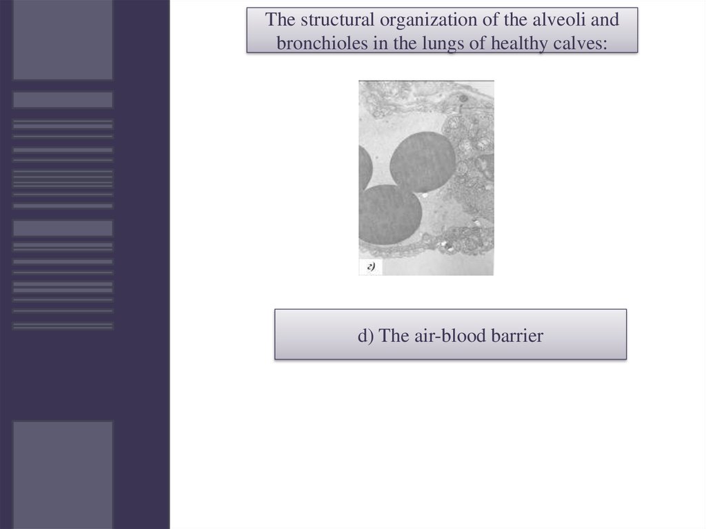
It can be traced to the ultrastructural level(Figure 1c). Blood barrier
was well formed. Its thickness throughout was approximately the same.
In capillaries it noted a moderate amount of red blood cell(Figure 1g).
Alveolocytes types I and II maintained a characteristic ultrastructure
(Figure 2a, b). In type II alveolocytes clearly identified osmiophil plate
calf with an average of 2-3 in each cell(Figure 2b).
7.
The structural organization of the alveoli andbronchioles in the lungs of healthy calves:
a)the bronchial tubes deep in the lung
tissue
8.
The structural organization of the alveoli andbronchioles in the lungs of healthy calves:
b)the terminal bronchioles
9.
The structural organization of the alveoli andbronchioles in the lungs of healthy calves:
c)Interalveolar partition
10.
The structural organization of the alveoli andbronchioles in the lungs of healthy calves:
d) The air-blood barrier
11.
CONCLUSIONSThus, the results of their own research and analysis of data in
the literature suggest that we have identified the cell type can be
assigned to alveolocytes type .
Histomorphological examination of the lungs of calves in the
very early stages of bronchial pneumonia has been found that in
addition to serous-catarrhal processes in the upper respiratory
tract (rhinitis, laryngitis, tracheitis) primary changes begin to
come to light in the end regions of the respiratory tract (bronchial
tubes of a different order), peribronchial tissue and in the lung
alveoli.
Moreover, the changes in these structures are developed at the
time when the disease is still clinically hardly seen. The main,
sometimes the only clinical sign in this stage can only be a serous
catarach of the upper respiratory tract. Therefore, this step can be
considered as subclinical pneumonia.
The features of pulmonary surfactant system and the
ultrastructural organization alveolocytes I and II types for
bronchopneumonia. For the first time in calves describes
pneumocyte type III. Elucidated pathogenetic mechanisms of
development of pneumonia in young cattle.
12.
REFERENCES:.
1 Belkin B.L. Impact of the microclimate on physiological functions of
calves//Veterinary. – 1998. - #7. - pages 52-54.
2. Sugaragchai Ts. Study of the microflora of upper airways and lungs of healthy
calves and those with pathologies: Author's thesis. PhD in veterinary science. - L.,
1956. - page 11.
3. Kovbasenko M.F. Etiology, pathogenesis, therapy and prevention of bronchial
pneumonia of calves: scientific notes. the Belotserkovskiy Agrarian Institute. - 1972. Vol. 20. - pages 69-73.
4. Velyamov M.T., Bakhtakhunov Yu.Kh. Viral mixed respiratory and gastrointestinal
infections in cattle // Vestnik (Herald). -1998. -#5. - pages 92-98.
5. Porokhov F.F., Buravova, Ivanenko I.T. – Prevention and emergency treatment of
calves suffering from bronchial pneumonia. Journal "Veterinary". 9, 56, 1983.
6. Sharabrin I.T. – Internal non-contagious diseases of farm animals. M. Kolos, 1976.
7. Muralinov K.K. Diagnostics, treatment and prevention of lung diseases in animals//
Mat. of the IVth International Scientific and Practical Conference, Ulan-Bataar, 2001,
pages 301-302.
8. Marchuk G.I., Verbentsova Z.P. Acute pneumonia, immunology, assessment of
severity, clinics, treatment/Science, M.: 1989, pages 63-68
9. Boroday G.P. Prevention of respiratory diseases in young animals on farms, - Kiev,
UkrNIINTI (the Ukrainian Scientific-Research Institute of Scientific and Technical
Information and Technical and Economical Research), 1978-pages 18-22.
10. Bilyalov E.E. Morphological and immunological indicators of the pathogenesis of
bronchial pneumonia in calves. Thesis, of the PhD in veterinary science, 2007- 87b.