


 biology
Similar presentations:


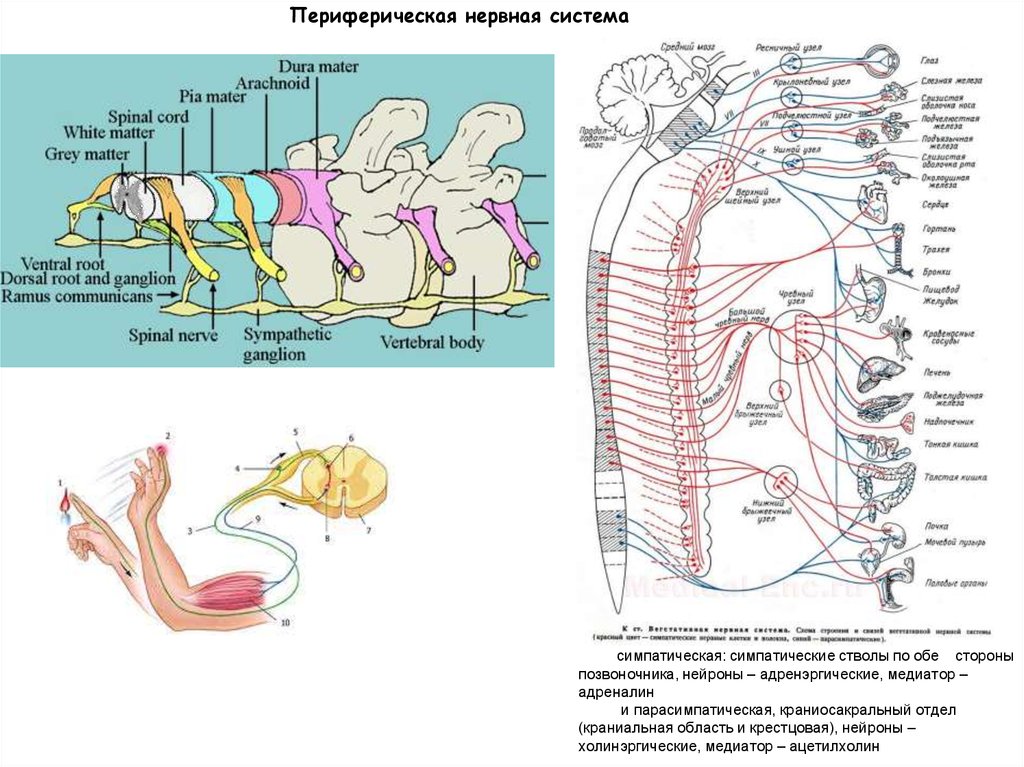
Периферическая нервная система
1.
2.
3.
Периферическая нервная системасимпатическая: симпатические стволы по обе стороны
позвоночника, нейроны – адренэргические, медиатор –
адреналин
и парасимпатическая, краниосакральный отдел
(краниальная область и крестцовая), нейроны –
холинэргические, медиатор – ацетилхолин
4.
Эпителиально-мезенхимный переход при формированиипроизводных нервного гребня
5.
Сроки выселения материала НГ вдоль передне-заднейоси у куриных зародышей:
Ст. 9 (7 пар сомитов, 29-33 ч) – граница prosencephalon и
mesencephalon
Ст. 11 (13 пар сомитов, 40-45 ч) – mes/met
Ст. 13 (19 пар сомитов, 48-52 ч) – met/myelen,
потом вдоль всего туловищного и хвостового отделов
зародыша
6.
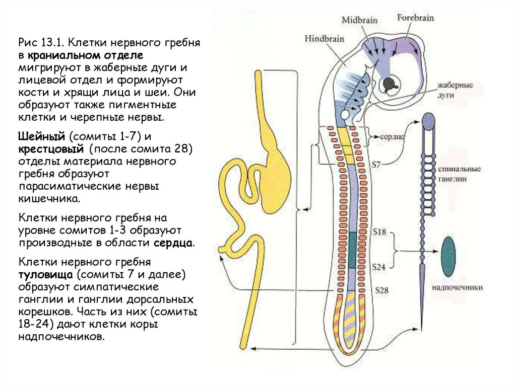
Рис 13.1. Клетки нервного гребняв краниальном отделе
мигрируют в жаберные дуги и
лицевой отдел и формируют
кости и хрящи лица и шеи. Они
образуют также пигментные
клетки и черепные нервы.
Шейный (сомиты 1-7) и
крестцовый (после сомита 28)
отделы материала нервного
гребня образуют
парасиматические нервы
кишечника.
Клетки нервного гребня на
уровне сомитов 1-3 образуют
производные в области сердца.
Клетки нервного гребня
туловища (сомиты 7 и далее)
образуют симпатические
ганглии и ганглии дорсальных
корешков. Часть из них (сомиты
18-24) дают клетки коры
надпочечников.
7.
Пути миграции разных популяций клеток туловищного отделанервного гребня
8.
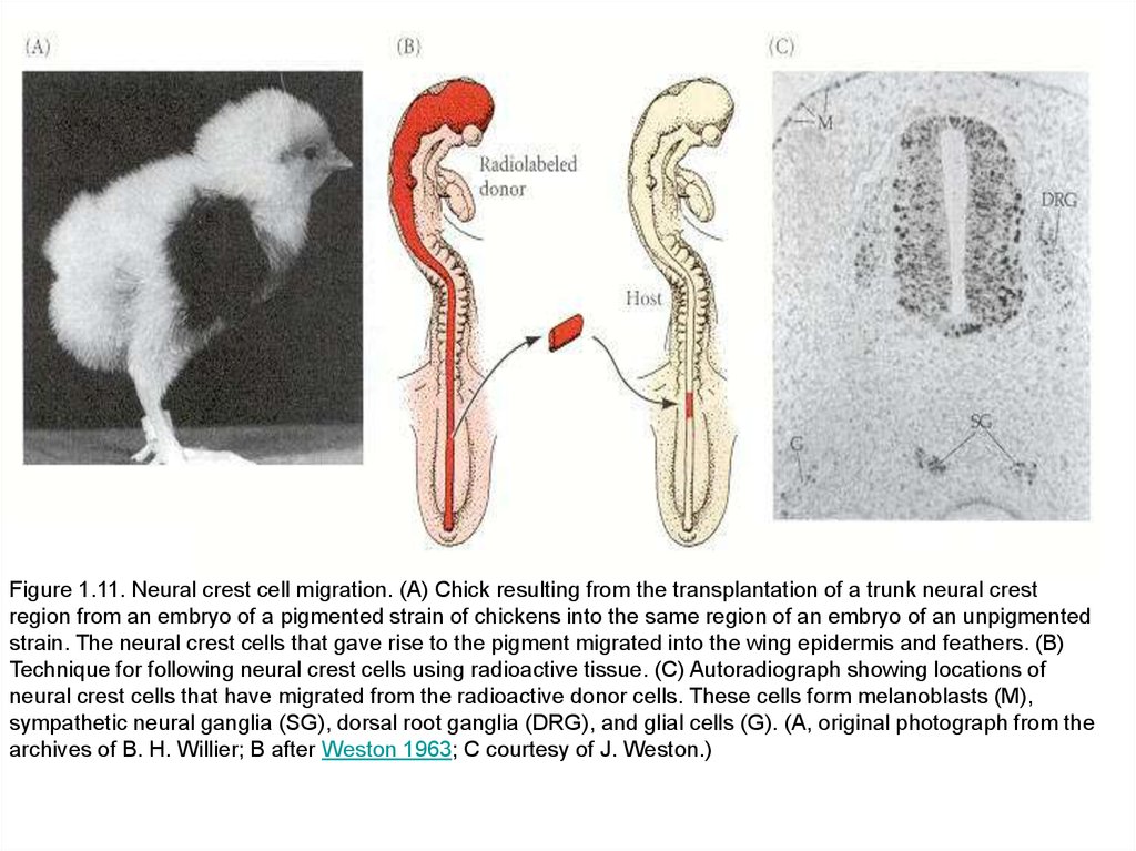
Figure 1.11. Neural crest cell migration. (A) Chick resulting from the transplantation of a trunk neural crestregion from an embryo of a pigmented strain of chickens into the same region of an embryo of an unpigmented
strain. The neural crest cells that gave rise to the pigment migrated into the wing epidermis and feathers. (B)
Technique for following neural crest cells using radioactive tissue. (C) Autoradiograph showing locations of
neural crest cells that have migrated from the radioactive donor cells. These cells form melanoblasts (M),
sympathetic neural ganglia (SG), dorsal root ganglia (DRG), and glial cells (G). (A, original photograph from the
archives of B. H. Willier; B after Weston 1963; C courtesy of J. Weston.)
9.
Fig. 8. A dynamic spatiotemporal fate map of NC derivatives.(A) Dorsal NT cells prior to emigration.
(B) Emigrating NC progenitors and, within the NT, dorsalward cell
relocation prior to migration.
(C) Organogenetic stage. Color coding represents relative positions of
NC progenitors in the NT in relation to their final homing sites. The
sequential and stereotypical ventral to dorsal order of colonization of
trunk NC derivatives is accounted for by an ordered emigration of
presumptive NC progenitors. Furthermore, continuous cell exit is
accounted for by a corresponding ventral to dorsal relocation of
epithelial progenitors towards the dorsalmost area of the NT, which
therefore acts as a transition zone for the progressive influx and
departure of cells. Sequential cell emigration thus causes a progressive
narrowing of the pre-migratory NC domain until its disappearance from the
dorsal NT and its concomitant replacement by the definitive RP.
NT – нервная трубка; NC – нервный гребень; RP – кроющая
пластинка; M – меланоциты; DRG – дорсальные ганглии; VR –
вентральные корешки; SG – симпатические ганглии.
Krispin et al., 2010.
10.
Figure 13.5. Pluripotency of trunk neural crest cells. (A) A single neural crest cell isinjected with highly fluorescent dextran shortly before migration of the neural crest
cells is initiated. The progeny of this cell will each receive some of these fluorescent
molecules. (B) Two days later, neural crest-derived tissues contain dextran-labeled
cells descended from the injected precursor. The figure summarizes data from two
different experiments (case 1 and case 2). (After Lumsden 1988a.)
11.
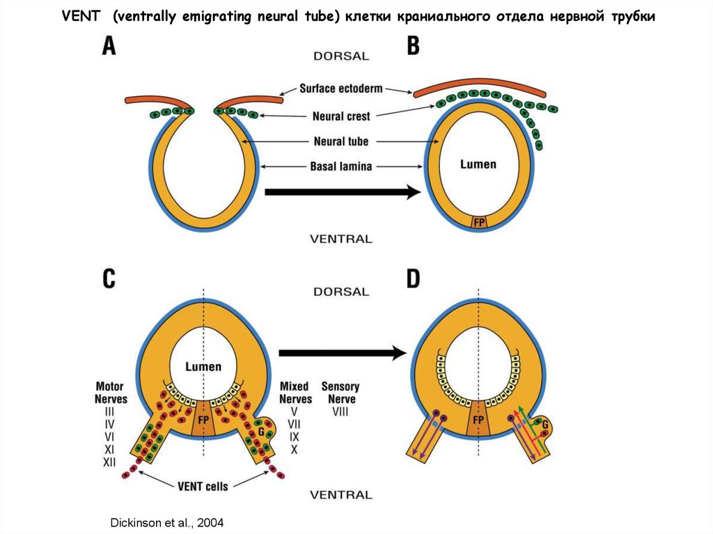
VENT (ventrally emigrating neural tube) клетки краниального отдела нервной трубкиDickinson et al., 2004