


![[18F]fluorodeoxyglucose PET for diagnosing Takayasu’s arteritis](https://cf.ppt-online.org/files/slide/m/MeodCX3bzB01chIVy4q9nmwOjxfaSTPQKJrYWp/slide-25.jpg "[18F]fluorodeoxyglucose PET for diagnosing Takayasu’s arteritis")


 medicine
Similar presentations:


Takayasu’s arteritis
1. TAKAYASU’S ARTERITIS
PREPARED BY: NURMAGAMBETOV SH. 462 GM2. EPIDEMIOLOGY
More case reports from Japan ,India, South-east Asia, MexicoNo geographic restriction
No race – immune
Incidence-2.6/million/year-N.America/Europe
The incidence in Asia is 1 case/1000-5000 women.
3.
AgeMc-2nd & 3rd decade
May range from infancy to middle age
Indian studies-age 3- 50 yrs
Gender diff
Japan-F:M=8-9:1
India-F:M ratio varies from -1:1 - 3:1
( Padmavati S, Aurora AP, Kasliwal RR Aortoarteritis in India. J
Assoc
Physicians India 1987)
India=F:M- 6.4:1 (Panja et al, 1997 JACC)
4. Genetics
Japan - HLA-B52 and B39Mexican and Colombian patients - HLADRB1*1301 and HLA-DRB1*1602
India-
HLA- B 5, -B 21
5. Histopathology
Idiopathic c/c infla arteritis of elastic arteriesresulting in occlusive &/ ectatic changes
Large vessels, esp, Aorta & its main branches
(brachiocephalic, carotid, SCL, vertebral,
RA)
+Coronary & PA
Ao valve –usually not beyond IMA
Multiple segs with dis & skipped nl areas
or diffuse involvement
6.
7.
GrossHistology
1)Gelatinous plaques-early
Panarteritis-granulomatous lesion with
giant cells
2)White plaques-collagen
3)Diffuse intimal thickening
1)
a/c phase
diffuse infil-mono
granulomatous infil
Superficial– deep scarring
circumferential
stenosis
4)Mural thrombus
5)2⁰ atheromatous changes
long standing,
HTN
2)c/c phase-coll rich fibrous tissueadventitia thicker than media
3)Healed phase-no infl cells, vas media
scarred
8.
Wall thickening, Fibrosis, Stenosis, & Thrombus formation →endorgan ischaemia
More a/c inflammation → destroys arterial media → Aneurysm
(fibrosis inadequate)
Stenotic lesions predominate & tend to be B/L
Nearly all pts with aneurysms also have stenoses
9.
Associated pathology-TB (LN)-55%Erthema multiforme
Bazins disease(eryt induratum)
churg strauss synd
reteroperitoneal fib
PAN,UC,CD etc
10. Clinical features
Early pre pulseless/genmanif
Fever,weight loss,headache,
fatigue,malaise,night sweats,
arthralgia
+/_ splenomegaly/ cervical,
axillary lymphadenopathy
Disappear partly/ completely in
3 months
Late ischemic phase
Sequel of occl of Ao arch/br
Diminished/absent pulses
(84–96%)
Bruits (80–94%)
Hypertension (33–83% )
RAS(28–75%) &
CCF(28%)
50% -no h/o acute phase
11.
CVS↓/− pulses (84–96%) -claudication & BP Diff ,Bruits (80–94%) carotids, subcl & abd vess.
HTN- (33–83%) –Mcc RAS (28–75%),↓Ao capacitance,atyp CoA,
barroreceptor reactivity
CHF-(28%)- HTN, AR, DCM-5%
AR-(7-24%) Ao root dil > valve inv, annuloaortic ectasia
Coronary & vascular involvement
CNS
Cerebral ischemia 2 ⁰ to obliterative arteritis, seizures etc
RENAL
RAS & Ischemic Nephropathy
SKIN
Erythema nodosum, Raynauds disease, leg& hand ulcers
PULMONARY
15-27%, stenosis/ occlusion of lobar/segmental pul art
UL>LL, R> L—INDIA (Panja et al 1997)
12. Coronary involvement in TA
Occurs in 10 30%Often fatal
Classified into 3 types
Type1:stenosis or occlu of coronary ostia
Type2:diffuse or focal coronary arteritis
Type3:coronary aneurysm
13. Occular involvement-Amaurosis fugax, pain behind eye, no real visual loss
Nonhypertensiveretinopathy
Hypertensive retinopathy
Commonest
UYAMA & ASAYAMA CLASS
Arteriosclerotic –art narrowing, stage 1- Dil of small vessels
av nipping,silver wiring
stage 2- Microaneurysm
Neuroretinopathy-exudates
stage 3- Art-ven anastomoses
and papilloedema
stage 4- Ocular complications
Direct opthalmoscopy
Mild -stage 1
Moderate -stage 2
Severe -stages 3 & 4
Flourescien angio sensitive
14.
15.
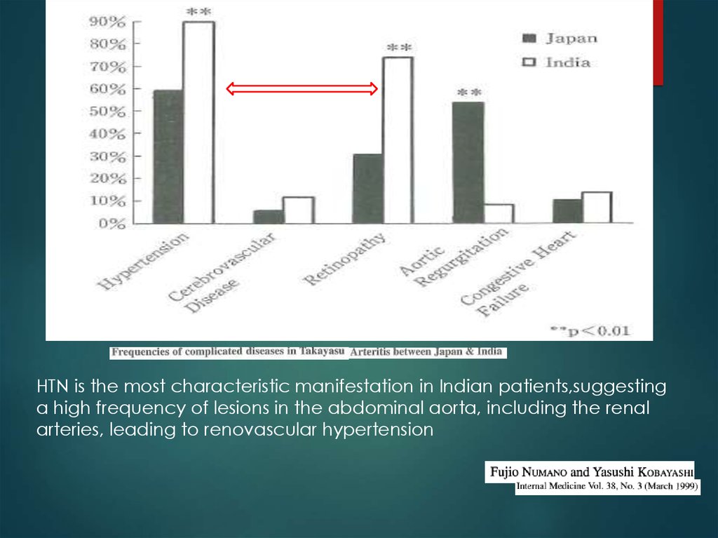
HTN is the most characteristic manifestation in Indian patients,suggestinga high frequency of lesions in the abdominal aorta, including the renal
arteries, leading to renovascular hypertension
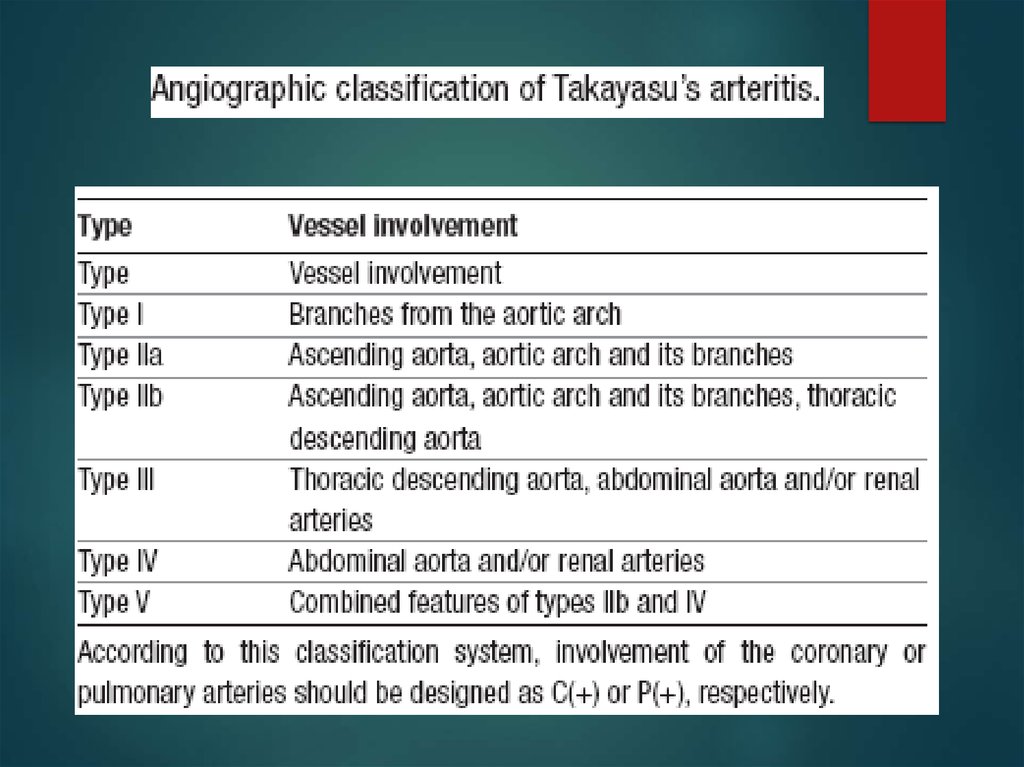
16. Ishikawa clinical classification of Takayasu arteritis 1978
4Complications
Retinopathy, Secondary HTN, AR, &
Aneurysm
17.
18.
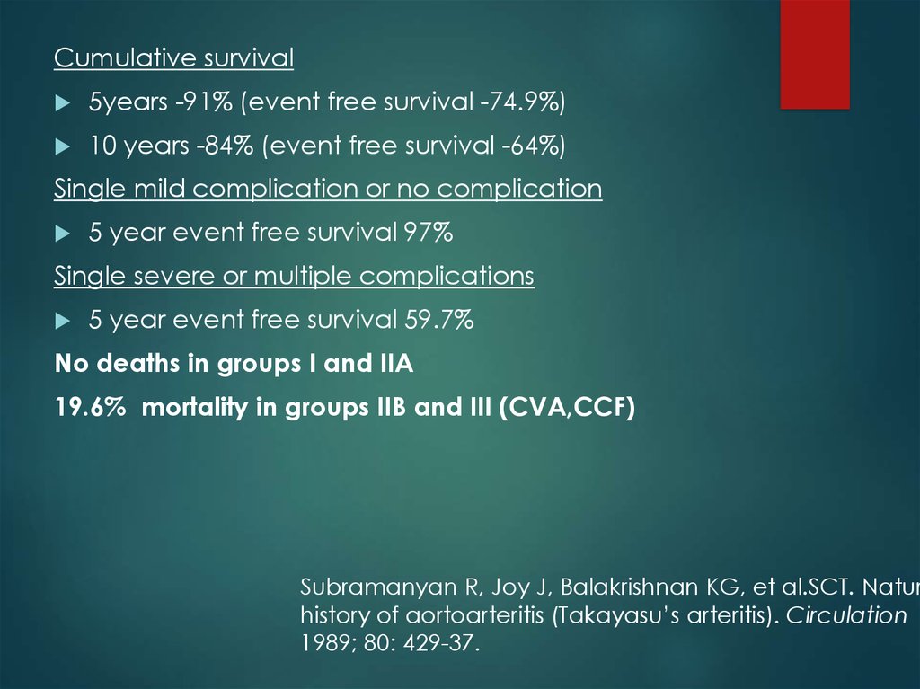
Cumulative survival5years -91% (event free survival -74.9%)
10 years -84% (event free survival -64%)
Single mild complication or no complication
5 year event free survival 97%
Single severe or multiple complications
5 year event free survival 59.7%
No deaths in groups I and IIA
19.6% mortality in groups IIB and III (CVA,CCF)
Subramanyan R, Joy J, Balakrishnan KG, et al.SCT. Natur
history of aortoarteritis (Takayasu’s arteritis). Circulation
1989; 80: 429-37.
19. 1990
20. 1995
21.
Sharma BK, Jain S, Suri S, Numano F. Diagnostic criteria foTakayasu arteritis. Int J Cardiol 1996; 54 : S141-S147
22.
23. nee
24.
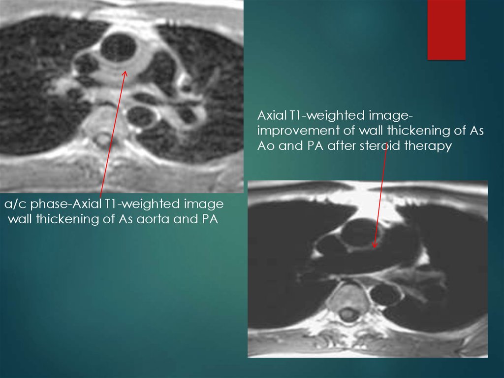
Axial T1-weighted imageimprovement of wall thickening of AsAo and PA after steroid therapy
a/c phase-Axial T1-weighted image
wall thickening of As aorta and PA
25.
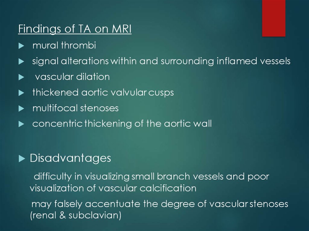
Findings of TA on MRImural thrombi
signal alterations within and surrounding inflamed vessels
vascular dilation
thickened aortic valvular cusps
multifocal stenoses
concentric thickening of the aortic wall
Disadvantages
difficulty in visualizing small branch vessels and poor
visualization of vascular calcification
may falsely accentuate the degree of vascular stenoses
(renal & subclavian)
26. [18F]fluorodeoxyglucose PET for diagnosing Takayasu’s arteritis
common [18F]FDG uptake pattern TAearly phase - linear and continuous
late phase-patchy rather than continuous ,linear
shown to identify more affected vascular regions than
morphologic imaging with MRI
does not provide any information about changes in the wall
structure or luminal blood flow
sensitivities of 83% and specificity 100%
( Meller Jet al. Value of F-18 FDG hybrid camera PET and MRI in
earlyTakayasu aortitis. Eur Radiol 2003)
Sensitivity of 92%, specificity of 100% and a diagnostic accuracy
of 94%
( Webb M et al. The role of 18F-FDG PET in characterising
disease activity in Takayasu arteritis. Eur J Nucl Med Imaging 2004
27.
remission after treatment28. Treatment of TA
Control of vasculitisSteroids
・
If uncontrolled
immunosuppressants
Cyclosporine,Cyclophosphamide,
Mtx,Mycophenolate mofetil
Symptomatic occlusion
angioplasty/surgery
thrombosis
Anti-platelet therapy low-dose Aspirin
29. Medical treatment
0.7-1 mg/kg/day –prednisolone for 1-3months
common tapering regimen once remission
↓ pred by 5 mg/week → 20 mg/day.
Thereafter, ↓by 2.5 mg/week → 10 mg/day
↓1 mg/day each week, as long as disease
does not become more active
Pulse iv corticosteroids - CNS symptoms- no
data to support
30.
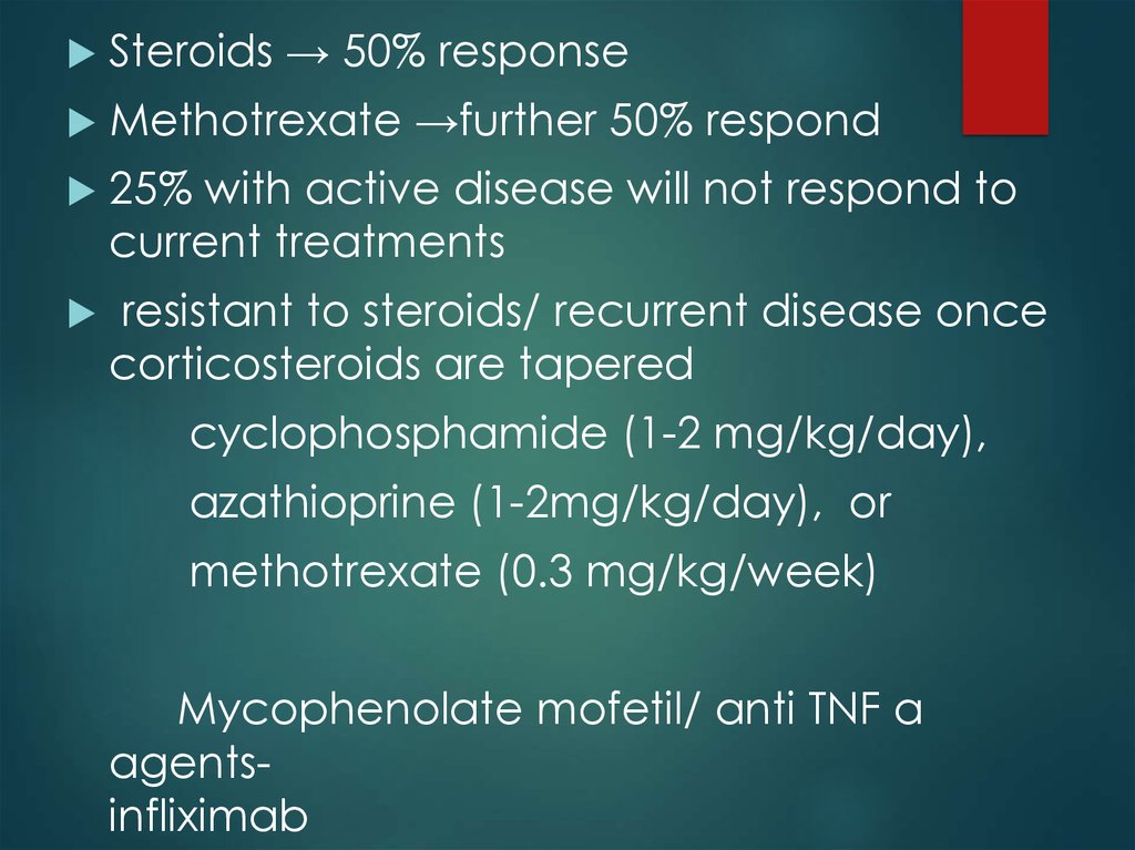
Steroids → 50% responseMethotrexate →further 50% respond
25% with active disease will not respond to
current treatments
resistant to steroids/ recurrent disease once
corticosteroids are tapered
cyclophosphamide (1-2 mg/kg/day),
azathioprine (1-2mg/kg/day), or
methotrexate (0.3 mg/kg/week)
Mycophenolate mofetil/ anti TNF α
agentsinfliximab
31.
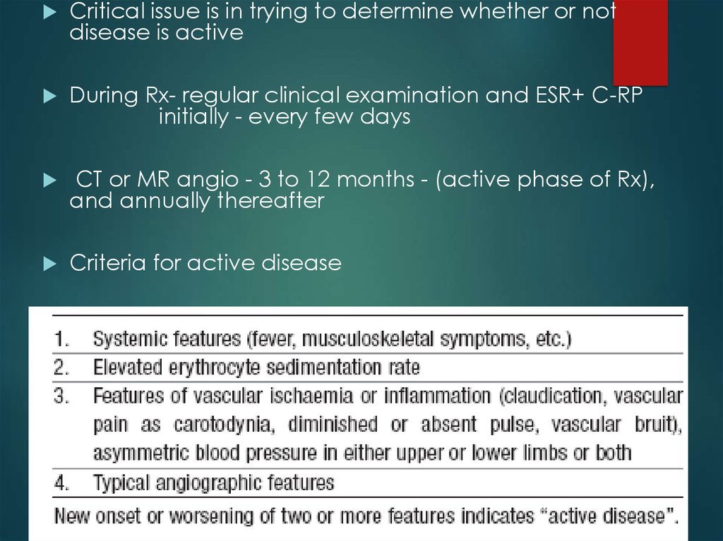
Critical issue is in trying to determine whether or notdisease is active
During Rx- regular clinical examination and ESR+ C-RP
initially - every few days
CT or MR angio - 3 to 12 months - (active phase of Rx),
and annually thereafter
Criteria for active disease
32.
chronic phase- persistent inflammationsteroids should be continued –
<1.0 mg/dL of s.C-RP and 20 mm/h of ESR
33. Surgical treatment
HTN with critical RASExtremity claudication limiting daily activities
Cerebrovascular ischaemia or critical stenoses of ≥3 cerebral
vessels
Moderate AR
Cardiac ischaemia with confirmed coronary involvement
Aneurysms
Recommended at quiescent state-avoids compli
(restenosis, anastamotic failure, thrombosis, haemorrhage, &
infection)
34.
Surgical techniquesCarry high morbidity & mortality
Steno /aneurysm -anastomotic points
Progressive nature of TA
Diffuse nature of TA
35. Renal artery involvement
Best treated by PTAStent placement following PTA
Ostial lesions
Long segment lesions
Incomplete relief of stenoses
Dissection
36.
ostial stenosis of the rightrenal artery
after deployment of a stent
37.
Renal PTA - 33 stenoses (20 pts)Indi-sev HTN,angio 70% stenosis with pr grad 20mm,
nl-ESR
Tech success -28 lesions (85%) clin success-14(82%)
Failures - Coexistent abd Ao disease & tight, prox RAS
Tech diffi - tough, noncompliant stenoses, difficult to
cross & resisted repeated, prolonged balloon
inflations - backache & ↓SBP during balloon inflation
Follow-up –mean (8/12) -restenosis in 6 (21%)
Renal PTA in TA -tech difficulties; Short-term results good, Complication rate-acceptable
Sharma s et al, AIIMS
Am J Roentgenol. 1992 Feb;158(2):41722
38. Aortoarteritic lesions
Balloon dilationsafe & reasonably effective
Can be performed repeatedly without any added risks
Balloon dilation diff from atherosclerotic lesions
Minimal intimal involvement –permits easy wiring and balloon
crossing
Resistance to dilation – high fibrotic element in the stenotic
lesion
restenosis> frequent in TA - diffuse and long
stenotic lesions
39.
Left subclavian angiograms95% stenosis with extensivecollaterals
Post angioplasty and stenting.
40.
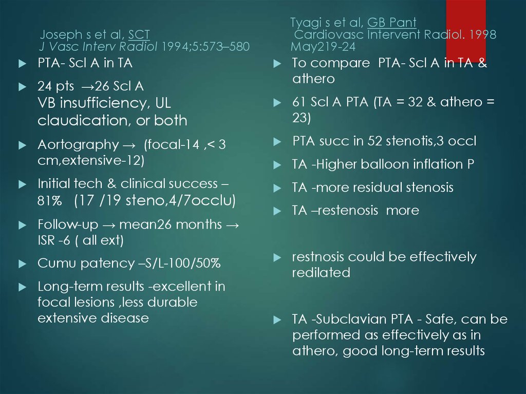
Tyagi s et al, GB PantCardiovasc Intervent Radiol. 1998
May219-24
Joseph s et al, SCT
J Vasc Interv Radiol 1994;5:573–580
PTA- Scl A in TA
24 pts →26 Scl A
To compare PTA- Scl A in TA &
athero
VB insufficiency, UL
claudication, or both
61 Scl A PTA (TA = 32 & athero =
23)
Aortography → (focal-14 ,< 3
cm,extensive-12)
PTA succ in 52 stenotis,3 occl
TA -Higher balloon inflation P
TA -more residual stenosis
TA –restenosis more
restnosis could be effectively
redilated
TA -Subclavian PTA - Safe, can be
performed as effectively as in
athero, good long-term results
Initial tech & clinical success –
81% (17 /19 steno,4/7occlu)
Follow-up → mean26 months →
ISR -6 ( all ext)
Cumu patency –S/L-100/50%
Long-term results -excellent in
focal lesions ,less durable
extensive disease
41. Aortoplasty and Stenting
PTA -desc thoracic and/or abd Ao (TA) stenosis16 pts (12+4)- HTN/severe b/l- LL claudication
Aortography – stenosis→ DTA-5, abd Ao-10, Both -1
Initial tech & clinical success -100%
patency rate of 67% in a 52-month follow-up
Follow-up (mean 21months)- Restenosis -3
PTA has a definite role in TA management
residual gradient < 20 mm -criterion for successful aortoplasty
long-segment disease, dissection or persistence of a grad > 20
mm Hg after PTBA- aortic stenting
al, SCT
Rao AS et
Radiology. 1993
42.
long-segment diffuse stenoticinvolvement of the DTA
after deployment of stents.
43. Treatment for cor A occulusion in TA
Surgery (CABG)-often not indicated
・IMA can’t be used often
occlu
of Innomi A / Scl A
calcification
of aorta
High incidence of restenosis:36
Angioplasty(PTCA)
・alternative to surgery
Very high incidence of restenosis:78
DES-effectiveness ?
44. Percutaneous Management of Aneurysmal Lesions
Percutaneous Management ofAneurysmal Lesions
Aneurysmal dilatation- isolation or together with
stenotic lesions
fusiform or saccular
one of the major complications related to the
prognosis in TA
Incidence of aneurysm rupture -low
Management - mainly surgical.
Covered stent-grafts may be useful