 physics
Similar presentations:

")
 Image Formation Processing")


")

")


Development of the Scanning Laser Doppler Microscope with Structured Illumination
1.
Development of the Scanning Laser DopplerMicroscope
with Structured Illumination
Victoria P. Fomicheva, Ivan V. Fedosov
Saratov State University, Saratov, Russia
Introduction
Signal spectrum
The report considers the development and production of
scanning laser Doppler microscope with a rotating
diffraction grating. The advantage of this scheme is its
high reliability and low requirements for collimation. The
high spatial resolution is provides by the independence
of the period of the interference bands in the measuring
volume from the wavelength of the laser radiation. The
disadvantage of the scheme is the large loss of laser
power when it passes through the optical system. This
disadvantage can be corrected by using a higher-power
light source.
Scheme of the microscope
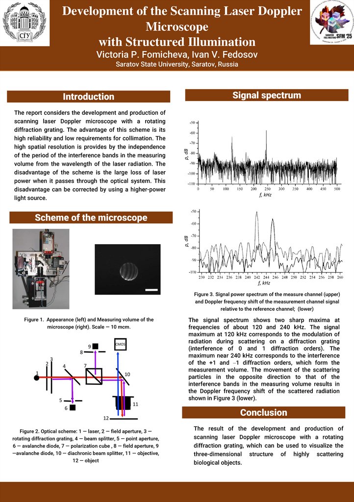
Figure 3. Signal power spectrum of the measure channel (upper)
and Doppler frequency shift of the measurement channel signal
relative to the reference channel; (lower)
Figure 1. Appearance (left) and Measuring volume of the
microscope (right). Scale — 10 mcm.
The signal spectrum shows two sharp maxima at
frequencies of about 120 and 240 kHz. The signal
maximum at 120 kHz corresponds to the modulation of
radiation during scattering on a diffraction grating
(interference of 0 and 1 diffraction orders). The
maximum near 240 kHz corresponds to the interference
of the +1 and –1 diffraction orders, which form the
measurement volume. The movement of the scattering
particles in the opposite direction to that of the
interference bands in the measuring volume results in
the Doppler frequency shift of the scattered radiation
shown in Figure 3 (lower).
Conclusion
Figure 2. Optical scheme: 1 — laser, 2 — field aperture, 3 —
rotating diffraction grating, 4 — beam splitter, 5 — point aperture,
6 — avalanche diode, 7 — polarization cube , 8 — field aperture, 9
—avalanche diode, 10 — diachronic beam splitter, 11 — objective,
12 — object
The result of the development and production of
scanning laser Doppler microscope with a rotating
diffraction grating, which can be used to visualize the
three-dimensional structure of highly scattering
biological objects.