


 biology
Similar presentations:


The sensorimotor system
1.
5The Sensorimotor System
2.
5.1 Receptor Cells Detect Various Forms of EnergyAll animals have sensory organs containing receptor cells that sense some stimuli but
not others.
Sensory organs are very diverse, but all senses use the same type of energy—action
potentials.
Labeled lines: The brain recognizes the senses as distinct because their action potentials
travel along separate nerve tracts.
Receptor potential—local change in membrane potential
Sensory transduction—the conversion of electrical energy from a stimulus into a change
in membrane potential in a receptor cell
Sensory events are encoded as streams of action potentials.
Some sensory systems employ multiple sensory receptor cells, each specializing in just
one part of a range of intensities.
The intensity of a stimulus can be represented by the number and thresholds of activated
cells.
3.
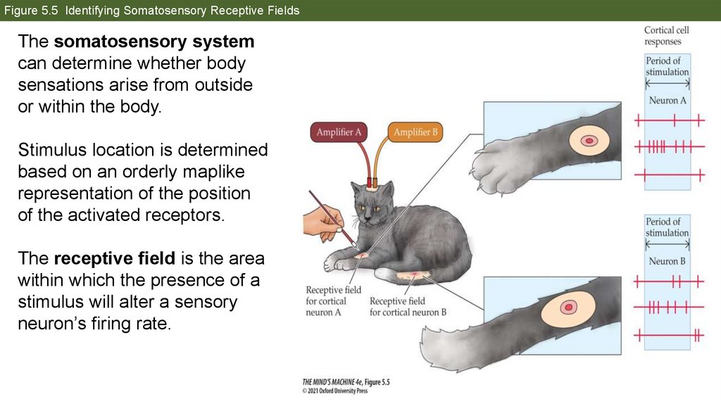
Figure 5.5 Identifying Somatosensory Receptive FieldsThe somatosensory system
can determine whether body
sensations arise from outside
or within the body.
Stimulus location is determined
based on an orderly maplike
representation of the position
of the activated receptors.
The receptive field is the area
within which the presence of a
stimulus will alter a sensory
neuron’s firing rate.
4.
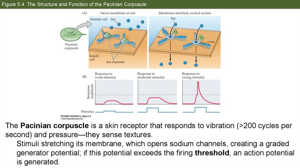
Figure 5.4 The Structure and Function of the Pacinian CorpuscleThe Pacinian corpuscle is a skin receptor that responds to vibration (>200 cycles per
second) and pressure—they sense textures.
Stimuli stretching its membrane, which opens sodium channels, creating a graded
generator potential; if this potential exceeds the firing threshold, an action potential
is generated.
5.
Figure 5.3 Receptors in SkinMeissner’s corpuscles respond to
changes in stimuli
Merkel’s discs respond to edges and
isolated points.
Ruffini corpuscles detect stretching of the
skin when we move fingers or limbs.
Free nerve endings in the skin respond to
pain, heat, and cold.
6.
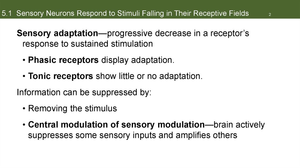
5.1 Sensory Neurons Respond to Stimuli Falling in Their Receptive FieldsSensory adaptation—progressive decrease in a receptor’s
response to sustained stimulation
• Phasic receptors display adaptation.
• Tonic receptors show little or no adaptation.
Information can be suppressed by:
• Removing the stimulus
• Central modulation of sensory modulation—brain actively
suppresses some sensory inputs and amplifies others
2
7.
5.1 Successive Levels of the CNS Process Sensory InformationEach sensory system has a distinct pathway from the periphery to
the central nervous system.
• The dorsal column system delivers touch information.
o
Receptors send axons via the dorsal spinal cord to synapse on
neurons in the brainstem.
o
Axons from those neurons cross the midline and go to the
thalamus.
o
Information about each sensory modality is sent to a different
region of the thalamus where it may be emphasized or
suppressed.
8.
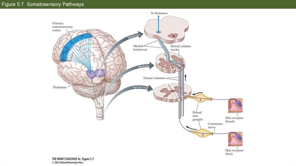
Figure 5.7 Somatosensory Pathways9.
Figure 5.8 Dermatomes10.
5.1 Successive Levels of the CNS Process Sensory Information• Primary sensory cortex—one exists for each modality
2
• Nonprimary sensory cortex, or secondary sensory cortex, receives direct
projections from the primary sensory cortex area for that modality.
Association areas in the brain process inputs from different modalities.
Polymodal neurons process input from different sensory systems.
Synesthesia is a condition in which a stimulus in one modality also creates a
sensation in another.
11.
5.2 Pain: The Body’s Emergency Signaling System2
Pain—the discomfort associated with tissue damage
Pain causes us to withdraw from its source, to engage in recuperative actions, and to warn
others.
The McGill Pain Questionnaire describes three aspects of pain:
1. Sensory-discriminative dimension
2. Motivational-affective (emotional) dimension
3. Overall cognitive-evaluative dimension
Nociceptors are peripheral receptors on free nerve endings that respond to painful stimuli.
The transient receptor potential vanilloid type1 (TRPV1), normally detects painful heat.
• This receptor also binds capsaicin, which evolved in chili peppers to ward off mammalian
predators.
12.
5.2 A Discrete Pain Pathway Projects from Body to Brain2
The transient receptor potential type M3 (TRPM3) receptor differs
from TRPV1:
• Detects even higher temperatures
• Does not respond to capsaicin
• Is found on A delta (Aδ) fibers—large myelinated axons that
register pain quickly
TRPV1 receptors are on thin, unmyelinated C fibers that conduct
more slowly, producing lasting pain.
13.
5.2 Special Neural PathwaysThe anterolateral, or spinothalamic,
system transmits the sensations of
pain and temperature to the brain.
• Nerve fibers send axons into the
dorsal horns of the spinal cord.
• They synapse on spinal neurons
that project across the midline
before ascending to the thalamus.
• Within the spinal cord, glutamate
and a peptide, substance P, are
released to boost pain signals
and remodel neurons.
14.
5.2 Pain Control Can Be DifficultThe gate control theory says that spinal “gates”—modulation sites—
control the signal that goes to the brain.
Analgesia—the absence of or reduction in pain
Opiate drugs and endogenous opioids, including the endorphins,
bind to specific receptors in the brain to reduce pain.
Epidural or intrathecal injections place opiates directly into the spinal
cord.
15.
Table 5.2 3 Types of Pain ReliefPHARMACOLOGICAL
Opiates
Bind to opioid receptors in periaqueductal gray and spinal cord
Spinal block
Blocks pain signals in spinal cord
Anti-inflammatory drugs
Block chemical inflammatory signals at the site of injury (see Figure 5.12)
Cannabinoids
Act in nociceptor endings, spinal cord, and brain
PSYCHOGENIC
Placebo
May activate endorphin-mediated pain control system
Hypnosis
Alters brain’s perception of pain
Stress
Uses both opioid and non-opioid mechanisms
Cognitive (learning, coping
strategies)
May activate endorphin-mediated pain control system
STIMULATION
TENS/mechanical
On large fibers, blocks or alters pain signal to brain
Acupuncture
Activates endogenous opioids and/or placebo-like effect, possibly modulating effect on
activity of peripheral pain pathways
Central gray
Electrically activates endorphin-mediated pain control systems, blocking pain signal in
spinal cord
16.
5.2 Pain Control Can Be Difficult2
Opiates bind to the receptors of endorphins and other endogenous
opioids.
Naloxone is an opioid antagonist.
Transcutaneous electrical nerve stimulation (TENS) relieves
pain by stimulating the nerves around the source of the pain—
nalaxone can block this analgesic effect.
The placebo effect is relief of a symptom even though the
treatment is an inert substance.
Acupuncture relieves pain by inducing endorphin release.
Stress can activate analgesia systems.
17.
5.3 Movement and the Motor System2
A reflex is a simple, stereotyped, and unlearned response to a
particular stimulus.
Acts are complex, sequential behaviors.
A motor plan, or motor program, is a set of muscle commands that
is established before the action occurs.
Electromyography (EMG) records the electrical activity of muscles.
18.
Figure 5.18 The Hierarchy of Movement Control19.
Figure 5.19 The Arrangement of Muscles around the ElbowMuscles and the skeleton work together to move the
body.
Tendons connect muscles to bone in a
reciprocal fashion.
When one muscle group contracts, it stretches
the other group—they are antagonists.
Muscles that act together to move a limb are
synergists.
Skeletal muscles are those used for movement of
the skeleton.
20.
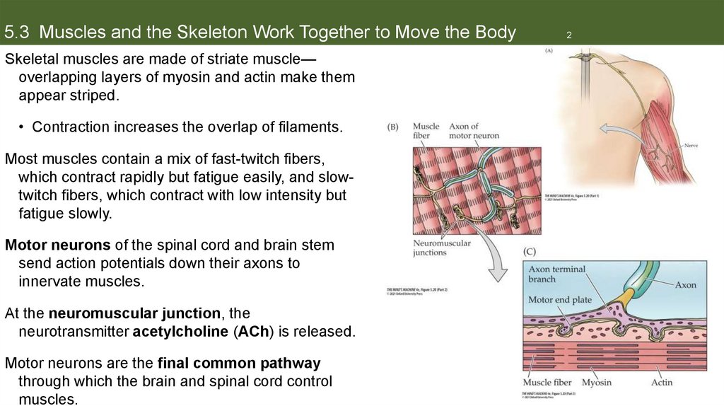
5.3 Muscles and the Skeleton Work Together to Move the BodySkeletal muscles are made of striate muscle—
overlapping layers of myosin and actin make them
appear striped.
• Contraction increases the overlap of filaments.
Most muscles contain a mix of fast-twitch fibers,
which contract rapidly but fatigue easily, and slowtwitch fibers, which contract with low intensity but
fatigue slowly.
Motor neurons of the spinal cord and brain stem
send action potentials down their axons to
innervate muscles.
At the neuromuscular junction, the
neurotransmitter acetylcholine (ACh) is released.
Motor neurons are the final common pathway
through which the brain and spinal cord control
muscles.
2
21.
5.3 Muscles and the Skeleton Work Together to Move the Body4
Proprioception is the collection of information about body movements and position.
Two kinds of proprioceptors:
• A muscle spindle is a capsule, buried in other muscle fibers, that contains intrafusal
fibers—it responds to stretch.
• Golgi tendon organs are sensitive to muscle tension.
22.
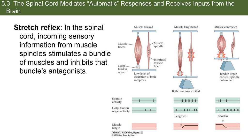
5.3 The Spinal Cord Mediates “Automatic” Responses and Receives Inputs from theBrain
Stretch reflex: In the spinal
cord, incoming sensory
information from muscle
spindles stimulates a bundle
of muscles and inhibits that
bundle’s antagonists.
23.
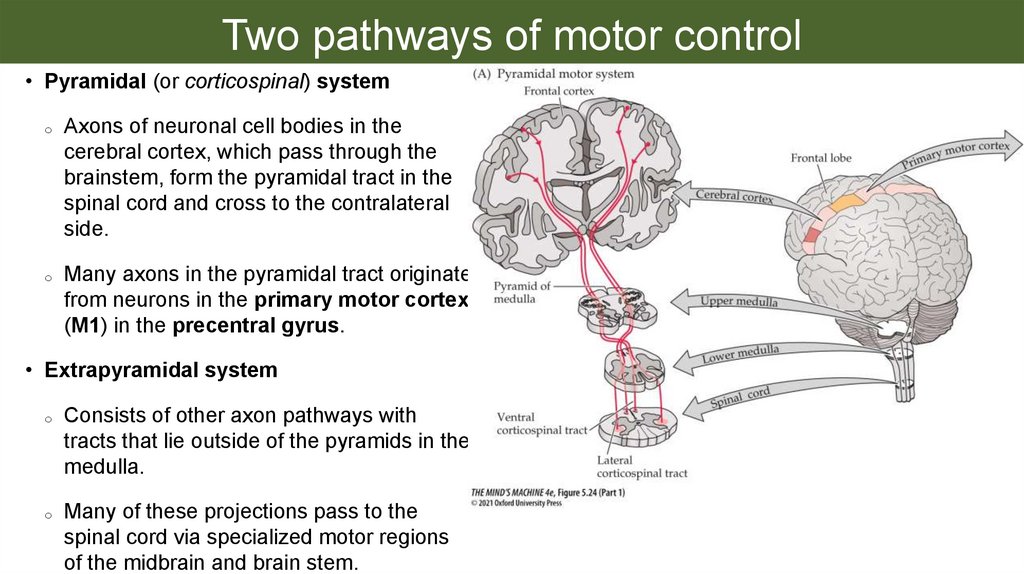
Two pathways of motor control• Pyramidal (or corticospinal) system
o
Axons of neuronal cell bodies in the
cerebral cortex, which pass through the
brainstem, form the pyramidal tract in the
spinal cord and cross to the contralateral
side.
o
Many axons in the pyramidal tract originate
from neurons in the primary motor cortex
(M1) in the precentral gyrus.
• Extrapyramidal system
o
Consists of other axon pathways with
tracts that lie outside of the pyramids in the
medulla.
o
Many of these projections pass to the
spinal cord via specialized motor regions
of the midbrain and brain stem.
24.
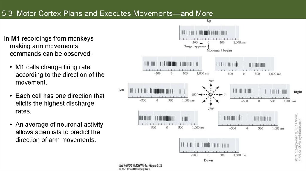
5.3 Motor Cortex Plans and Executes Movements—and MoreIn M1 recordings from monkeys
making arm movements,
commands can be observed:
• M1 cells change firing rate
according to the direction of the
movement.
• Each cell has one direction that
elicits the highest discharge
rates.
• An average of neuronal activity
allows scientists to predict the
direction of arm movements.
25.
5.3 Motor Cortex Plans and Executes Movements—and More2
Nonprimary motor cortex has two main regions:
• Supplementary motor area (SMA)
o
Medial, and important for initiation of movement sequences, especially
preplanned
• Premotor cortex
o
Anterior to M1, and activated when motor sequences are guided by
external events
Motor cortex damage can cause plegia or paresis of voluntary movements.
Damage to nonmotor zones produces complicated changes in motor control,
such as apraxia.
26.
Figure 5.28 Mirror Neurons (Part 1)A subregion of premotor
cortex (F5) contains
cells called mirror
neurons.
The same neurons fire
before making a
movement as when
observing another
individual make the
same movement.
27.
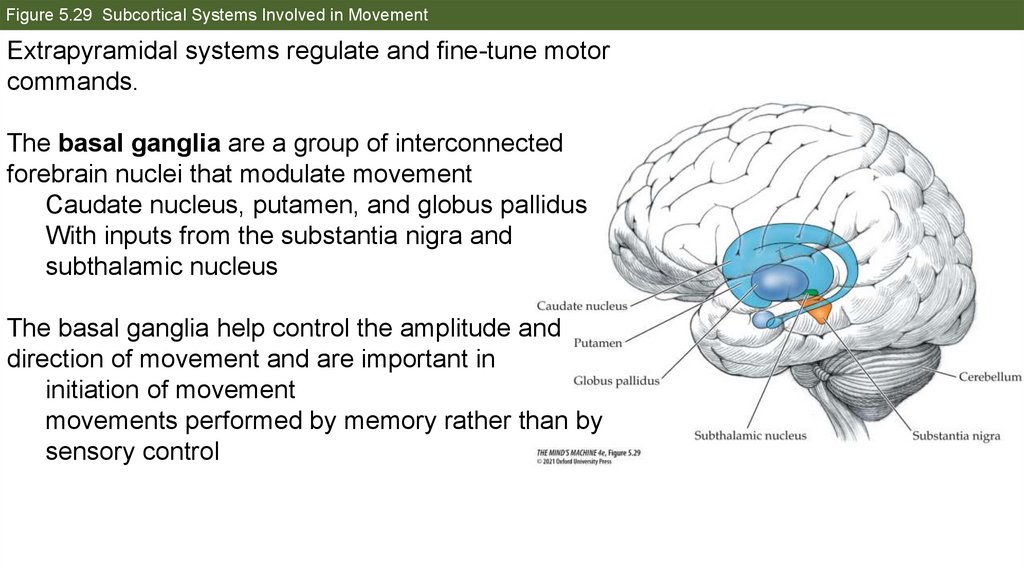
Figure 5.29 Subcortical Systems Involved in MovementExtrapyramidal systems regulate and fine-tune motor
commands.
The basal ganglia are a group of interconnected
forebrain nuclei that modulate movement
Caudate nucleus, putamen, and globus pallidus
With inputs from the substantia nigra and
subthalamic nucleus
The basal ganglia help control the amplitude and
direction of movement and are important in
initiation of movement
movements performed by memory rather than by
sensory control
28.
The cerebellum receives inputs from sensory sources and other brain motor systems.• Guides movement through inhibition
• Helps fine-tune skilled movements
29.
5.3 Extrapyramidal Systems Regulate and Fine-Tune Motor Commands4
Damage to extrapyramidal systems impairs movement.
Common symptoms of cerebellar damage include abnormal gait and
posture, especially ataxia (loss of coordination) of the legs.
Decomposition of movement describes gestures that are broken into
segments instead of being executed smoothly.
Parkinson’s disease is caused by progressive loss of dopaminergic cells in
the substantia nigra, which results in slowed movement, tremors in the
hands and face, rigid posture, and reduced facial expression.
Huntington’s disease is caused by progressive damage to the basal
ganglia, especially the caudate and putamen, which results in increasingly
excessive movement, beginning with clumsiness and twitches of the
fingers and face.