


 biology
Similar presentations:


Physiology of Bacteria
1.
Physiology of Bacteria2.
Microbial Metabolism• The primary function of all living cells is to grow
and reproduce
• Growth & reproduction rely on the outcome of
chemical reactions in the cells
• The sum of all cellular chemical reactions is
referred to as metabolism
3.
Microbial Metabolism• The metabolic process that involves the
degradation of chemical components is called
catabolism
• The synthesis of chemical components is called
anabolism or biosynthesis
4.
• Most metabolic processes in the cell would takeforever if it were not for enzymes
• Enzymes are proteins that have molecular
weights ranging from 600 to 12 000
▫ Their function is to speed up the various chemical
reactions that occur in the cell
▫ Molecules that speed up chemical reactions are
called catalysts
▫ Enzymes often cannot function alone and require
additional molecules, called cofactors, to enhance
activity
5.
Classification of enzymes• Oxidoreductases are involved in electron (
hydrogen) transfer reactions
• Transferases transfer specific groups such as
aldehydes or phosphates from one substrate to
another
• Hydrolyses add water across chemical bonds
to be cleaved or hydrolyzed
6.
Classification of enzymes• Lyases remove chemical groups from
substrates, forming double bonds, or add
chemical groups to double bonds
• Isomerases rearrange certain compounds to
produce molecules having the same groups of
atoms, but in different arrangements
• Ligases produce bonds accompanied by the
cleavage of ATP
7.
Classification of enzymes• Enzymes synthesized by the cell remain within
the cell to carry out specific reactions and are
called endoenzymes
• Enzymes relased from the cell into the
surrounding environment and are called
exoenzymes
8.
Classification of enzymes• Pathogenicity enzymes – are enzymes that
damage cells and tissues
▫ Coagulase – enables the organisms to clot plasma
to form a sticky coat of fibrin around themselves
for protection from phagocytes and other body
defense machanisms (Staphylococcus)
▫ Kinases – reffered to as fibrinolysin, kinase has
opposite effect of coagulase. Streptokinase, for
example, lyses fibrin clots, thus enabling
streptococci to invade and spread throughout the
body
9.
Classification of enzymes• Hyaluronidase – enables pathogens to spread through
connective tissue by breaking down hyaluronic acid, the
“cement” that holds tissue cells together
(Staphylococcus, Streptococcus and Clostridium)
• Collagenase – This enzyme breaks down collagen, the
supportive protein founding tendons, cartilage and
bones. Cl. perfringens a major cause of gas gangrene,
spreads deeply within the body by secreting both
collagenase and hyaluronidase
10.
Classification of enzymes• Hemolysin – enzyme that cause damage to the
host’s red blood cells. In the laboratory,
hemolysis of the red blood cells in the blood agar
is useful for identifying types of Staphylococcus
and Streptococcus
• Lecithinase – one of the toxins produced by
Staphylococcus aureus, which breaks down
phospholipids collectively referred to as lecithin
• Leukocidin – enzyme secreted some
Staphylococcus aureus causes destruction
11.
Classification of enzymes• Hyaluronidase – enables pathogens to spread through
connective tissue by breaking down hyaluronic acid, the
“cement” that holds tissue cells together
(Staphylococcus, Streptococcus and Clostridium).
• Collagenase – This enzyme breaks down collagen, the
supportive protein founding tendons, cartilage and
bones. Cl. perfringens a major cause of gas gangrene,
spreads deeply within the body by secreting both
collagenase and hyaluronidase.
12.
Growth & Multiplication of Bacteria• Bacteria divide by binary fission
• Bacterial cell divides to form two daughter
cells
• Nuclear division precedes cell division & in
a growing population many cells carrying
two nuclear bodies can be seen
13.
14.
• The interval of time between two cell division, orthe time required for a bacterium to give rise to
two daughter cells under optimum conditions, is
known as the generation time or population
doubling time
15.
Growth & Multiplication of Bacteria• Bacteria divide by binary fission
• Bacterial cell divides to form two daughter
cells
• Nuclear division precedes cell division & in
a growing population many cells carrying
two nuclear bodies can be seen
16.
17.
• In many medically important bacteria, thegeneration time is about 20 minutes
• Some bacteria are slow-growing
Tubercle bacilli the generation time is about 20 hours
Lepra bacilli about 20 days
• Bacteria reproduce so rapidly & by geometric
progression, a single bacterial cell can
theoretically give rise to 1021 progeny in 24
hours, with a mass of approximately 4,000
tones!
18.
• When bacteria are grown in a vessel of liquid medium(batch culture), multiplication is arrested after a few cell
divisions due to depletion of nutrients or accumulation
of toxic products
• When pathogenic bacteria multiply in host tissues, the
situation may be intermediate between a batch culture &
a continuous culture
• Bacteria growing on solid media form colonies
• Each colony represents a clone of cells derived from a
single parent cell
• In liquid media, growth is diffuse
19.
Bacterial cell Growth Curve• A- Lag phase
▫ Immediately following the seeding of a culture medium
▫ This initial period is the time required for adaptation to
the new environment
▫ There is no increase in numbers, though there may be
an increase in the size of the cells
• B- Log (logarithmic) or exponential phase
▫ The cells start dividing & their numbers increase
exponentially or by geometric progression
20.
Bacterial cell Growth Curve• C- Stationary phase
▫ After a period of exponential growth, cell
division stops due to depletion of nutrients &
accumulation of toxic products
▫ The viable count remains stationary as an
equilibrium exists between the dying cells and
the newly formed cells
• D- Phase of Decline
▫ Population decreases due to cell death
21.
Bacterial cell Growth Curve22.
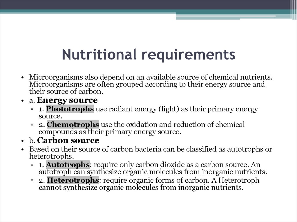
Nutritional requirements• Microorganisms also depend on an available source of chemical nutrients.
Microorganisms are often grouped according to their energy source and
their source of carbon.
• a. Energy source
▫ 1. Phototrophs use radiant energy (light) as their primary energy
source.
▫ 2. Chemotrophs use the oxidation and reduction of chemical
compounds as their primary energy source.
• b. Carbon source
• Based on their source of carbon bacteria can be classified as autotrophs or
heterotrophs.
▫ 1. Autotrophs: require only carbon dioxide as a carbon source. An
autotroph can synthesize organic molecules from inorganic nutrients.
▫ 2. Heterotrophs: require organic forms of carbon. A Heterotroph
cannot synthesize organic molecules from inorganic nutrients.
23.
Nutritional types in bacterial metabolismNutritional type
Phototrophs
Lithotrophs
Organotrophs
Source of
energy
Source of carbon
Sunlight
Organic compounds
(photoheterotrophs)
or carbon fixation
(photoautotrophs)
Examples
Cyanobacteria,
Green sulfur
bacteria, Chloroflexi,
or Purple bacteria
Inorganic
compounds
Organic compounds
Thermodesulfobacteria,
(lithoheterotrophs) or
Hydrogenophilaceae,
carbon fixation
or Nitrospirae
(lithoautotrophs)
Organic
compounds
Organic compounds
(chemoheterotrophs)
or carbon fixation
(chemoautotrophs)
Bacillus, Clostridium or
Enterobacteriaceae
24.
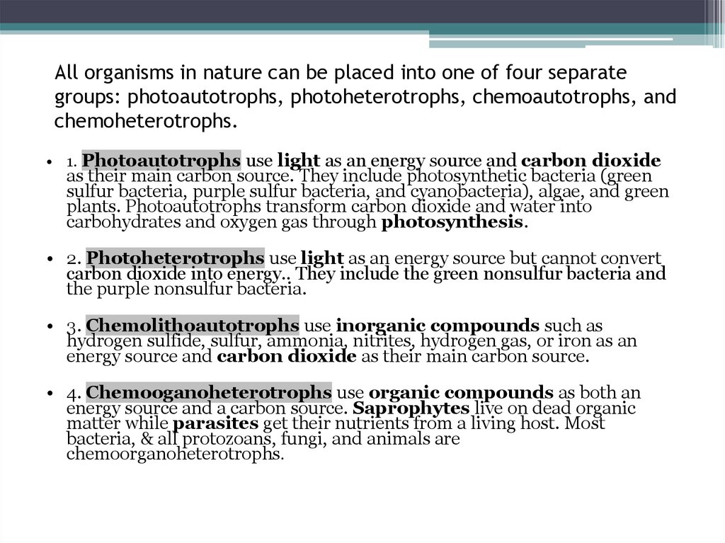
All organisms in nature can be placed into one of four separategroups: photoautotrophs, photoheterotrophs, chemoautotrophs, and
chemoheterotrophs.
• 1. Photoautotrophs use light as an energy source and carbon dioxide
as their main carbon source. They include photosynthetic bacteria (green
sulfur bacteria, purple sulfur bacteria, and cyanobacteria), algae, and green
plants. Photoautotrophs transform carbon dioxide and water into
carbohydrates and oxygen gas through photosynthesis.
• 2. Photoheterotrophs use light as an energy source but cannot convert
carbon dioxide into energy.. They include the green nonsulfur bacteria and
the purple nonsulfur bacteria.
• 3. Chemolithoautotrophs use inorganic compounds such as
hydrogen sulfide, sulfur, ammonia, nitrites, hydrogen gas, or iron as an
energy source and carbon dioxide as their main carbon source.
• 4. Chemooganoheterotrophs use organic compounds as both an
energy source and a carbon source. Saprophytes live on dead organic
matter while parasites get their nutrients from a living host. Most
bacteria, & all protozoans, fungi, and animals are
chemoorganoheterotrophs.
25.
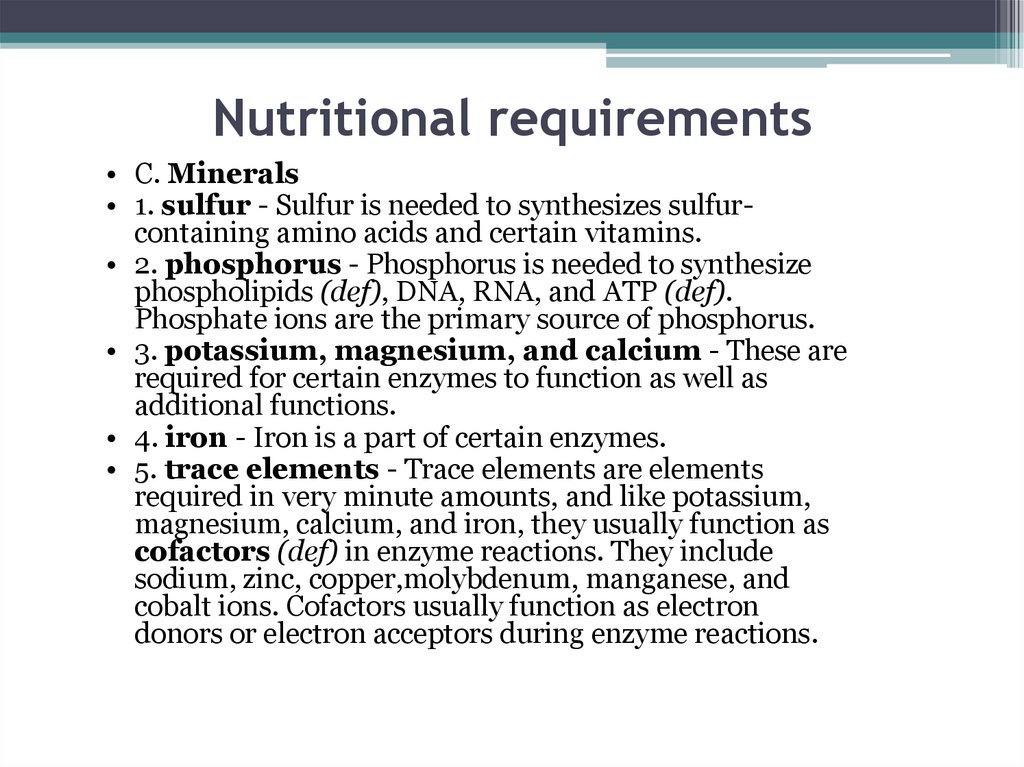
Nutritional requirements• C. Minerals
• 1. sulfur - Sulfur is needed to synthesizes sulfurcontaining amino acids and certain vitamins.
• 2. phosphorus - Phosphorus is needed to synthesize
phospholipids (def), DNA, RNA, and ATP (def).
Phosphate ions are the primary source of phosphorus.
• 3. potassium, magnesium, and calcium - These are
required for certain enzymes to function as well as
additional functions.
• 4. iron - Iron is a part of certain enzymes.
• 5. trace elements - Trace elements are elements
required in very minute amounts, and like potassium,
magnesium, calcium, and iron, they usually function as
cofactors (def) in enzyme reactions. They include
sodium, zinc, copper,molybdenum, manganese, and
cobalt ions. Cofactors usually function as electron
donors or electron acceptors during enzyme reactions.
26.
Nutritional requirements• D. Water
• E. Growth factors
Growth factors are organic compounds such as amino
acids (def), purines (def), pyrimidines (def), and
vitamins (def) that a cell must have for growth but
cannot synthesize itself. Organisms having complex
nutritional requirements and needing many growth
factors are said to be fastidious.
27.
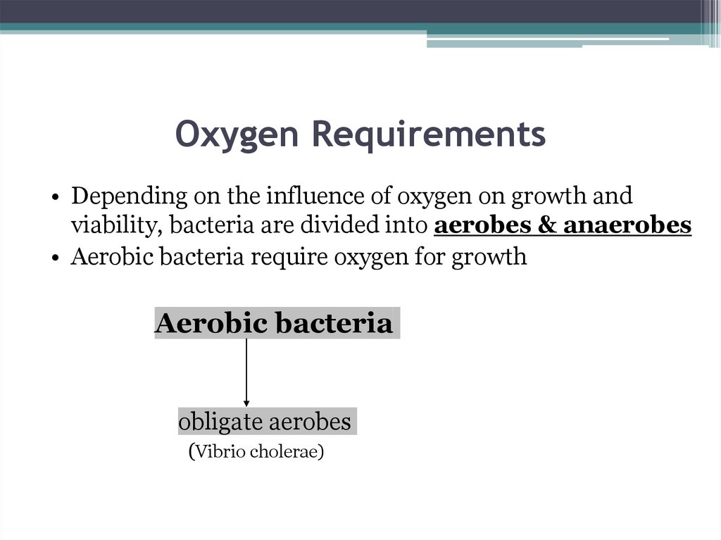
Oxygen Requirements• Depending on the influence of oxygen on growth and
viability, bacteria are divided into aerobes & anaerobes
• Aerobic bacteria require oxygen for growth
Aerobic bacteria
obligate aerobes
(Vibrio cholerae)
28.
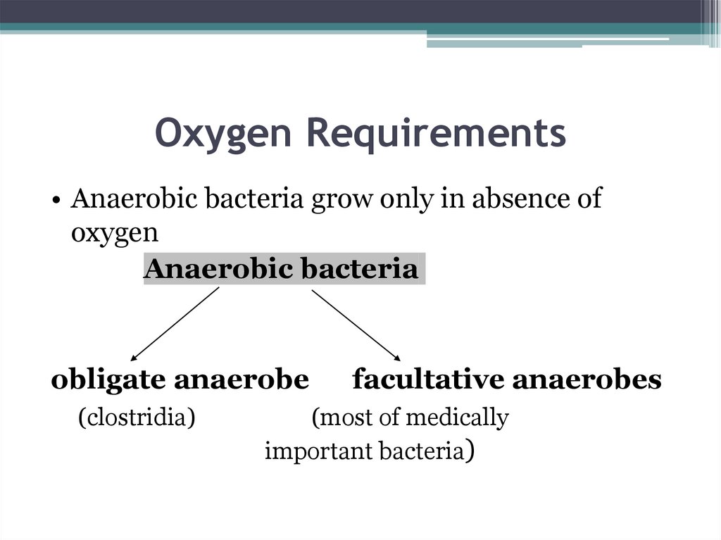
Oxygen Requirements• Anaerobic bacteria grow only in absence of
oxygen
Anaerobic bacteria
obligate anaerobe
(clostridia)
facultative anaerobes
(most of medically
important bacteria)
29.
Oxygen requirements can beclassified
• Obligate aerobes — which can grow only in
the presence of oxygen (e.g., P. aeruginosa)
• Obligate anaerobes are organisms that grow
only in the absence of oxygen and, in fact, are
often inhibited or killed by its presence. They
obtain their energy through anaerobic
respiration or fermentation. (e.g., Clostridium
botulinum Clostridium tetani, etc.)
• Facultative anaerobes which are ordinary
aerobes but can also grow without oxygen (e.g.,
E. coli). Most of the pathogenic bacteria are
facultative aerobes.
30.
Oxygen requirements can beclassified
• Microaerophiles are organisms that require a
low concentration of oxygen (2% to 10%) for
growth, but higher concentrations are inhibitory.
They obtain their energy through aerobic
respiration. (e.g., Campylobacter jejuni).
• Aerotolerant anaerobes like obligate
anaerobes, cannot use oxygen to transform
energy but can grow in its presence. They obtain
energy only by fermentation and are known as
obligate fermenters.
31.
Physical requirements• Temperature
▫ 1. Psychrophiles are cold-loving bacteria. Their
optimum growth temperature is between -5C and
15C. They are usually found in the Arctic and
Antarctic regions and in streams fed by glaciers.
▫ 2. Mesophiles are bacteria that grow best at
moderate temperatures. Their optimum growth
temperature is between 25C and 45C. Most
bacteria are mesophilic and include common soil
bacteria and bacteria that live in and on the body.
32.
• pH• Microorganisms can be placed in one of
the following groups based on their
optimum pH requirements:
• 1. Neutrophiles grow best at a pH range of
5 to 8.
• 2. Acidophiles grow best at a pH below
5.5.
• 3. Allaliphiles grow best at a pH above 8.5.
33.
Culture Media• A growth medium or culture medium is a
substance in which microorganisms or cells can
grow
• There are two major types of growth media:
those used for cell culture, which use specific cell
types derived from plants or animals, and
microbiological culture, which are used for
growing microorganisms, such as bacteria or
yeast
34.
Types of Growth Media• The most common growth media for
microorganisms are nutrient broths (liquid
nutrient medium) or Lysogeny broth (LB
medium). Bacteria grown in liquid cultures often
form colloidal suspensions.
• Liquid mediums are often mixed with agar and
poured into petri dishes to solidify. These agar
plates provide a solid medium on which
microbes may be cultured.
35.
Types of Growth Media• Nutrient media
Undefined media (also known as basal or
complex media)
Defined media (also known as chemical
defined media)
Differential medium some sort of indicator,
typically a dye, is added, that allows for the
differentiation of particular chemical reactions
occurring during growth
36.
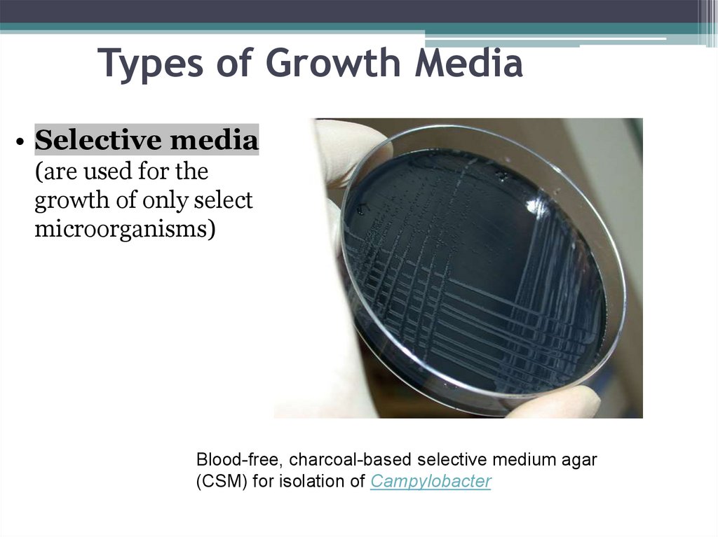
Types of Growth Media• Selective media
(are used for the
growth of only select
microorganisms)
Blood-free, charcoal-based selective medium agar
(CSM) for isolation of Campylobacter
37.
Types of Growth Media• Differential media or indicator media
distinguish one microorganism type from
another growing on the same media
(MacConkey’s, Nagler’s medium)
This type of media uses the biochemical
characteristics of a microorganism growing in
the presence of specific nutrients or indicators
(such as neutral red, phenol red, eosin y,
or methylene blue)
38.
Types of Growth Media39.
Types of Growth Media• Enriched media contain the nutrients
required to support the growth of a wide variety
of organisms
▫ Blood agar is an enriched medium in which
nutritionally rich whole blood supplements the
basic nutrients.
▫ Chocolate agar is enriched with heat-treated
blood (40-45°C), which turns brown and gives the
medium the color for which it is named.
40.
Blood agar plates are often used to diagnose infection. On the rightis a positive Staphylococcus infection; on the left a positive
Streptococcus culture.
41.
Types of Growth Media• Transport media used for the temporary
storage of specimens being transported to the
laboratory for cultivation. Transport media
typically contain only buffers and salt (Stuart’s
medium for gonococci, buffeerd glycerol saline
for enteric bacilli ).
• Indicator media contain an indicator which
chainges colour when a bacterium grows in them
(Bismuth sulphite media(S.typhi), potassium
tellurite(diphteria bacilli).
42.
Types of Growth Media• Sugar Media used for sugar fermentation
(Hiss’serum sugars)
▫ The sugar media consist of 1% of the sugar in peptone
water along with an appropriate indicator
▫ Durham’s tube is kept inverted in the sugar tube to
detect gas production
• Anaerobic media are used to grow anaerobic
organisms (Robertson’s cooked meat medium)
43.
• Isolation of bacteria forms a very significantstep in the diagnosis and management of the
illness.
• Isolation of bacteria involves various steps –
• z Specimen collection
• z Preservation and transportation of specimen
• z Microscopic examination of sample
• z Various methods used for isolation of
bacteria
44.
• Common specimens include urine,faeces, wound swabs, throat swabs,
vaginal swabs, sputum, and blood.
Less common, but important
specimens include cerebrospinal
fluid, pleural fluid, joint aspirates,
tissue, bone and prosthetic
material (e.g. line tips).
45.
• It is preferred to obtain thesamples for bacteriological culture
before antibiotic therapy is started.
This maximizes the sensitivity of
the investigations and reduces
false-negative results.
46.
• Specimens must be accuratelylabelled and accompanied by a
properly completed requisition
form, indicating the nature of the
specimen, the date of sample
collection,
relevant
clinical
information, the investigations
required, and details of antibiotic
therapy, if any.
47.
• Specimens should be transported as soon aspossible to the laboratory. In case a delay is
anticipated the specimen should be stored
at 4° C.
• Immediate transport is necessary in order
to:
• (i) Preserve the viability of the ‘delicate’
bacteria, such as Streptococcus pneumoniae
or Haemophilus influenzae (delays in
processing can cause false-negative culture
results);
48.
• (ii) Minimize the multiplication of bacteria(e.g. coliforms) within specimens before
they reach the laboratory. In particular
urine and other specimens that utilize a
semiquantitative culture technique for
their detection, as delays in transport can
give rise to falsely high bacterial counts
when the specimen is processed.
49.
CULTURE ON SOLID MEDIA• The principal method for the
detection of bacteria from clinical
specimens is by culture on solid
culture media. Bacteria grow on the
surface of culture media to produce
distinct colonies.
50.
• Different bacteria produce differentbut characteristic colonies, allowing
for early presumptive identification
and easy identification of mixed
cultures.
• There are many different types of
culture media
51.
Types of Growth Media52.
Blood agar plates are often used to diagnose infection. On the rightis a positive Staphylococcus infection; on the left a positive
Streptococcus culture.
53.
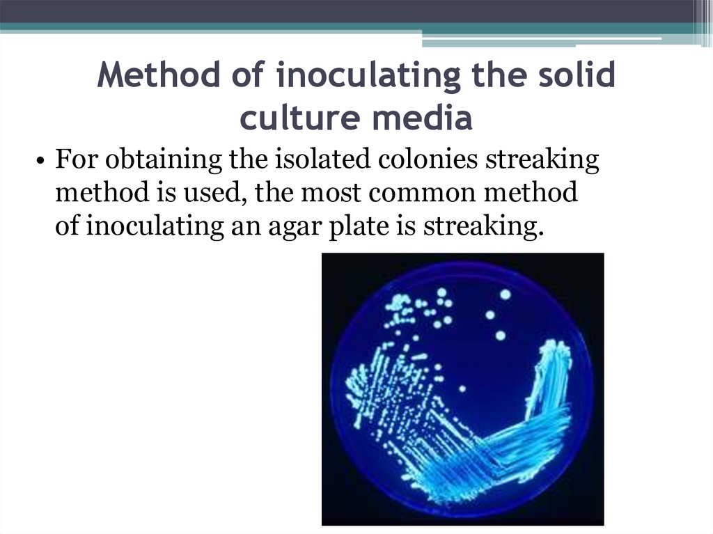
Method of inoculating the solidculture media
• For obtaining the isolated colonies streaking
method is used, the most common method
of inoculating an agar plate is streaking.
54.
• In this method single bacterial cellsget isolated by the streaking, and
when the plate is incubated, forming
discrete colonies that will have
started from just one bacterium each
55.
Colony Morphology of Bacteria• Bacteria grow on solid media as colonies. A
colony is defined as a visible mass of
microorganisms all originating from a single
mother cell. Key features of these bacterial
colonies serve as an important criteria for their
identification.
56.
57.
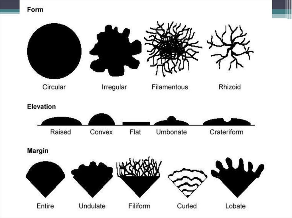
1.Form of the bacterial colony: – Theform refers to the shape of the colony.
These forms represent the most common
colony shapes you are likely to encounter.
e.g. Circular, Irregular, Filamentous,
Rhizoid etc.
2.Elevation of bacterial colony: This
describes the “side view” of a colony. These
are the most common. e.g. Flat, raised,
umbonate (having a knobby
protuberance), Crateriform, Convex,
Pulvinate (Cushion-shaped)
58.
• Margin of bacterial colony: The marginor edge of a colony may be an important
characteristic
in
identifying
an
organisms. Common examples are Entire
(smooth),
irregular,
Undulate
(wavy), Lobate, Curled, Filiform etc.
Colonies that are irregular in shape and/or
have irregular margins are likely to be
motile organisms.
59.
1.Size of the bacterial colony: The size of thecolony can be a useful characteristic for
identification. The diameter of a representative
colony may be measured in millimeters or
described in relative terms such as pin point,
small, medium, large. Colonies larger than
about 5 mm are likely to be motile organisms.
60.
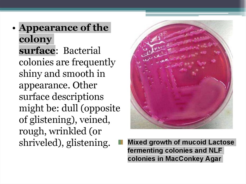
• Appearance of thecolony
surface: Bacterial
colonies are frequently
shiny and smooth in
appearance. Other
surface descriptions
might be: dull (opposite
of glistening), veined,
rough, wrinkled (or
shriveled), glistening.
Mixed growth of mucoid Lactose
fermenting colonies and NLF
colonies in MacConkey Agar
61.
• Color of the colonies (pigmentation):Some bacteria produce pigment when they
grow in the medium e.g., green pigment
produces by Pseudomonas aeruginosa,
buff colored colonies of Mycobacterium
tuberculosis in L.J medium, red colored
colonies of Serratia marcescens.
62.
• Opacity of the bacterial colony: Isthe colony transparent (clear),
opaque (not transparent or clear),
translucent (almost clear, but
distorted vision–like looking
through frosted glass), iridescent
(changing colors in reflected
light).