


 medicine
 biology
Similar presentations:


")

Blood
1.
PhD Inna A. DemyanenkoHistology department MA CFU Simferopol
Blood
2.
3.
Blood is a specialized connective tissue that consistsof formed elements
(erythrocytes, leukocytes, and platelets) and a fluid
component called
plasma.
4.
5.
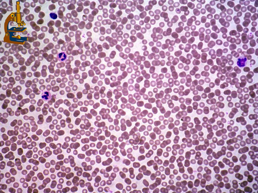
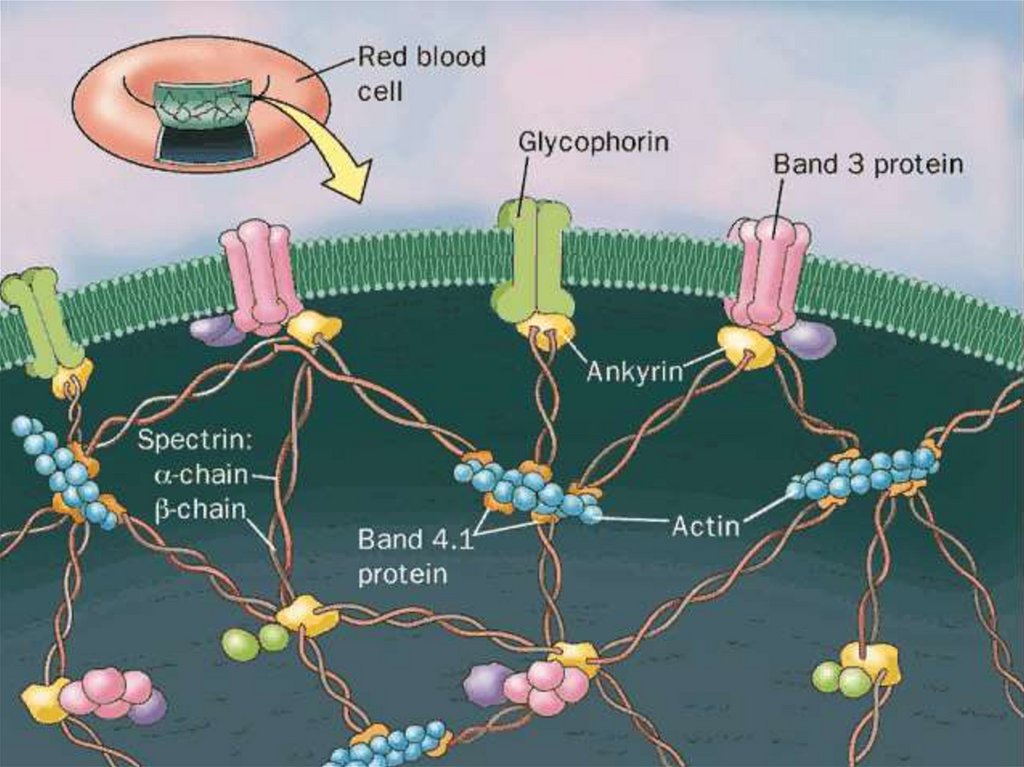
Erythrocytes [red blood cells (RBCs)]General features:
1. RBCs are round, anucleate, biconcave cells that stain
with Romanovsky-Giemsa method.
2. Mature erythrocytes possess no organelles but are filled
with hemoglobin (Hb).
3. Several cytoskeletal proteins (ankyrin, band 4.1 and band
3 proteins, spectrin, and actin) function in maintaining the
shape of RBCs
Carbohydrate determinants for the A, B, and 0 blood groups
are located on the external surface of their plasmalemmae.
6.
7.
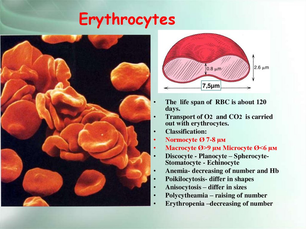
Erythrocytes7,5μm
The life span of RBC is about 120
days.
Transport of O2 and CO2 is carried
out with erythrocytes.
Classification:
Normocyte Ø 7-8 µм
Macrocyte Ø>9 µм Microcyte Ø<6 µм
Discocyte - Planocyte – SpherocyteStomatocyte - Echinocyte
Anemia- decreasing of number and Hb
Poikilocytosis- differ in shapes
Anisocytosis – differ in sizes
Polycytheamia – raising of number
Erythropenia –decreasing of number
8.
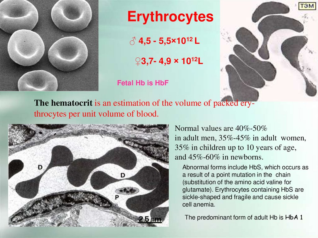
Erythrocytes♂ 4,5 - 5,5×10¹² L
♀3,7- 4,9 × 10¹²L
Fetal Hb is HbF
The hematocrit is an estimation of the volume of packed erythrocytes per unit volume of blood.
Normal values are 40%-50%
in adult men, 35%-45% in adult women,
35% in children up to 10 years of age,
and 45%-60% in newborns.
Abnormal forms include HbS, which occurs as
a result of a point mutation in the chain
(substitution of the amino acid valine for
glutamate). Erythrocytes containing HbS are
sickle-shaped and fragile and cause sickle
cell anemia.
The predominant form of adult Hb is HbA 1
9.
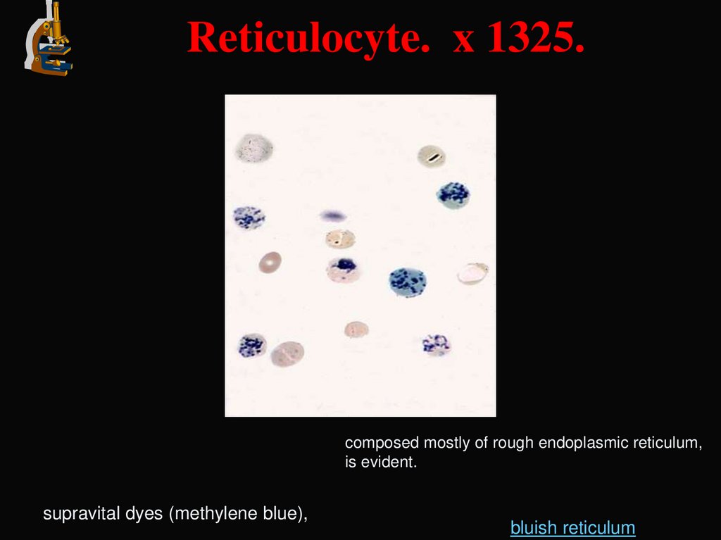
Reticulocyte. x 1325.composed mostly of rough endoplasmic reticulum,
is evident.
supravital dyes (methylene blue),
bluish reticulum
10.
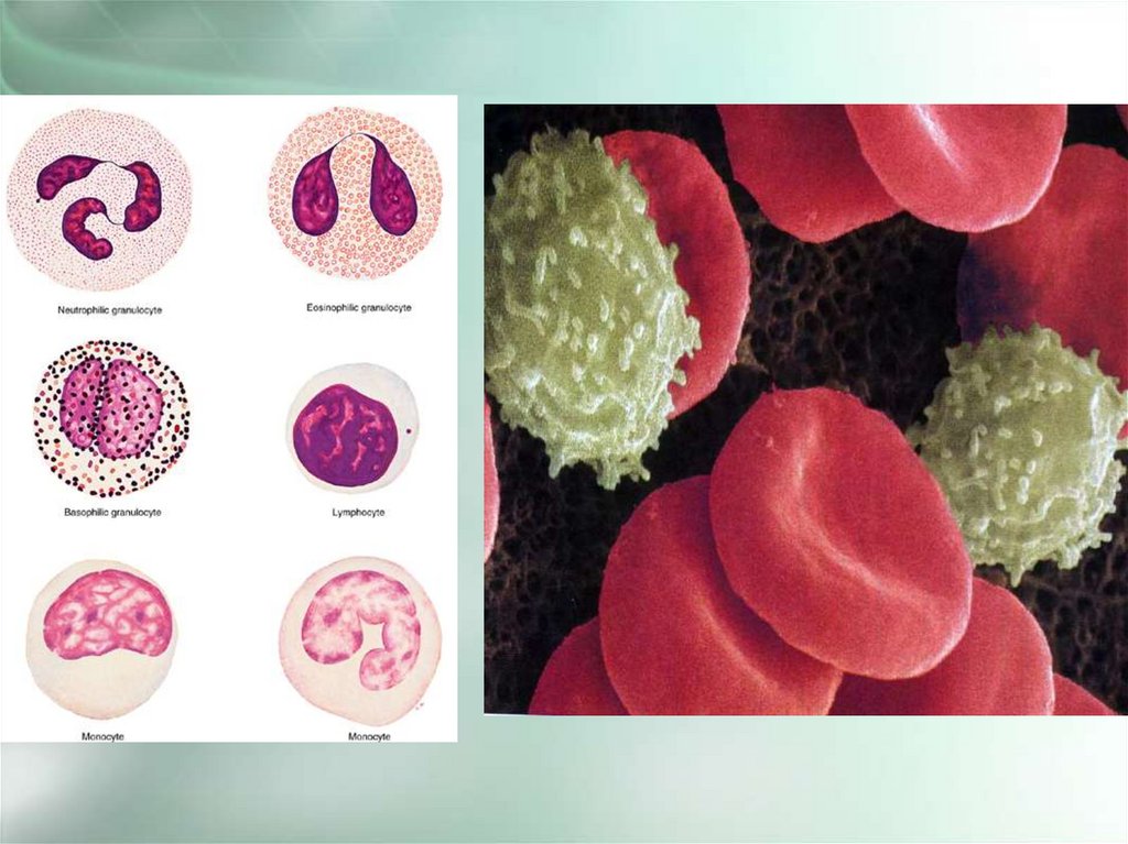
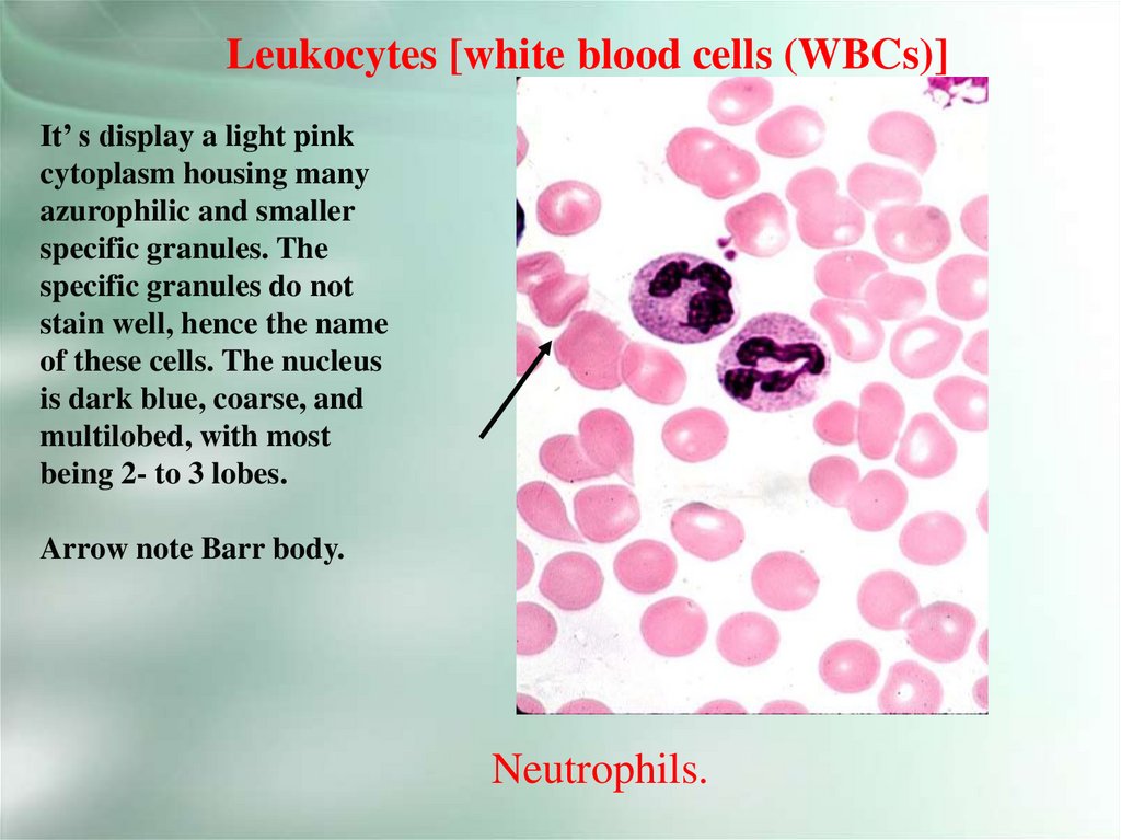
Leukocytes [white blood cells (WBCs)]It’ s display a light pink
cytoplasm housing many
azurophilic and smaller
specific granules. The
specific granules do not
stain well, hence the name
of these cells. The nucleus
is dark blue, coarse, and
multilobed, with most
being 2- to 3 lobes.
Arrow note Barr body.
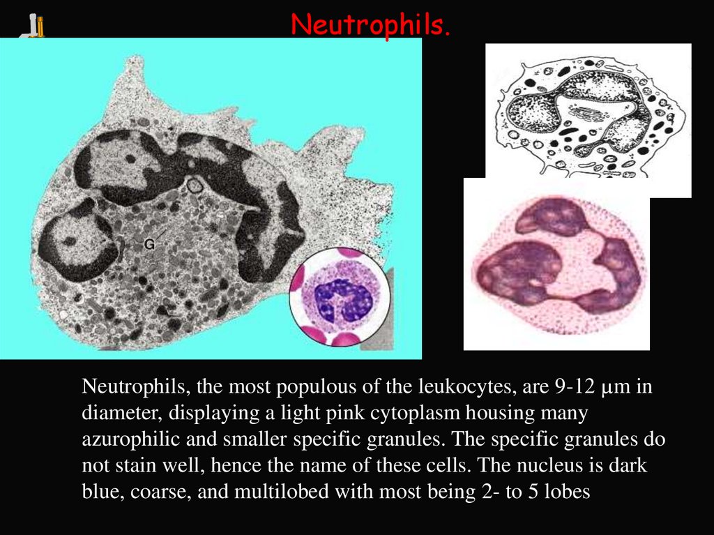
Neutrophils.
11.
Neutrophils.Neutrophils, the most populous of the leukocytes, are 9-12 µm in
diameter, displaying a light pink cytoplasm housing many
azurophilic and smaller specific granules. The specific granules do
not stain well, hence the name of these cells. The nucleus is dark
blue, coarse, and multilobed with most being 2- to 5 lobes
12.
Neutrophils.Young (bean-shaped nucleus) -0,5%; band -1-6% (S- or horseshoe- shaped nucleus), segmented (multilobed nucleus) - 47-72%
13.
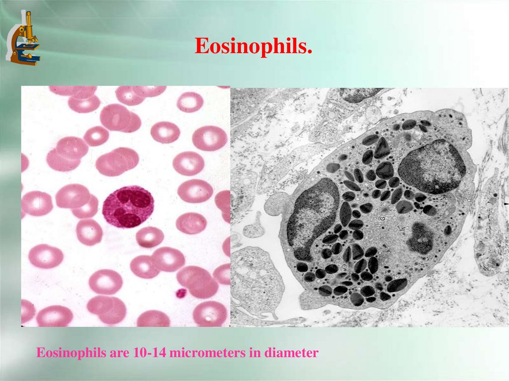
Eosinophils.Eosinophils are 10-14 micrometers in diameter
14.
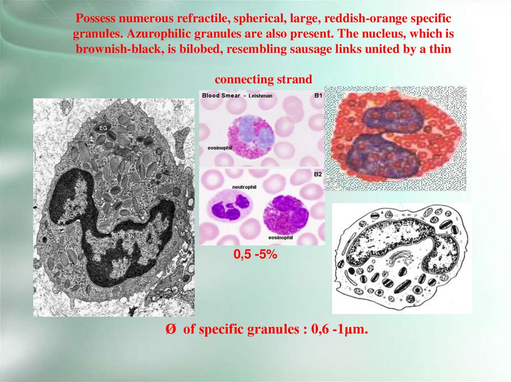
Possess numerous refractile, spherical, large, reddish-orange specificgranules. Azurophilic granules are also present. The nucleus, which is
brownish-black, is bilobed, resembling sausage links united by a thin
connecting strand
0,5 -5%
Ø of specific granules : 0,6 -1μm.
15.
Basophils.16.
Basophils contein large metachromatic granulesØ of specific granules : 0,5 -1.2μm
0 – 1%
Size – 11-12 μm. Participate inflammation, specific granules :presence of
heparin,histamine,proteases + azurophilic granules – lysosomes.
17.
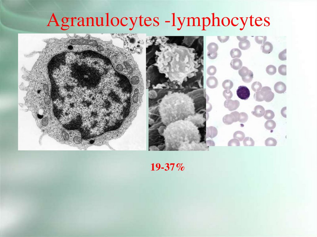
Agranulocytes -lymphocytes19-37%
18.
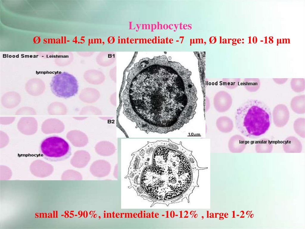
LymphocytesØ small- 4.5 μm, Ø intermediate -7 μm, Ø large: 10 -18 μm
small -85-90%, intermediate -10-12% , large 1-2%
19.
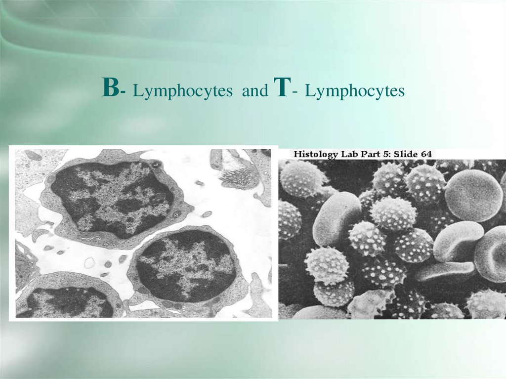
В- Lymphocytesand Т- Lymphocytes
20.
Presence of Ig-receptors on the plasmolemma note onВ- lymphocyte (from the left) , аbsence of them - on
Т- lymphocyte (from the right).
SEM of lymphocytes
21.
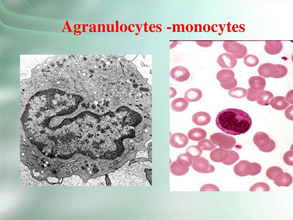
Agranulocytes -monocytes22.
MonocytesØ
18-20 μm
3-11% in blood
23.
24.
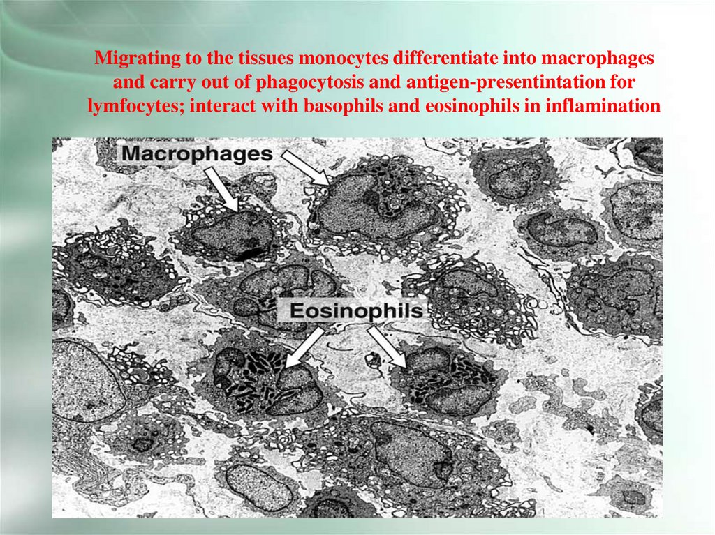
Migrating to the tissues monocytes differentiate into macrophagesand carry out of phagocytosis and antigen-presentintation for
lymfocytes; interact with basophils and eosinophils in inflamination
25.
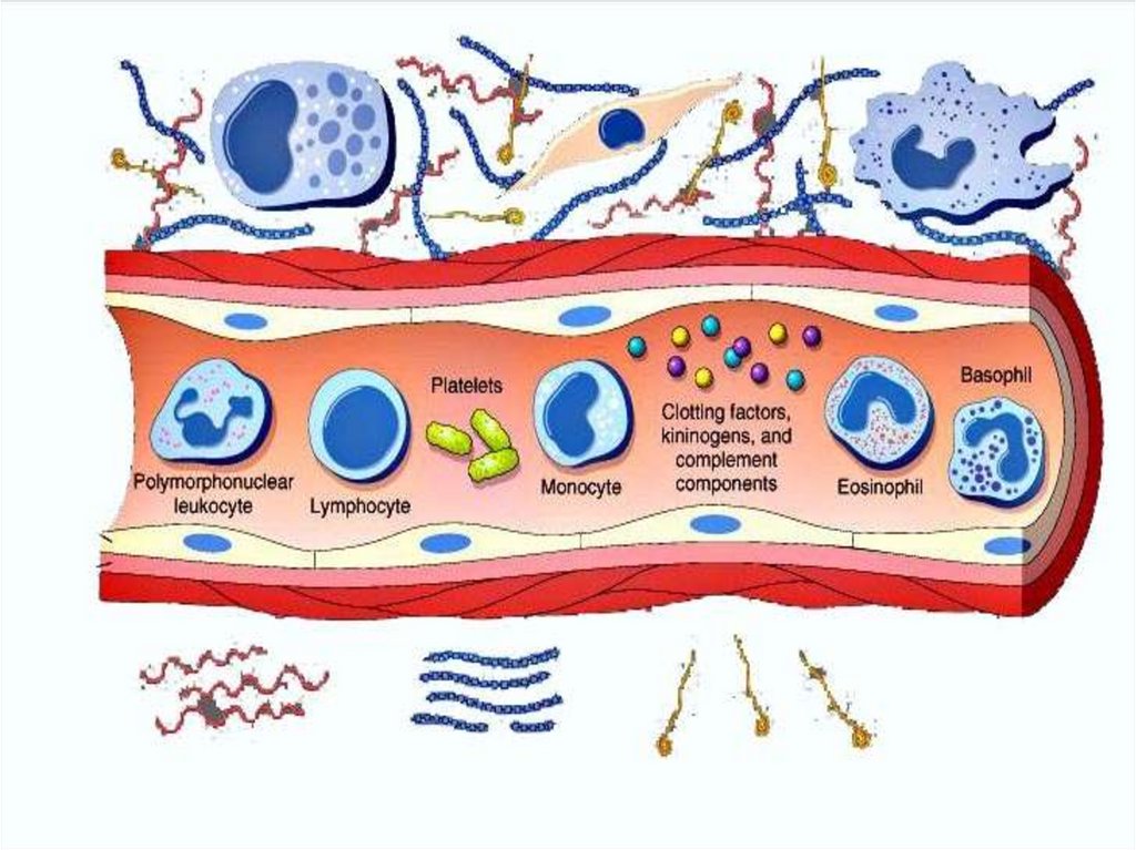
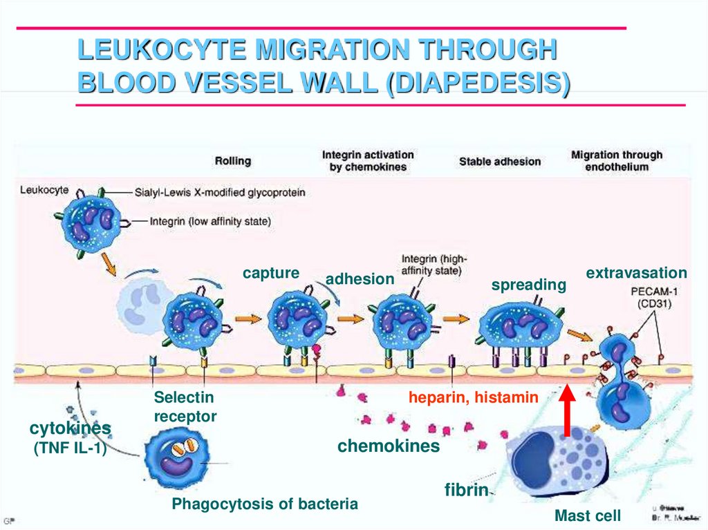
LEUKOCYTE MIGRATION THROUGHBLOOD VESSEL WALL (DIAPEDESIS)
capture
cytokines
(TNF IL-1)
adhesion
Selectin
receptor
spreading
extravasation
heparin, histamin
chemokines
Phagocytosis of bacteria
fibrin
Mast cell
26.
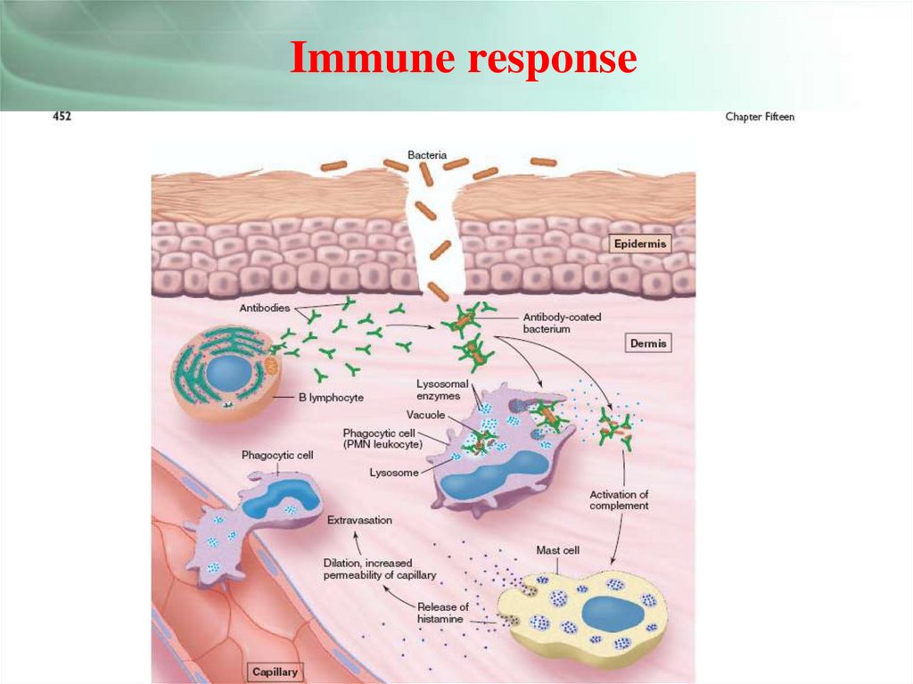
Immune response27.
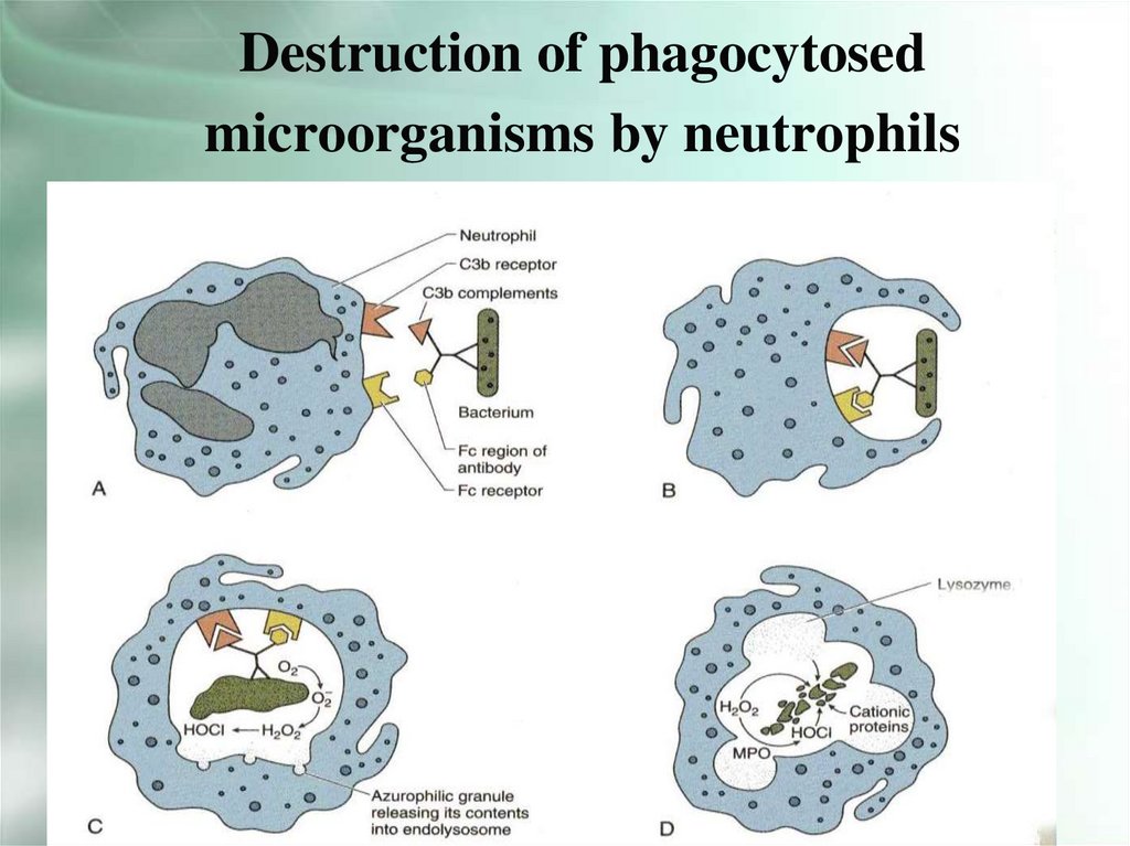
Destruction of phagocytosedmicroorganisms by neutrophils
28.
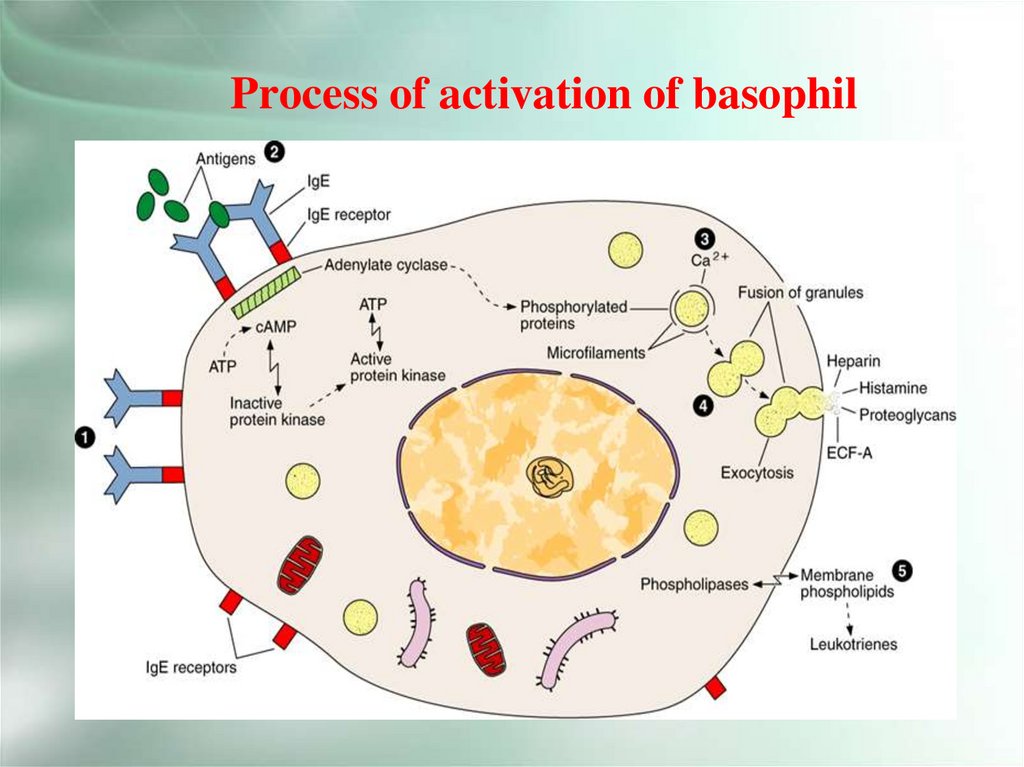
Process of activation of basophil29.
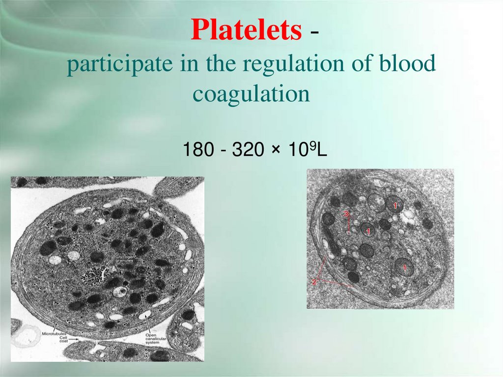
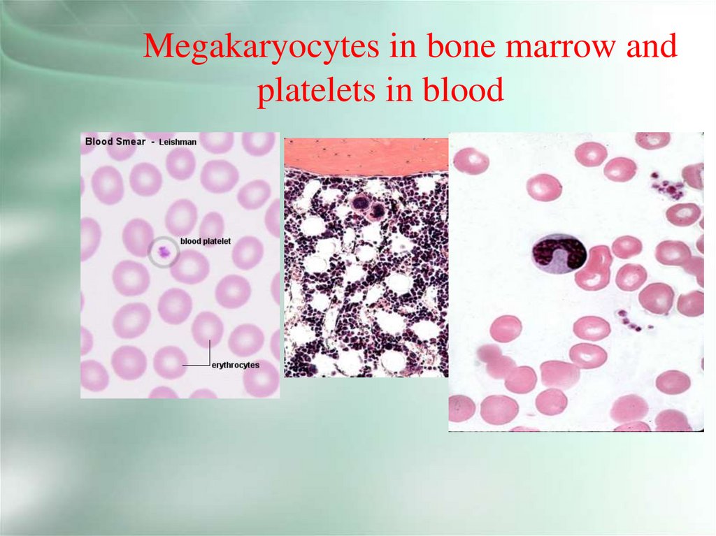
Platelets participate in the regulation of bloodcoagulation
180 - 320 × 109L
30.
31.
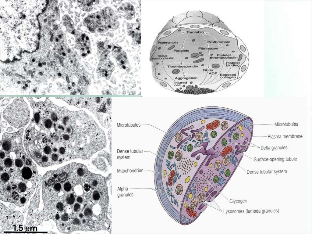
Platelets structure• The cytoplasm consists of a hyalomer and a
granulomer.
• 1) The hyalomer contains microtubules, actin
and myosin microfilaments.
• 2) In the granulomer 3 types of spec. granules:
α (alpha granules) - large, contain proteins and
glycoproteins involved in blood coagulation;
• δ (delta granules) contain serotonin, histamine,
ATP.
• λ (lambda granules) contain lytic enzymes.
32.
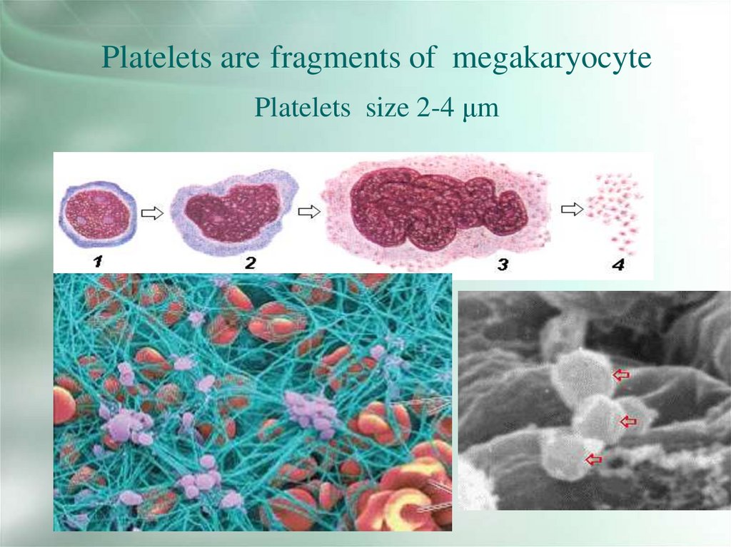
Platelets are fragments of megakaryocytePlatelets size 2-4 μm
33.
Megakaryocytes from bone marrow sendprocesses into the vessel and platelets are
split offaccording to demarcational canals
34.
Megakaryocytes in bone marrow andplatelets in blood