


")
")


 medicine
Similar presentations:

")


 identification")


Tertiary, visceral syphilis, neurosyphilis
1.
Tertiary, visceralsyphilis,
neurosyphilis
Zaporozhye 2016
2. General progress of tertiary syphilis. Classification of tertiary syphilis
1.It develops only in a very small number of patients:
- who has had bad treatment or has not been treated at all
- in childhood or in old age
- in people with physical, psychic, medicamental traumas
- with chronic diseases and intoxication (alcoholism, drug-addiction).
2.
Clinical features of tertiary syphilis develop after 3-4 years and more.
3.
Signs of tertiary syphilis:
- on the skin (tubercular syphilid)
deep nodes or gummas.
4. Small number of T. pallidum in the tissue fluid.
5. Small number of foci of infection and asymmetrical localization.
6. Tendency of the tertiary infiltrate towards destruction with the
development of ulcers and scars.
7. Possible infection of any tissue and organ. Characterized by serious
destruction and sclerosis.
3. General progress of tertiary syphilis. Classification of tertiary syphilis
8. Localization in essential organs can lead to lethaloutcome.
9. Without treatment healing is very slow.
10. treated with iodine preparations and salts of heavy
metals.
11. In 25-30% of cases classic serologic reaction is
negative.
12.The main reaction of imobilization of T. pallidum
(100%).
13. Active tertiary syphilis should be differentiated from
latent tertiary syphilis
4. Tubercular syphilid
1.grouped
2. serpiginous (creeping)
3. platform (field)
4.dwarf (small)
5. Diagnosis of tubercular and tubercular-ulcer syphilid is based on the following characteristic features
Diagnosis of tubercular and tubercularulcer syphilid is based on the followingcharacteristic features
a)
dense infiltrate of the tubercle;
b) copper-red colour with blue tint;
c) hemispheric shape, equal size;
d) even borders;
e) cherry-stone in size,
f)asymmetrically grouped or crawling;
g) the tubercles develop "mosaic" cicatrix;
h) subjective feelings are absent.
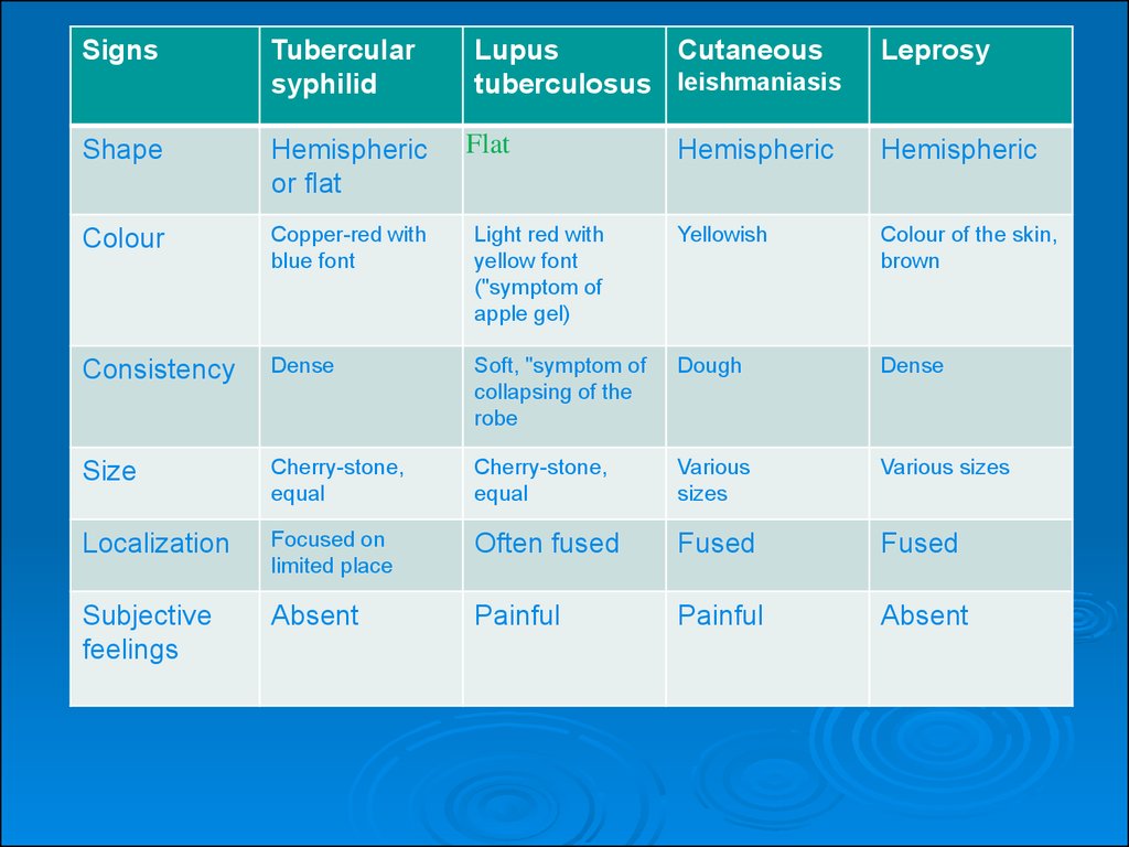
6.
SignsTubercular
syphilid
Lupus
Cutaneous
tuberculosus leishmaniasis
Leprosy
Shape
Hemispheric
or flat
Flat
Hemispheric
Hemispheric
Colour
Copper-red with
blue font
Light red with
yellow font
("symptom of
apple gel)
Yellowish
Colour of the skin,
brown
Consistency
Dense
Soft, "symptom of
collapsing of the
robe
Dough
Dense
Size
Cherry-stone,
equal
Cherry-stone,
equal
Various
sizes
Various sizes
Localization
Focused on
limited place
Often fused
Fused
Fused
Subjective
feelings
Absent
Painful
Painful
Absent
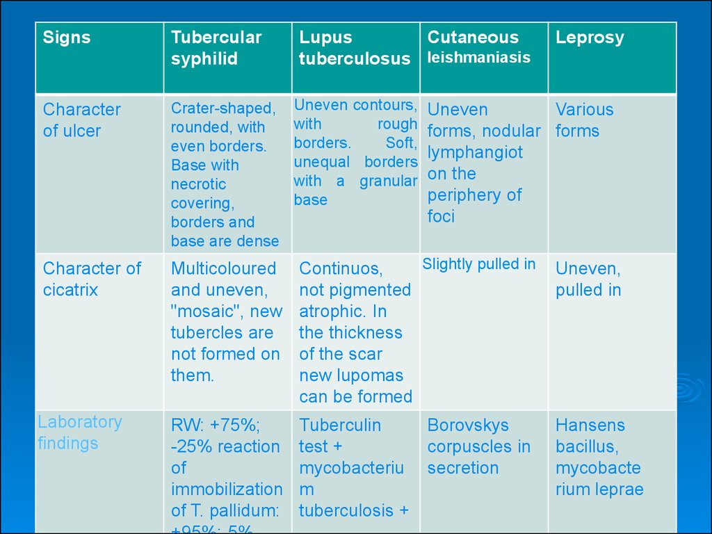
7.
SignsTubercular
syphilid
Lupus
Cutaneous
tuberculosus leishmaniasis
Character
of ulcer
Crater-shaped,
rounded, with
even borders.
Base with
necrotic
covering,
borders and
base are dense
Character of
cicatrix
Multicoloured
and uneven,
"mosaic", new
tubercles are
not formed on
them.
Slightly pulled in
Continuos,
not pigmented
atrophic. In
the thickness
of the scar
new lupomas
can be formed
Uneven,
pulled in
Laboratory
findings
RW: +75%;
-25% reaction
of
immobilization
of T. pallidum:
Tuberculin
test +
mycobacteriu
m
tuberculosis +
Hansens
bacillus,
mycobacte
rium leprae
Uneven contours,
with
rough
borders.
Soft,
unequal borders
with a granular
base
Leprosy
Uneven
Various
forms, nodular forms
lymphangiot
on the
periphery of
foci
Borovskys
corpuscles in
secretion
8. Gummatous syphilid
gummais a sharply separated dense
spherical or flat node.
lying deeply under the skin.
The epicenter of the development:
- subcutaneous fatty tissue
- lymphatic nodes
- Periosteum
9. Gummatous syphilid
Size:- pea-sized,
- slowly increases to size of a nut;
- painless;
- movable: the skin on it is not changed.
the node softens in the central part, takes a reddish-blue
colour.
Fluctuation develops and the node opens, secreting gumlike fluid.
The opening increases, an ulcer with dense and opening
borders can be seen.
10. Gummatous syphilid
On the base of the ulcer of dead tissue(gummatous shaft),
the ulcer undergoes cicatrization in a form of
deep star-shaped scars.
situated on the anterior surface of the crus,
forehead, and forearms.
Single gumma (solitary), gummatous infiltrate,
nodular gummas around the joints (fibrous
gumma).
11. Latent syphilis (syphilis latens)
The following information may be of assistance in the diagnosis of this form ofsyphilis:
the medical history which should be taken thoroughly with proper attention focused
on a past history of erosive and ulcerous efflorescence on the genitals and in the mouth,
various eruptions on the skin; on antibiotic therapy, on treatment of gonorrhea, etc.;
the results of confrontation;
revealing a scar or induration at the site of primary syphiloma and enlarged lymph
nodes corresponding clinically to regional scleradenitis;
high reagin titre in sharply positive results of all the serological tests;
a temperature exacerbation reaction at the beginning of penicillin therapy;
rapid drop in the reagin titre as early as during the first course of specific treatment;
the serological reactions are reversed to negative by the end of the first to the second
course of treatment;
sharply positive immunofluorescence test in these patients, although the T. pallidum
immobilization test may still be negative in some of them;
the patient's age;
the cerebrospinal fluid may be normal.
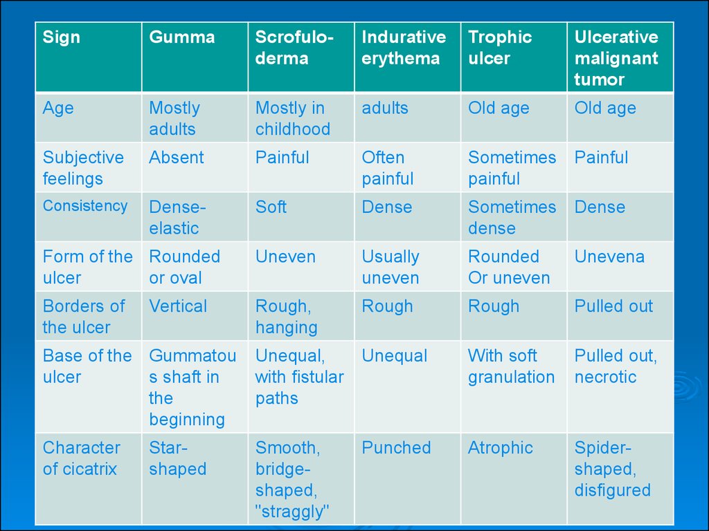
12.
SignGumma
Scrofuloderma
Indurative
erythema
Trophic
ulcer
Ulcerative
malignant
tumor
Age
Mostly
adults
Mostly in
childhood
adults
Old age
Old age
Subjective
feelings
Absent
Painful
Often
painful
Sometimes
painful
Painful
Consistency
Denseelastic
Soft
Dense
Sometimes
dense
Dense
Form of the Rounded
ulcer
or oval
Uneven
Usually
uneven
Rounded
Or uneven
Unevena
Borders of
the ulcer
Rough,
hanging
Rough
Rough
Pulled out
Base of the Gummatou
ulcer
s shaft in
the
beginning
Unequal,
with fistular
paths
Unequal
With soft
granulation
Pulled out,
necrotic
Character
of cicatrix
Smooth,
bridgeshaped,
"straggly"
Punched
Atrophic
Spidershaped,
disfigured
Vertical
Starshaped
13. Latent syphilis (syphilis latens)
The following information facilitates the diagnosis of late latentsyphilis:
the medical history;
low reagin titre in sharply
positive results of the classical serological
test (CST) or weakly positive results of CST;
reversal of serological reactions to negative by the middle or end of
specific treatment and the frequent absence of negative reversal of CST,
IFT and TPI despite vigorous antisyphilitic treatment and the use of nonspecific agents;
absence of the exacerbation reaction at the beginning of penicillin
therapy; it is preferable to begin treatment of such patients with
preparatory agents such as iodine preparations and bioquinol;
abnormalities in the cerebrospinal fluid which are encountered more
often in these patients than in those with early latent syphilis and are
corrected very slowly. Moreover, the sex partners may also have late
latent syphilis or they may have no manifestations of the syphilitic
infection.
14. Congenital syphilis
Transmission of congenital syphilisThe most common theory is that the only way of transmission
of congenital syphilis is from the mother to the fetus,
through syphilis of the placenta. The possible transmission
of syphilis from the father is now rejected. The
transmission of syphilis to the fetus may occur in three
ways:
1. Carrying of T. pallidum through the vena umbilicalis in
the organism of the baby.
2. Penetration of T. pallidum through lymphatic clefts of the
umbilical vessels.
3. Entering of T. pallidum into the fetus in the maternal
blood through damaged placenta.
15. Syphilis of the placenta
Pay attention to the size, weight, colour of theplacenta. Explain the results of histologic
examination in the infection of placenta.
Relation of weight of the placenta to the
weight of fetus is 1:3 (normally 1:5 – 1:6).
16. Syphilis of the fetus
The infection of the parenchymal organs and fetus has a character ofinterstitial process. Histologic examination of the infiltrate in the
parenchymal organ shows, that they are made of lymphocytes,
histiocytes, plasmatic cells, and sometimes miliary or solitary gumma.
There are many T. pallidum in the internal organs. List the characteristic
features of the changes in the liver and the lungs.
The infection of the locomotor system of the fetus: the development of
osteochondritis. Pay attention to the character of infection of blood
vessels, spread of infection, which is the main reason for intrauterine
death of the fetus. In small vessels, different stages of endarteritis, up to
obliteration may be observed.
Infection of endocrine glands, changes in the central nervous system:
productive mytomeningitis, meningoencephalitis and sclerosis of vessels.
As the death of the fetus is not rare in toxoplasmosis, the diagnosis of
syphilis of the fetus should be made on the basis of clinical, serologic,
pathological examinations and roentgenologic diagnosis of long tubular
bones. The diagnosis is confirmed by examining the infection of the
organs for the pathogen, positive STS in the mother.
17. Syphilis of the infants
The development of congenital syphilis has a uniquecharacter. Syphilis in children born from untreated mothers
with active elements of secondary syphilis, is a serious
disease. Almost all visceral organs are affected, locomotor
system and specific infection of the skin and mucous
membrane are observed. One of the earliest pathologic
infection of the skin in congenital syphilis of the infants is
syphilitic pemphigus (Fig. 1, 2) (2-23% of newborn patients
have this form). The most common sign of congenital syphilis
of the infants is diffused papular infiltrate of the skin, first
described by Gochzinger (Fig. 3). It is found in 60-65%,
mostly during the 8-10th week of the life of the baby.
18. Syphilis of the infants
Fig. 1, 2. Syphilitic pemphigusFig. 3. Diffused papular infiltrate Gochzingers.
19. Syphilis of the infants
Syphilis of locomotor system is one of thebasic and important signs of congenital
syphilis. It may explained by the following:
between the epiphysis and the diaphysis of
long tubular bones there is a high blood
circulation and hyperemia, which creates
favorable conditions for the reproduction of
T. pallidum. There are 3 stages of
osteochondritis. In the third stage the
epiphysis may detach from the diaphysis,
there appears intraepiphysal fracture, and
false paralytic condition develops.
Periostitis is observed in 45-50% of cases at
birth or during the first months after birth.
20. Syphilis of the early childhood
At the beginning of the secondhalf of the first year after birth
already, the syphilitic signs take
the form characteristic for
congenital syphilis of early
childhood, i.e., children at the age
of 1-4 years.
It should be noted that the
papular elements on different
places of the skin do not differ by
their form, localization and
evolution from papules in
secondary relapsing stage of
acquired syphilis.
21. Syphilis of the early childhood
Pay attention to the predominationof erosive, vegetating papule in the
region of the anus and inguinal
plicae. They also appear on the
mucous membrane of the oral
cavity, tonsils.
The changes in the bone,
particularly, in the tibia, have a
character of periostitis or
osteoperiostitis.
Serologic examinations are of great
importance in the diagnosis of
congenital syphilis.
22. Late congenital syphilis
Signs of the late congenital syphilis are divided intounconditional and accessory signs.
The unconditional signs include Hutchinson’s triad:
•parenchymal keratitis,
•labyrinthine deafness,
•Hutchinson’s teeth.
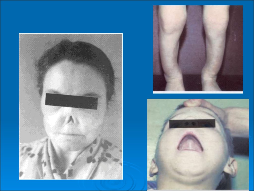
23. Late congenital syphilis
Accessory signs include:dystrophy of teeth;
infection of locomotor system (saber shin crus), natiform
skull, saddle nose, Avsitideisky’s symptom (thickening of
sternoclavicular joint, Gothic hard palate, axiphoidia,
Dubois’ infant little finger, racket shaped little finger.
tubercle-ulcerative and gummatous infection of the skin
and the mucous membrane does not differ from the infection
in tertiary acquired syphilis in its manifestations and
progress. Specific changes of the visceral organs in late
congenital syphilis are rarer than those in syphilis of infants.
pathologic changes in central nervous system: tabes
dorsalis, jacksonian epilepsy, atrophy of optic nerve, psychic
retardation.
24.
25. Late congenital syphilis
The following stigmata of late congenital syphilis are the mostsignificant:
1) Avsitidiisky's sign;
2) Gothic hard palate;
3) infantile little finger
4) axiphoidia, i.e. absence of the sternal xiphoid process;
5) Carabelli cusp, the presence of a fifth auxiliary cusp on the
masticatory surface of the first upper molar;
6) diastema: gaps between the upper incisors;
7) hypertrichosis in children and growth of hair on the forehead
almost to the eyebrows;
8) dystrophy of the skull bones, bossing of the frontal and parietal
eminences but without a separating groove.
26. Prevention of Congenital Syphilis
The timely detection and proper treatment of syphilis in women is thebasis of prevention of congenital syphilis. The role of examination of
pregnant women is particularly important because they must be treated
promptly. According to the valid authoritative instructions in our
country, antenatal clinics are obliged to register all pregnant women and
subject them to clinical and serological examination for syphilis.
Serological examination is carried out twice, during the first and second
periods of pregnancy. If the active or latent form of syphilis is found in
the pregnant woman, specific treatment (only with antibiotics) is
conducted. If the woman had been ill with syphilis earlier and had
completed antisyphilitic treatment, specific treatment is nonetheless
conducted, which in this event is called preventive, for the purpose of
ensuring the birth of a healthy offspring.
One or two weeks prior to childbirth, non-specific, false positive
serological reactions may appear. Therefore, if they are detected two
weeks before childbirth, the expectant mother is not given specific
treatment, but two weeks later she and her child are examined again. If
the diagnosis of syphilis is confirmed, antisyphilitic treatment is
prescribed for both mother and child. The newborns, whose mothers
have been sick with syphilis and have received proper treatment prior to
and during pregnancy, are subject to thorough and comprehensive