Similar presentations:
Calcium-binding proteins immunoreactivity in not yet differentiated retina of the fetal guinea pig
1. Calcium-binding proteins immunoreactivity in not yet differentiated retina of the fetal guinea pig (Cavia porcellus) in the
CALCIUM-BINDING PROTEINS IMMUNOREACTIVITY IN NOT YET DIFFERENTIATED RETINA OFTHE FETAL GUINEA PIG (CAVIA PORCELLUS) IN THE SECOND TRIMESTER OF GESTATION
Sandra Gąsiorowska, Jakub Kosma Frydrych, Edyta Dej
Department of Animal Anatomy and Physiology, University of Warmia and Mazury in Olsztyn, Olsztyn
Student Neuroanatomy Research Group – NEURON, sci. supervisor Anna Robak, Assoc. Prof.
Introduction
Calcium-binding proteins (CaBPs) i.e. calbindin D28k (CB), calretinin (CR) and parvalbumin (PV) are specific markers of human retina
neurons (Kantor 2016). The guinea pig unlike other rodents (rat, mouse) shows many features similar to human development
(precocial species) and their retinas structure are similar prior to birth (Spira, 1975; Kantor, 2016).
Although CaBPs during ontogeny take part in multiple cell processes such as cell migration, differentiation and synaptogenesis to
serve buffering or sensing proteins functions (Loeliger & Rees, 2005) and they were examined in retina development in many
vertebrates, data obtained in the guinea pig are incomplete (Hamano et al. 1990; Loeliger & Rees, 2005). Thus, the aim of the study
was to examine distribution and immunoreactivity of CB, CR, PV in not yet full laminated retina at midgestation in the guinea pig.
Material and methods
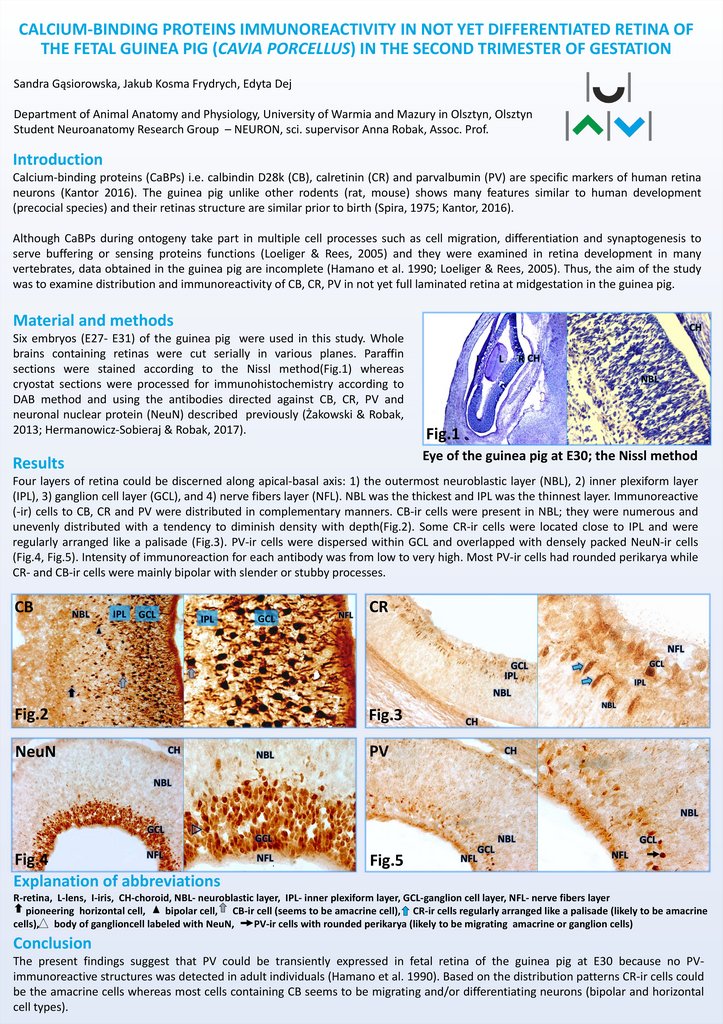
Six embryos (E27- E31) of the guinea pig were used in this study. Whole
brains containing retinas were cut serially in various planes. Paraffin
sections were stained according to the Nissl method(Fig.1) whereas
cryostat sections were processed for immunohistochemistry according to
DAB method and using the antibodies directed against CB, CR, PV and
neuronal nuclear protein (NeuN) described previously (Żakowski & Robak,
2013; Hermanowicz-Sobieraj & Robak, 2017).
I
NBL
Fig.1
Eye of the guinea pig at E30; the Nissl method
Results
Four layers of retina could be discerned along apical-basal axis: 1) the outermost neuroblastic layer (NBL), 2) inner plexiform layer
(IPL), 3) ganglion cell layer (GCL), and 4) nerve fibers layer (NFL). NBL was the thickest and IPL was the thinnest layer. Immunoreactive
(-ir) cells to CB, CR and PV were distributed in complementary manners. CB-ir cells were present in NBL; they were numerous and
unevenly distributed with a tendency to diminish density with depth(Fig.2). Some CR-ir cells were located close to IPL and were
regularly arranged like a palisade (Fig.3). PV-ir cells were dispersed within GCL and overlapped with densely packed NeuN-ir cells
(Fig.4, Fig.5). Intensity of immunoreaction for each antibody was from low to very high. Most PV-ir cells had rounded perikarya while
CR- and CB-ir cells were mainly bipolar with slender or stubby processes.
CB
NBL
IPL
GCL
IPL
GCL
CR
Fig.2
Fig.3
NeuN
PV
Fig.4
Explanation of abbreviations
Fig.5
R-retina, L-lens, I-iris, CH-choroid, NBL- neuroblastic layer, IPL- inner plexiform layer, GCL-ganglion cell layer, NFL- nerve fibers layer
pioneering horizontal cell,
bipolar cell, CB-ir cell (seems to be amacrine cell), CR-ir cells regularly arranged like a palisade (likely to be amacrine
cells), body of ganglioncell labeled with NeuN,
PV-ir cells with rounded perikarya (likely to be migrating amacrine or ganglion cells)
Conclusion
The present findings suggest that PV could be transiently expressed in fetal retina of the guinea pig at E30 because no PVimmunoreactive structures was detected in adult individuals (Hamano et al. 1990). Based on the distribution patterns CR-ir cells could
be the amacrine cells whereas most cells containing CB seems to be migrating and/or differentiating neurons (bipolar and horizontal
cell types).

 medicine
