


 english
Similar presentations:


Erysipelas
1.
ФГБОУ ВО «Казанский государственныймедицинский университет» МЗ РФ
Кафедра инфекционных болезней
Erysipelas
Prof., MD, PhD Fazylov V.Kh.
Kazan 2018
2.
Definition of the Disease■Erysipelas
– human infectious disease of streptococcal
etiology, with acute and chronic forms and is
characterised by intoxication syndrome and local
changes looking like circumscribed locus of serous
hemorrhagic inflammation of skin (rarely mucosa)
3.
Etiologic Peculiarities■1.
In primary and recurring erysipelas with exogenic route
of transmission the cause of infection is beta-haemolytic
streptococcus group A with its aggression factors:
= antigenous substrates Т-, R-, М- proteins (М-protein
and associated antigens – lipoteichoic acid, opalescence
factor lipoproteinase,polysaccaride peptidoglicane etc)
= extracellular substrates (exotoxins or locally applicable
toxins: erythrogenous toxin А, В, С, streptolysine О,
streptolysine S, streptokinase, hyaluronidase, proteinase)
4.
Etiologic Peculiarities (continued)■2.
In relapsing and particularly frequently relapsing
erysipelas the etiological cause are the L-forms of
streptococcus, which form after primary erysipelas as a
result of inadequate etiotropic therapy.
■3.
At the high point of disease from the local focus of
inflammation different opportunistic species are released
(staphylococcus,
hemolysing
E.coli,
proteus,
Ps.aeruginosa etc). This indicates the opportunistic flora
activation in patients with immunodeficiency (often
secondary).
5.
Laboratory diagnosis■1.
Detection of antigenemia:
= А-polysaccaride (А-PSC)
= protein-ribosomal antigens (PR-Аg)
= L-form antigens
2. Detection of antibodies:
= AB to А-PSC in ELISA
= AB to О-SL (АSL-О)
= АB to DNA-ase
6.
Epidemiological peculiarities■1.
In primary, recurrent, rarely relapsing erysipelas the source of
infection are patients with different forms of streptococcal infection:
tonsillitis, scarlet fever, streptodermia; healthy carriers. Patients with
erysipelas are not very contagious, but theoretically the transmission
is possible.
■The main route of transmission – percutaneous (through the defects in
skin and mucosa). Aerogenic mechanism of streptococcus
transmission has a certain significance with primary infection of
nasopharynx and subsequent lymphogenous and hematogenous
dissemination to the local focus (mostly in erysipelas of the skin).
■So, these forms are acute cyclic infectious process developing as a
result of exogenous infection and the incubation period can be
counted about 24-48 hours.
7.
Epidemiological peculiarities (continued)■2.
In frequently relapsing erysipelas - only endogenous
mechanism of infection – reversion of the L-forms of
streptococcus, that persist in the scar tissue, along small
blood and lymphatic vessels, lymph nodes, bone marrow.
That’s why FRE is considered as chronic form. In this
form of erysipelas the incubation period can be counted
according
to
provocative
factors:
hypothermia,
hyperthermia, insolation, emotional stress, hurts, blunt
trauma etc.
8.
Epidemiological peculiarities (continued)■3.
Vulnerability depends on basic immune status condition
and the virulence of the bacteria.
■4.
Season is generally summer-autumn, but relapsing
forms have no typical season.
■5.
After infection with streptococcus the disease develops
only in those who have congenital or
acquired
predilection. Women get sick more often than men,
especially the relapsing form.
■6.
Most of the patients are 40-60 years old and older.
9.
Pathogenesis■1.
■2.
Permeation of the streptococcus into the skin.
Reproduction of bacteria in the lymphatic capillaries of
derma.
■3.The toxins of the streptococcus get into the blood stream
(toxemia).
■4. Forming of the inflammatory locus.
■5. Forming of the locuses of chronic streptococcal infection
(providing relapses of the disease)
OR
Elimination of vegetative forms of streptococcus with
phagocytosis and other immune mechanisms (recovery).
10.
Pathogenic features of frequently relapsing erysipelas:■1.
Forming of the resistant locus of streptococcal infection in the
body (L-forms)
■2. Dramatic decrease of phagocytosis and bactericidic activity of the
skin
■3. Depression of cellular immunity: decrease of the T-cells, CD4+,
CD8+ subpopulations
■4. Decrease of humoral immunity: low level of immunoglobulines
class А and anti-streptococcal antibodies (ASL-О, ASG, ASK) in
the serum
■5. Extremely high degree of allergisation to streptococcus
■6. Autoimmune reactions against skin and thymus antigens
■7. Disbalance in hormonal regulation: deficiency of glucocorticoids
and redundancy of mineralcorticoids (increase of edema)
■8. Stable alterations of lymph- and blood circulation with the
development of disseminated microthrombosis (DIC syndrome)
11.
Predisposing factors■1.
Concomitant diseases – plantar mycosis, diabetes
mellitus,
obesity,
chronic
venous
insufficiency,
lymphostasis, trophic ulcers, eczema etc.
■2. Professional factor – jobs connected with constant
dirtying and microtraumatization of the skin, wearing
rubber shoes etc.
■3. Locuses of chronic streptococcal infection as tonsillitis,
sinusitis, caries (erysipelas of the face), osteomielitis,
thrombophlebitis, ulcers (erysipelas of lower extremities)
etc.
12.
There are two main components in thepathogenesis of erysipelas:
■1.
Infectious-toxic (toxins, transient bacteriemia, secretion
of biologically active substances), causing fever and
intoxication;
■2.
Infectious-allergic, responsible for the local inflammation
13.
Clinical classification of erysipelas■1.
By frequency:
= Primary
= Recurrent
= Relapsing
2. By the character of local changes:
= Erythematous
= Erythematous-bullous
= Erythematous-hemorrhagic
= Bullous-hemorrhagic
14.
Clinical classification of erysipelas (continued)■3.
By severity:
= mild
= moderate
= severe
4. By localisation:
= lower extremities (55-60% - PE, 75-80% - RR)
= face (25-30%)
= upper extremities, trunk (5-12%)
15.
Examples of the clinical diagnosis■1.
■2.
Primary erysipelas of the left shank erythematous form moderate
severity.
Relapsing erysipelas (1st early relapse) of the right shank and foot
bullous-hemorrhagic severe form
Complication: phlegmon of the right shank soft tissues.
16.
Evolution of the erysipelas clinical features■1.
■2.
More older people (60 year old and older) – 55,8%
More lower extremities involvement – 66,5% less
frequent face involvement – 25%
■3. More relapsing forms – up to 45-50% of all cases
■4. More patients with hemorrhagic manifestations from
10-12% to 43,8%
■5.More frequent allergic reactions on antibiotics,
sulphanilamids and other drugs, especially among
patients with relapsing form.
17.
Clinical features of erysipelas■1. Acute onset
■2. Intoxication
of the disease.
syndrome is usually ahead of other
symptoms for practically 18-24 hours and is
characterised by high fever, chills, headache,
sometimes
nausea,
vomiting,
myalgias,
arthralgias.
Patients complain of malaise, weakness, body
pain, sleep problem, loss of appetite.
18.
Clinical features of erysipelas (continued)■3.
Early signs of the disease before the local changes can
be:
■а) regional lymphadenitis and lymphangitis (in lower
extremities erysipelas) characterized by pains in the
projection of regional lymphnodes (especially inguinal)
and along the lymphatic vessels (medial side of the
thigh),
■b) burning pain in erysipelas of the face that starts 5-6
hours before the local inflammatory focus forms.
19.
Clinical features of erysipelas (continued)■3.
Local process is characterised by sharply
circumscribed hyperemia with the peripheral
inflammatory wall, edge
painfullness, local
temperature reaction (erythematous form)
■On the background of hyperemia appearance of other
elements is possible:
■= hemorrhagias (erythematous-hemorrhagic form),
■=
bullas – bubbles filled with serous fluid
(erythematous-bullous form),
■= bubbles filled with serous-hemorrhagic fluid (bulloushemorrhagic form).
20.
Clinical features of erysipelas (continued)■4.
Local process is associated with lymphatic edema
of various degree depending on the character of
local process.
■5.
In case of paired organ involvement usually
unilateral process.
■6.
On the face is typically limited on the border of hairy
part of the head.
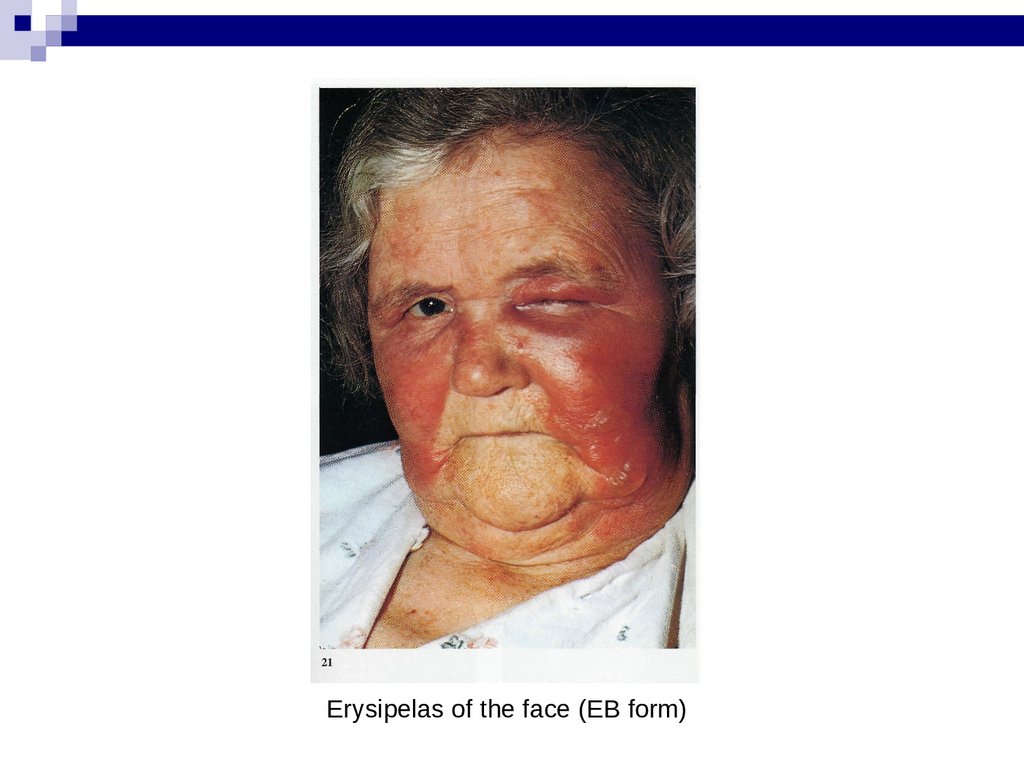
21.
Erysipelas of the face (EB form)22.
After recovery (peeling)Erysipelas of the face (Erythem. form)
23.
Erythematous-bullous formPigmentation and peeling (recovery)
24.
Elephantiasis (outcome)25.
Diagnosis■Main
method – clinical and anamnesis.
■Differential
■With
diagnosis:
infectious diseases (skin form of anthrax, erysipeloid
etc)
■With
dermatologic
diseases
(streptodermia,
staphylodermia, allergic dermatitis etc)
■With surgical diseases (abscess, phlegmon, acute and
relapse of chronic thrombophlebitis etc)
26.
Ethiotropic therapy■1.
In primary and recurrent erysipelas penicillin is the antibiotic of
choice – 5-6 mln Un in 24 hours IM, mild forms – 7 days, moderate
forms – 10 days, severe forms – 12-14 days.
■2. In relapsing erysipelas – semisynthetic penicillins (ampicillin,
oxacillin, ampiox, amoxicillin, augmentin etc) - 4 g in 24 hours.
■3. In frequently relapsing erysipelas – antibiotics of choice are
cephalosporines (1-4 generations) 2-4 g in 24 hours; linkomicin 1,22,4 g in 24 hours.
■(in persistent relapses – 2 course treatment)
■4. For out-patients – macrolides (spiramicin 6 mln IU/24 hours),
tetracyclines (doxycycline 0,2 g./24 hours)
27.
Pathogenetic therapy■1.
Detoxication therapy – oral (enterodez, regidron etc.);
parenteral – crystalloids (polyionic solutions: trisol,
acesol, chlosol, kvartasol, 5% glucose etc.), low- and
medimolecular colloids (reopolyglukine, reogluman,
reomakrodex etc) counted 1:1
■2. Desensibilisation therapy – antihistamine drugs
(dimedrol, suprastin, pipolfen, tavegil, claritin etc)
■3. Correction of the hemostasis alterations according to the
coagulogram control : desaggregants – dicinon, trental,
kurantil; dimephosphone etc.; direct acting anticoagulants
- heparin, fraxiparin, calciparin etc, indirect acting –
sinkumar, kumadin, pelentan etc.
28.
Pathogenetic therapy (continued)■4.
Immunocorrection with the control of the immune status
(immunoglobulines IV and IM, interferones, thymal drugs
– timalin, timogen; pirimidines – methyluracil, natrium
nucleinate,
ksimedon;
dimephosphone;
bacterial
polysaccarides – pirogenal, prodigiosan; herbal
adaptogens – eleuterococcus, ginseng, aralia and others.
■5. Physiotherapy in acute period - UV in suberythemal
doses №5 and UHF №5; projectional distant exposure of
low intensive laser, in reconvalescence period –
potassium iodide, lidase, ronidase electrophoresis;
indirect action – laser puncture
29.
Local treatment■1.
■2.
Do not touch (!) erythematous forms.
Vishnevsky ointment and ichtiolic ointment are strictly
forbidden.
■3.
■1
Is only recommended for bullous forms in 2 steps:
step – applications with antiseptic solutions (furacillin
1:1000, rivanol 1:1000, chlorfillipt, dimexid, 15%
dimephosphone etc.)
■2 step – emulsions and ointments (lanolin cream, sea
buckthorn oil, 10% methyluracilic ointment, aecol etc.)
30.
Ambulatory monitoring■1.
Finishing treatment
■2.
Sanation of the chronic focuses of infection
■3. Relapse prophylaxis:
■Bicillin-5 1,5 mln. Un IM once a month –
■In relapsing forms – every month for at least
2 years since
the last relapse;
■In primary, recurrent forms – for 6 months after the
disease.