


")


 medicine
Similar presentations:
")


Pathophysiology of diabetes mellitus. Specific forms of diabetes
1.
JSC «Astana Medical University»Department of Internal Medicine №1
Done by Abduova L.M., 445 GM
Checked by Baidurin S.A.
1
2. Plan
Introduction
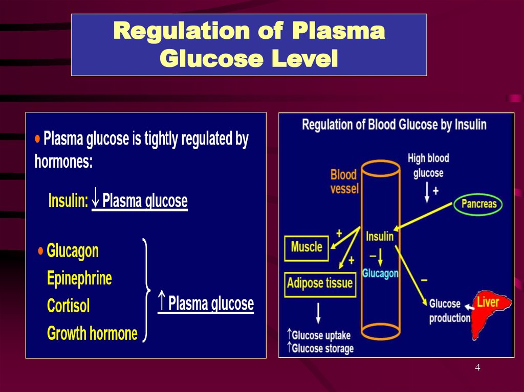
Regulation of Plasma Glucose Level
Classification of DM
Etiology
Risk factors
Pathophysiology
Clinical presentation
Gestational diabetes
Other types of DM
Bibliography
2
3. Introduction. What is diabetes?
Diabetes mellitus (DM) is a chronic conditionthat is characterised by raised blood glucose
levels (Hyperglycemia).
3
4.
Regulation of PlasmaGlucose Level
4
5.
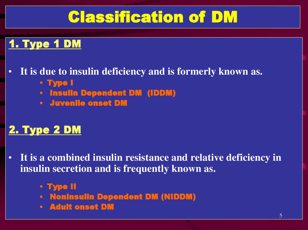
Classification of DM1. Type 1 DM
• It is due to insulin deficiency and is formerly known as.
• Type I
• Insulin Dependent DM (IDDM)
• Juvenile onset DM
2. Type 2 DM
• It is a combined insulin resistance and relative deficiency in
insulin secretion and is frequently known as.
• Type II
• Noninsulin Dependent DM (NIDDM)
• Adult onset DM
5
6.
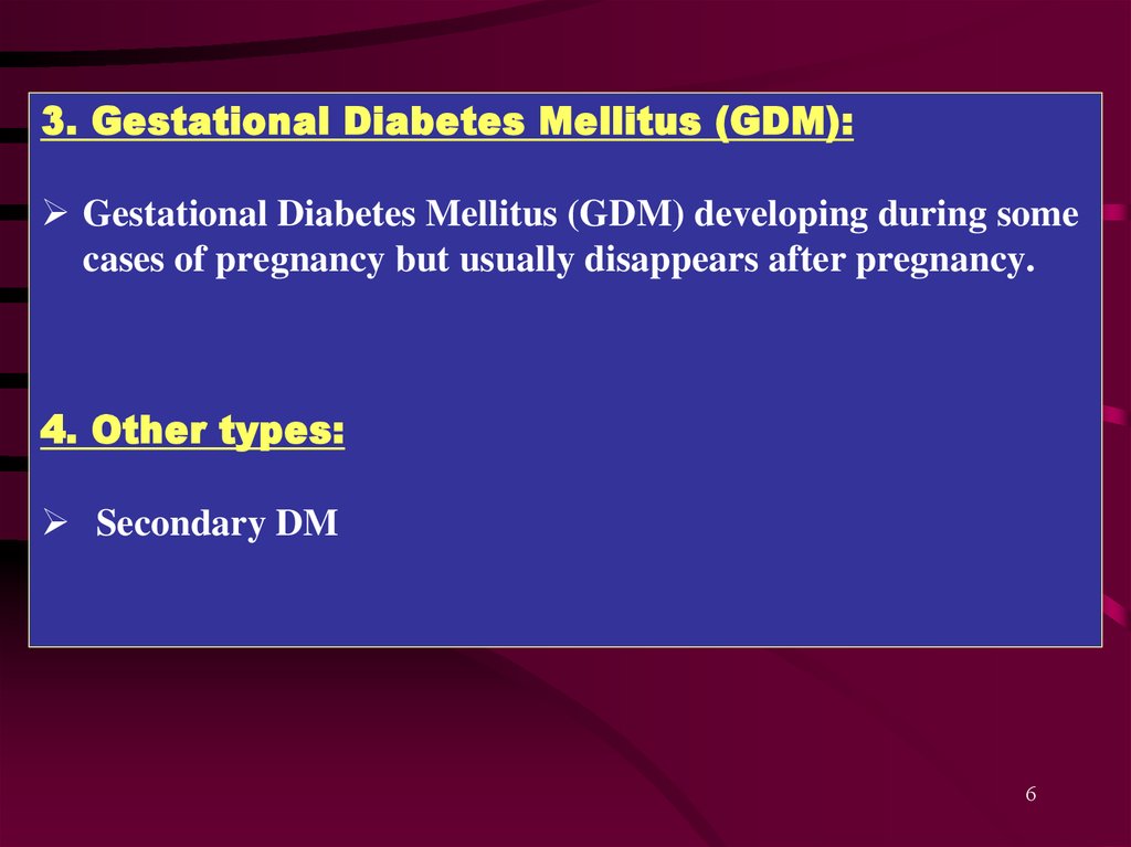
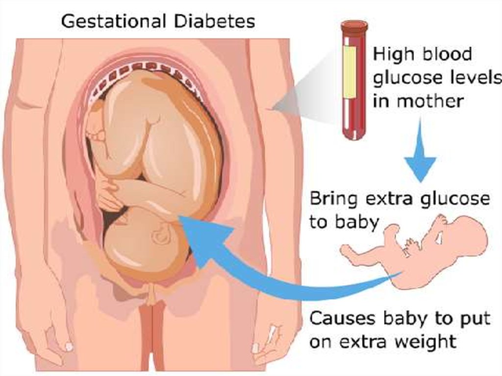
3. Gestational Diabetes Mellitus (GDM):Gestational Diabetes Mellitus (GDM) developing during some
cases of pregnancy but usually disappears after pregnancy.
4. Other types:
Secondary DM
6
7. Etiology
1. Etiology of Type 1 Diabetes7
8.
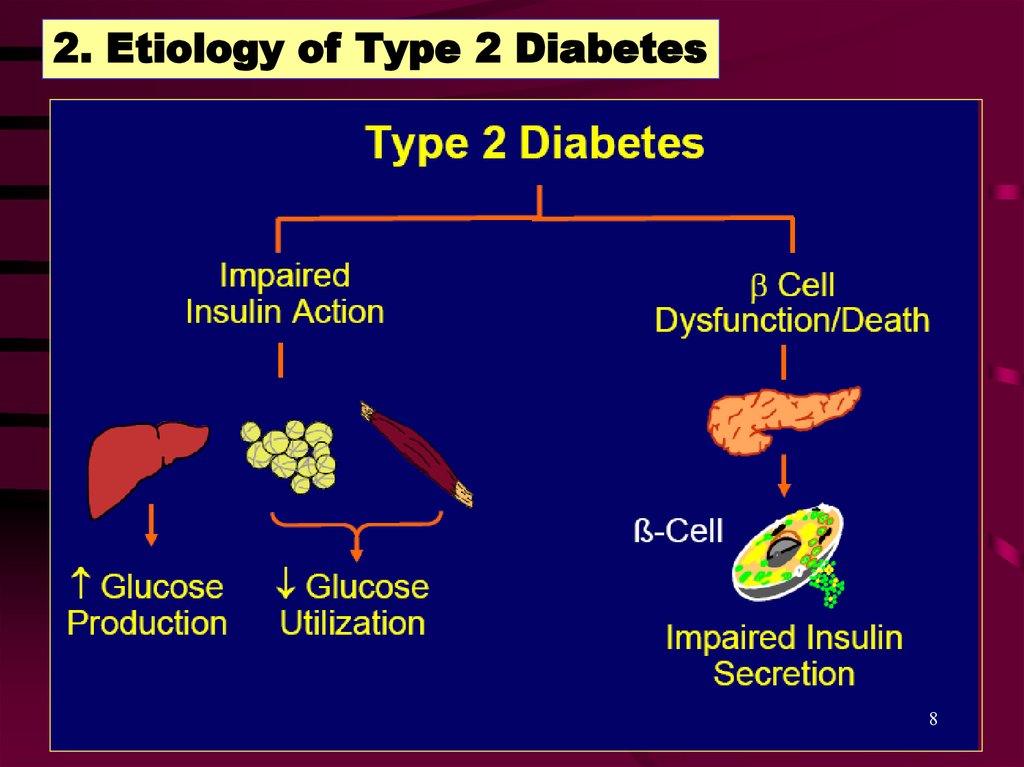
2. Etiology of Type 2 Diabetes8
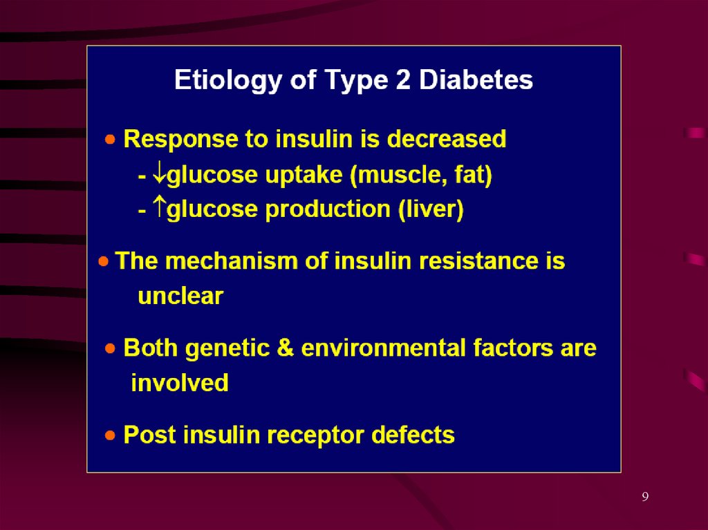
9.
910.
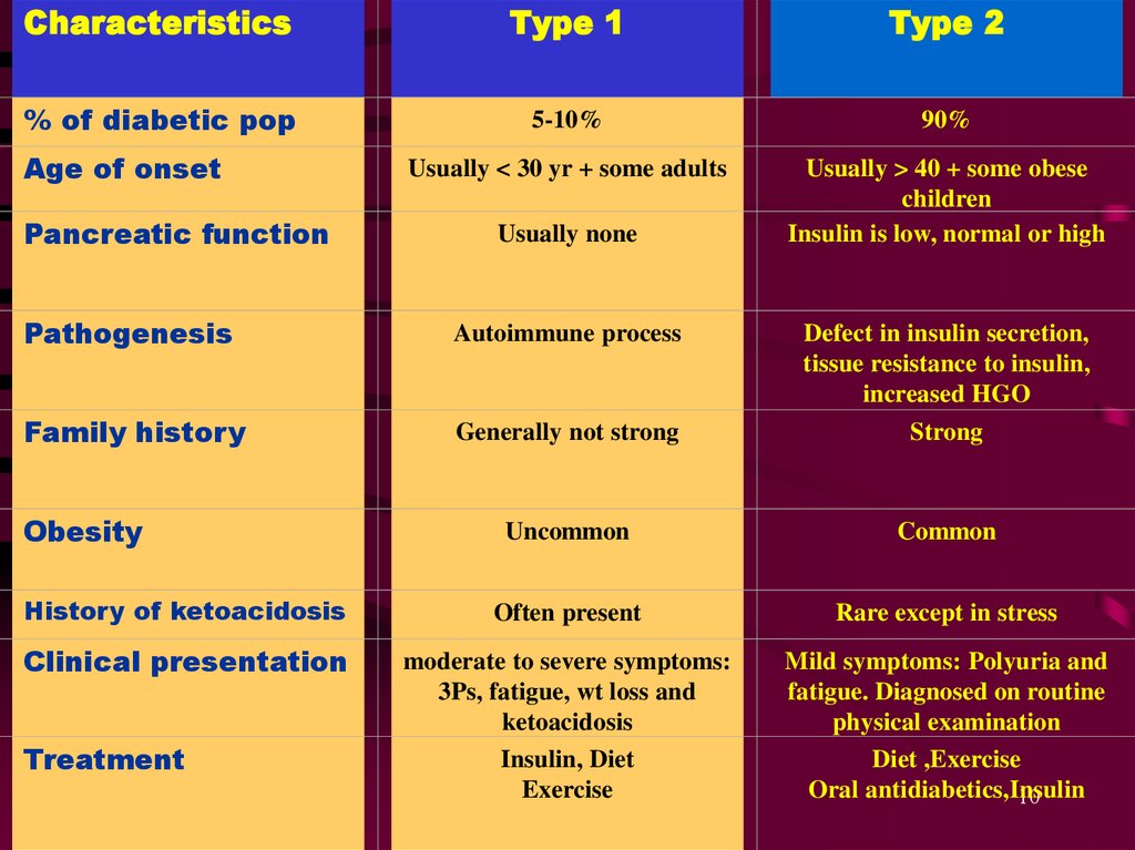
CharacteristicsType 1
Type 2
% of diabetic pop
5-10%
90%
Usually < 30 yr + some adults
Usually > 40 + some obese
children
Insulin is low, normal or high
Age of onset
Pancreatic function
Usually none
Pathogenesis
Autoimmune process
Defect in insulin secretion,
tissue resistance to insulin,
increased HGO
Family history
Generally not strong
Strong
Uncommon
Common
History of ketoacidosis
Often present
Rare except in stress
Clinical presentation
moderate to severe symptoms:
3Ps, fatigue, wt loss and
ketoacidosis
Insulin, Diet
Exercise
Mild symptoms: Polyuria and
fatigue. Diagnosed on routine
physical examination
Diet ,Exercise
Oral antidiabetics,Insulin
10
Obesity
Treatment
11. Risk Factors
• Type 1 DM–
Genetic predisposition
• In an individual with a genetic
predisposition, an event such as virus
or toxin triggers autoimmune
destruction of b-cells probably over a
period of several years.
11
12. Risk Factors
• Type 2 DM– Family History
–
–
–
–
–
Obesity
Habitual physical inactivity
Previously identified impaired glucose tolerance
(IGT) or impaired fasting glucose (IFG)
Hypertension
Hyperlipidemia
12
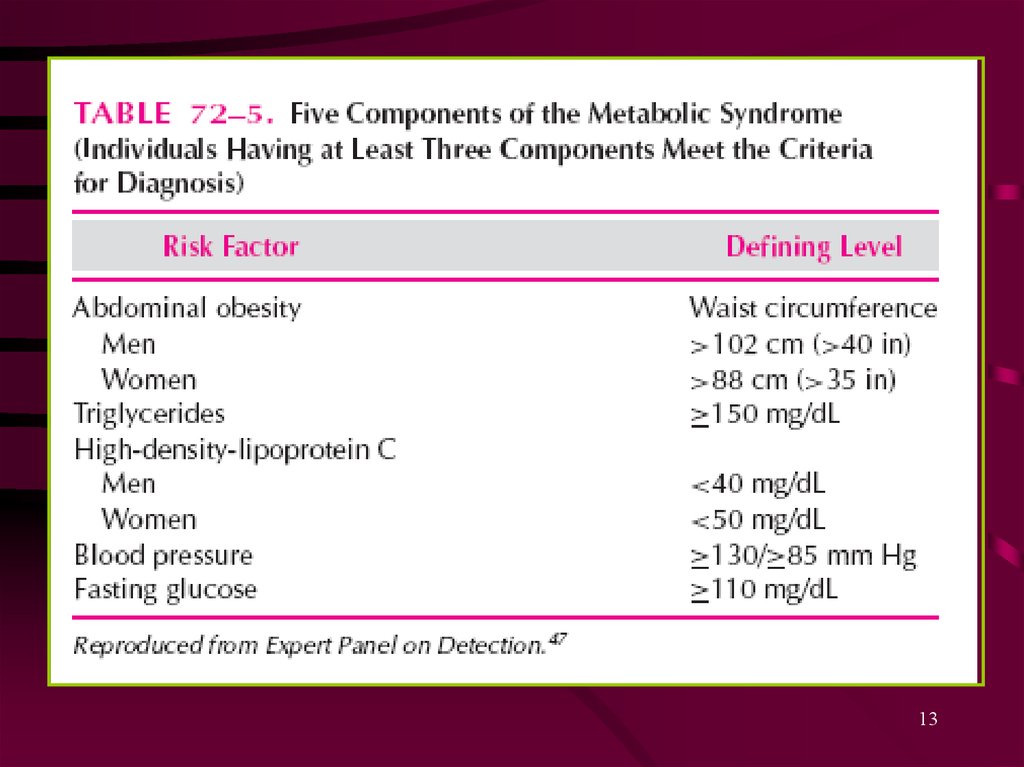
13.
1314. Pathophysiology
• Type 1 DM– Type 1 DM is characterized by an absolute
deficiency of insulin due to immune- mediated
destruction of the pancreatic b-cells
– In rare cases the b-cell destruction is not due to
immune mediated reaction (idiopathic type 1 DM)
14
15. Pathophysiology
Type 1 DM
There are four stages in the development of
Type 1 DM:
1.
Preclinical period with positive b-cell antibodies
2.
Hyperglycemia when 80-90% of the
β- cells are destroyed.
3.
Transient remission (honeymoon phase).
3.
Establishment of the disease
15
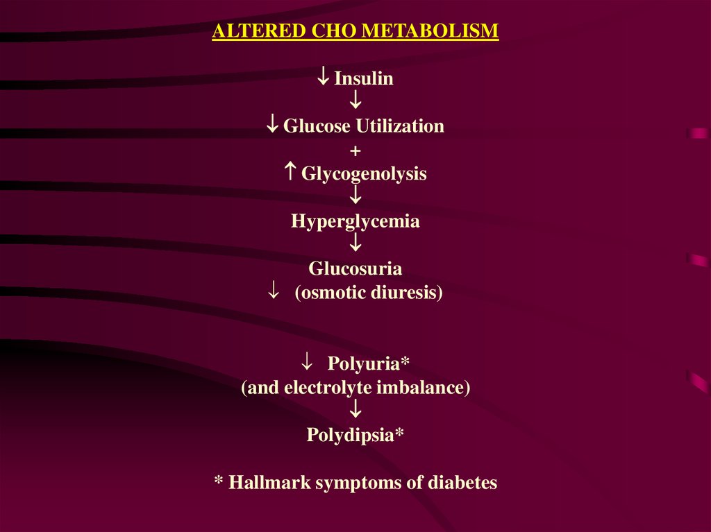
16.
ALTERED CHO METABOLISMInsulin
Glucose Utilization
+
Glycogenolysis
Hyperglycemia
Glucosuria
(osmotic diuresis)
Polyuria*
(and electrolyte imbalance)
Polydipsia*
* Hallmark symptoms of diabetes
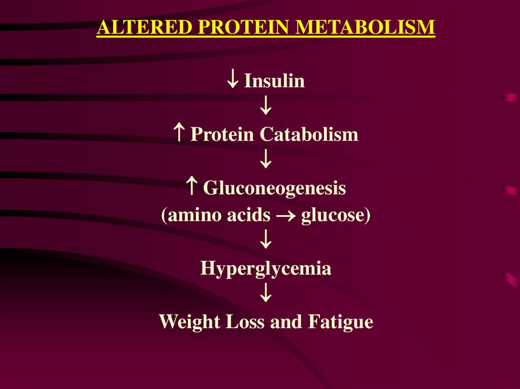
17.
ALTERED PROTEIN METABOLISMInsulin
Protein Catabolism
Gluconeogenesis
(amino acids glucose)
Hyperglycemia
Weight Loss and Fatigue
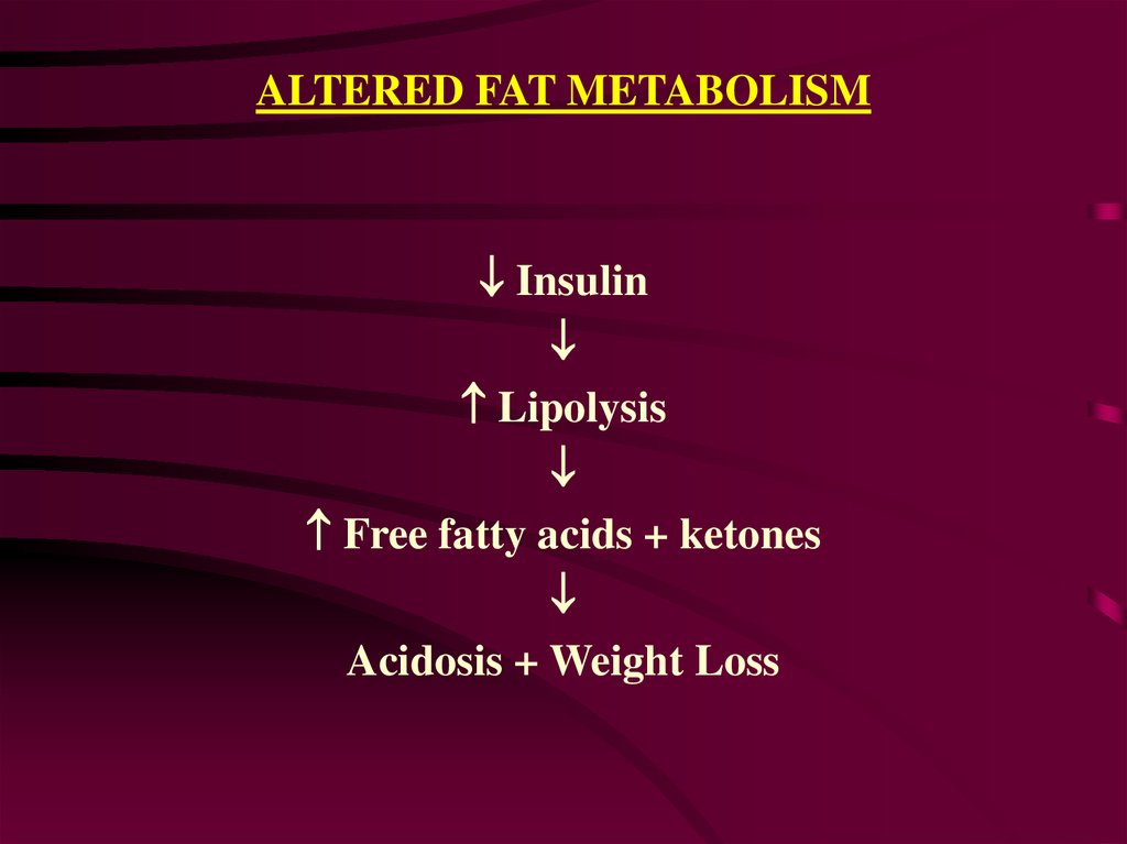
18.
ALTERED FAT METABOLISMInsulin
Lipolysis
Free fatty acids + ketones
Acidosis + Weight Loss
19. Type 1 Diabetes
Inflammation• Beta cell destruction
FasL
IFNg
TNFa
– Usually leading to
absolute
insulin deficiency
T cell
• Immune mediated
• Idiopathic
Autoimmune Reaction
Macrophage
Class I
MHC
TNFa
Class II
MHC
IL-1
Beta cell
NO
CD8+ T cell
Dendritic cell
Beta cell Destruction
Maahs DM, et al. Endocrinol Metab Clin North Am. 2010;39:481-497.
20. Pathophysiology of T1DM
• Chronic autoimmune disorder occurring in geneticallysusceptible individuals
– May be precipitated by environmental factors
• Immune system is triggered to develop an autoimmune
response against
– Altered pancreatic beta cell antigens
– Molecules in beta cells that resemble a viral protein
• ~ 85% of T1DM patients have circulating islet cell antibodies
– Majority also have detectable anti-insulin antibodies
• Most islet cell antibodies are directed against glutamic acid
decarboxylase (GAD) within pancreatic beta cells
Maahs DM, et al. Endocrinol Metab Clin North Am. 2010;39:481-497.
21. Pathophysiology
BirthTime (years)
21
22. Models for Pathogenesis of T1DM
van Belle TL, et al. Physiol Rev. 2011;91:79-118.23. Models for Pathogenesis of T1DM Fertile Field Hypothesis
van Belle TL, et al. Physiol Rev. 2011;91:79-118.24. How Type 1 Diabetes Might Arise
van Belle TL, et al. Physiol Rev. 2011;91:79-118.25. Major Metabolic Effects of Insulin and Consequences of Insulin Deficiency
Insulin effects: inhibits breakdown of triglycerides (lipolysis) inadipose tissue
• Consequences of insulin deficiency: elevated FFA levels
Insulin effects: Inhibits ketogenesis
• Consequences of insulin deficiency: ketoacidosis, production of
ketone bodies
Insulin effects in muscle: stimulates amino acid uptake and protein
synthesis, inhibits protein degradation, regulates gene transcription
• Consequences of insulin deficiency: muscle wasting
26. Pathophysiology
• Type 2 DM– Type 2 DM is characterized by the presence of both insulin
resistance (tissue insensitivity) and some degree of insulin
deficiency or b- cell dysfunction
– Type 2 DM occurs when a diabetogenic lifestyle (excessive
calories, inadequate caloric expenditure and obesity) is
superimposed upon a susceptible genotype
26
27. Pathophysiology
• Type 2 DMGlucose
mg/dL
300
250
200
150
100
50
0
Relative
b- cell
Function
%
250
200
150
100
50
0
Fasting blood glucose
Post-meal glucose
b-cell failure
Years of diabetes
27
28.
2829.
2930.
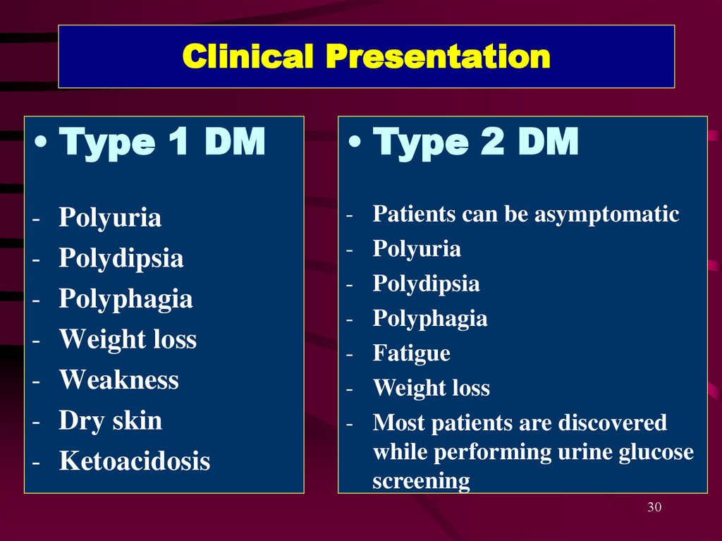
Clinical Presentation• Type 1 DM
• Type 2 DM
-
-
Polyuria
Polydipsia
Polyphagia
Weight loss
Weakness
Dry skin
Ketoacidosis
Patients can be asymptomatic
Polyuria
Polydipsia
Polyphagia
Fatigue
Weight loss
Most patients are discovered
while performing urine glucose
screening
30
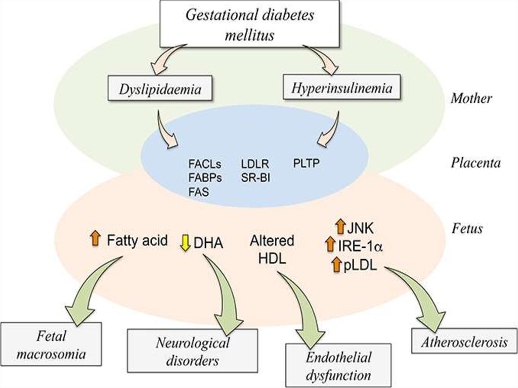
31. Gestational diabetes
A form of glucose intolerance that is diagnosed in some womenduring pregnancy.
Gestational diabetes occurs more frequently among African
Americans, Hispanic/Latino Americans, and American Indians.
It is also more common among obese women and women with a
family history of diabetes.
During pregnancy, gestational diabetes requires treatment to
normalize maternal blood glucose levels to avoid complications
in the infant.
After pregnancy, 5% to 10% of women with gestational
diabetes are found to have type 2 diabetes.
Women who have had gestational diabetes have a 20% to 50%
chance of developing diabetes in the next 5-10 years.
32.
3233.
3334.
3435. Other types of DM
• Other specific types of diabetes resultfrom specific genetic conditions (such as
maturity-onset diabetes of youth),
surgery, drugs, malnutrition, infections,
and other illnesses.
• Such types of diabetes may account for
1% to 5% of all diagnosed cases of
diabetes.
36.
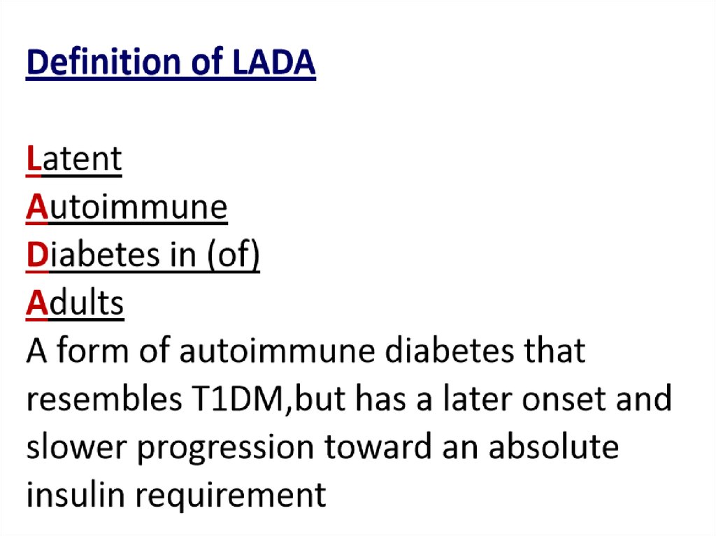
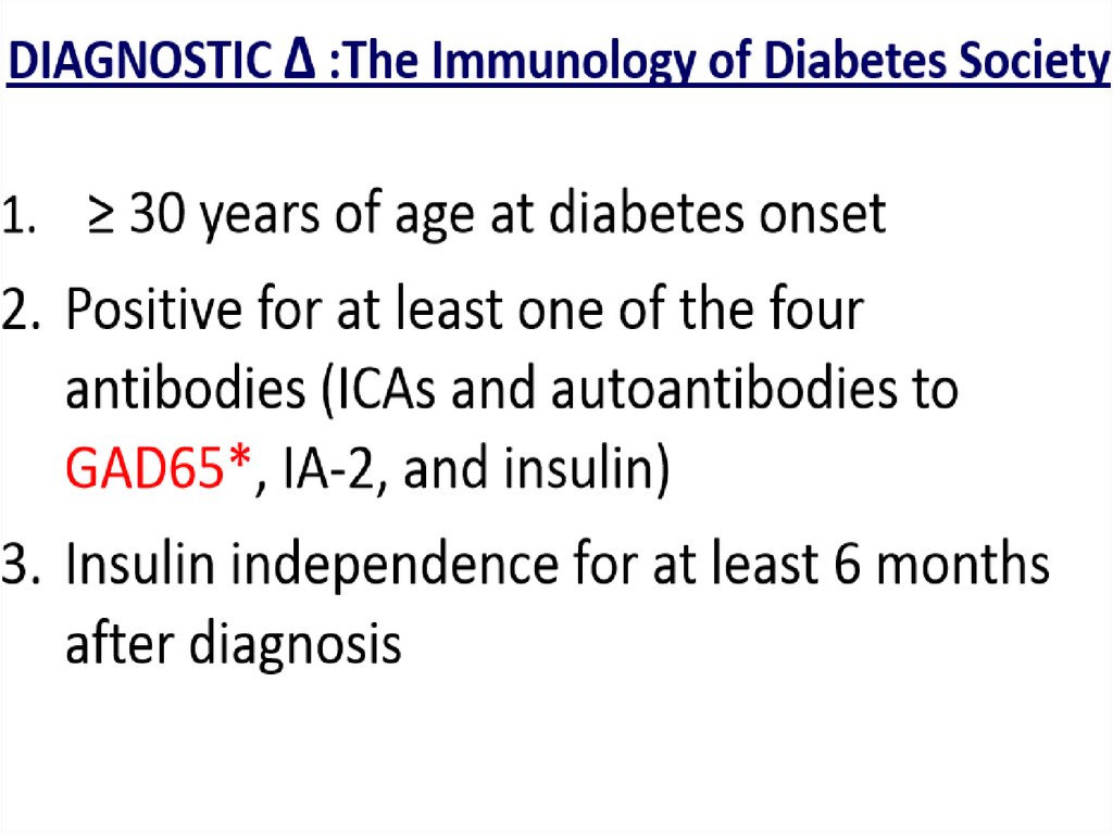
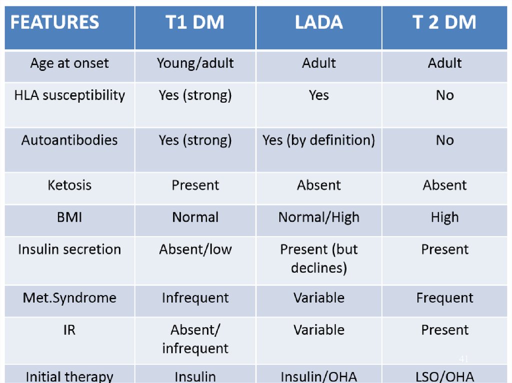
3637. LADA
Latent Autoimmune Diabetes in Adults (LADA) isa form of autoimmune (type 1 diabetes) which is
diagnosed in individuals who are older than the
usual age of onset of type 1 diabetes.
Alternate terms that have been used for "LADA"
include Late-onset Autoimmune Diabetes of
Adulthood, "Slow Onset Type 1" diabetes, and
sometimes also "Type 1.5
Often, patients with LADA are mistakenly thought
to have type 2 diabetes, based on their age at the
time of diagnosis.
38.
3839. LADA (cont.)
40. Clinical Types
• LADA-type 1 :Multiple antibodies or hightiters of GADAb. More resembles T1DM
• LADA-type 2 :Single antibody positivity in
low titers. More resembles T2DM
40
41.
4142.
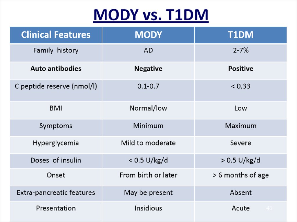
4243. MODY
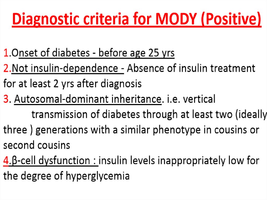
MODY – Maturity Onset Diabetes of the YoungMODY is a monogenic form of diabetes with an autosomal
dominant mode of inheritance:
◦ Mutations in any one of several transcription factors or in the enzyme
glucokinase lead to insufficient insulin release from pancreatic ß-cells,
causing MODY.
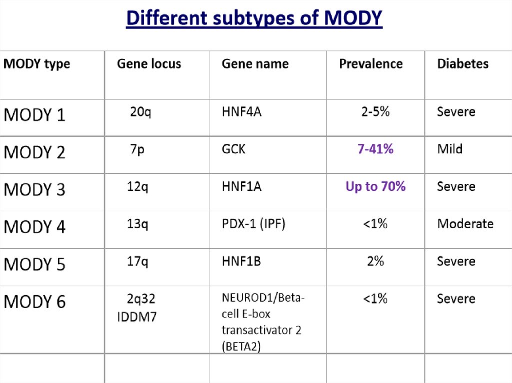
◦ Different subtypes of MODY are identified based on the mutated gene.
Originally, diagnosis of MODY was based on presence of nonketotic hyperglycemia in adolescents or young adults in conjunction
with a family history of diabetes.
However, genetic testing has shown that MODY can occur at any
age and that a family history of diabetes is not always obvious.
44.
4445.
4546.
4647. Secondary DM
Secondary causes of Diabetes mellitus include:Acromegaly,
Cushing syndrome,
Thyrotoxicosis,
Pheochromocytoma
Chronic pancreatitis,
Cancer
Drug induced hyperglycemia:
◦
◦
◦
◦
◦
◦
◦
◦
◦
Atypical Antipsychotics - Alter receptor binding characteristics, leading to increased insulin
resistance.
Beta-blockers - Inhibit insulin secretion.
Calcium Channel Blockers - Inhibits secretion of insulin by interfering with cytosolic calcium
release.
Corticosteroids - Cause peripheral insulin resistance and gluconeogensis.
Fluoroquinolones - Inhibits insulin secretion by blocking ATP sensitive potassium channels.
Naicin - They cause increased insulin resistance due to increased free fatty acid mobilization.
Phenothiazines - Inhibit insulin secretion.
Protease Inhibitors - Inhibit the conversion of proinsulin to insulin.
Thiazide Diuretics - Inhibit insulin secretion due to hypokalemia. They also cause increased
insulin resistance due to increased free fatty acid mobilization.
48. Bibliography
Протоколы заседаний Объединенной комиссии по качеству медицинских услуг МЗ РК, 20171)
American Diabetes Association. Standards of medical care in diabetes - 2017. DiabetesCare, 2017,
Volume 40 (Supplement 1). 2) World Health Organization. Definition, Diagnosis, and Classification
of Diabetes Mellitus and its Complicatios: Report of a WHO consultation. Part 1: Diagnosis and
Classification of Diabetes Mellitus. Geneva, World Health Organization, 1999
(WHO/NCD/NCS/99.2). 3) Алгоритмы специализированной медицинской помощи больным
сахарным диабетом. Под ред. И.И. Дедова, М.В. Шестаковой, А.Ю. Майорова, 8-йвыпуск.
Москва, 2017. 4) World Health Organization. Use of Glycated Haemoglobin (HbAlc) in the
Diagnosis of Diabetes Mellitus. Abbreviated Report of a WHO Consultation. World Health
Organization, 2011 (WHO/NMH/CHP/CPM/11.1). 5) БазарбековаР.Б., НурбековаА.А.,
ДаньяроваЛ.Б., ДосановаА.К. Консенсус по диагностике и лечению сахарного диабета.
Алматы, 2016. 6) Deutsche Diabetes Gesellschaft und Deutsche Vereinte Gesellschaftfür Klinische
Chemie und Labormedizin, 2016. 7) Pickup J., Phil B. Insulin Pump Therapy for Type 1 Diabetes
Mellitus, N Engl Med 2012; 366:1616-24. 8) Zhang M, Zhang L, Wu B, Song H, An Z, Li S.
Dapagliflozin treatment for type 2 diabetes: a systematic review and meta-analysis of randomized
controlled trials.Diabetes Metab Res Rev. 2014 Mar;30(3):204-21. 9) RaskinP.Sodium-glucose
cotransporter inhibition: therapeutic potential for the treatment of type 2 diabetes mellitus. Diabetes
Metab Res Rev. 2013 Jul;29(5):347-56. 10) Grempler R, Thomas L, EckhardtM,et al. Empagliflozin,
a novel selective sodium glucose cotransporter-2 (SGLT-2) inhibitor:characterisation and comparison
with other SGLT-2 inhibitors. Diabetes ObesMetab 2012; 14: 83-90.
48