


")


 medicine
Similar presentations:


Апоневрозы нижних конечностей
1. Апоневрозы нижних конечностей
2.
3. Ягодичные апоневрозы
Fasciae of the Low er LimbЯгодичные
апоневрозы
External oblique
muscle with its
fascia
Gluteus medius
fascia
Gluteus maximus
with its fascia
FIGURE 8.20 Posterolateral view of the gluteal region. The gluteus medius fascia has an aponeurotic aspect, while the fasciae of the gluteus
maximus and exter nal oblique muscles have epymisial featur es.
310
4. Мышечно-нервная и сосудистая лакуны паховой области
1 – lacuna musculorum2 - arcus iliopectineus
3 – lig. Inguinale
4 – a. femoralis
5 – v. femoralis
6 – lacuna vazorum
7 – anulus femoralis
8 – глубокий лимфатический
паховый узел
9 – lig.lacunare (Gimbernate)
10 – funiculus spermaticus
11 – m.pectineus
12 – n.a.v. obturatoriae
13 – n.femoralis
14 – m. ileopsoas
5. Треугольник Скарпа
6. Широкая фасция бедра
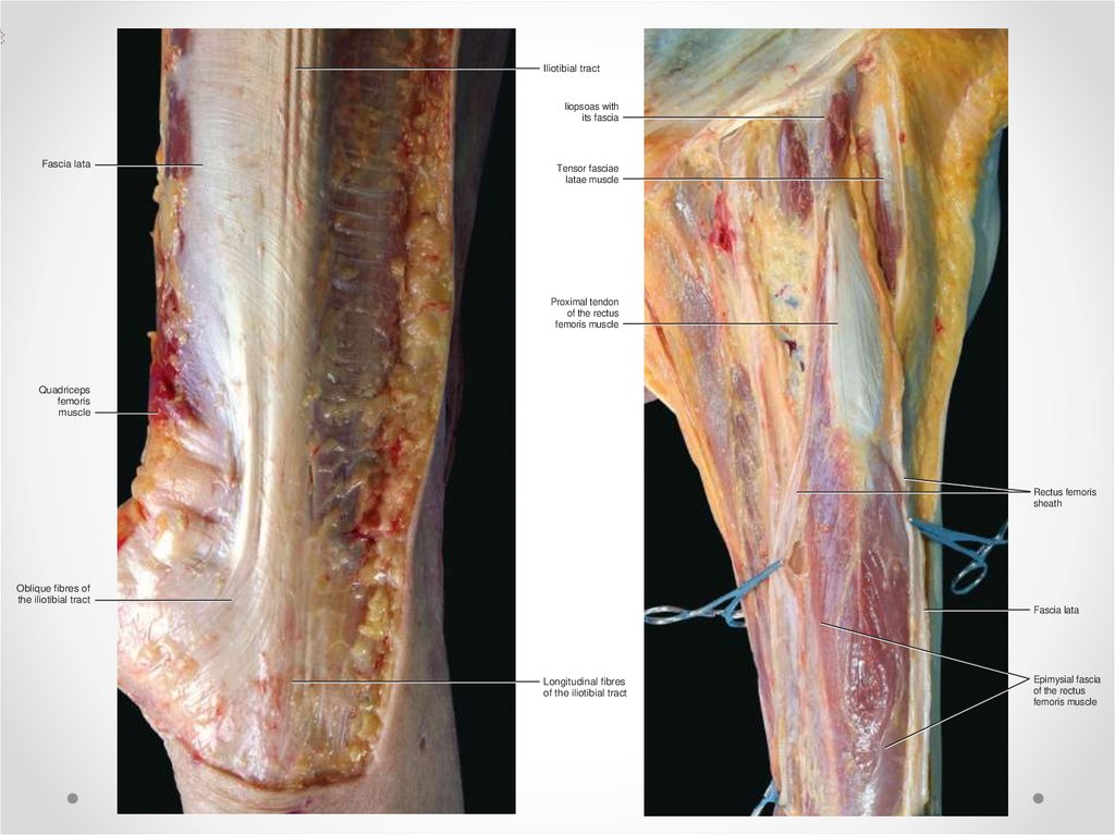
7.
Iliotibial tractFascia lata
Fasciae of the Low er Limb
liopsoas with
its fascia
Tensor fasciae
latae muscle
Proximal tendon
of the rectus
femoris muscle
Quadriceps
femoris
muscle
Rectus femoris
sheath
Oblique fibres of
the iliotibial tract
Fascia lata
Longitudinal fibres
of the iliotibial tract
Epimysial fascia
of the rectus
femoris muscle
FIGURE 8.26 Lateral view of the thigh. Near the knee, two main components of the iliotibial tract are shown: one vertical,FIGURE
one oblique.
The latter
8.24 Anterior
view of the thigh. The fascia lata is cut to show the rectus femoris. Note the continuity between the rectus femoris sheath
8. Фасциальные ложа бедра (средняя треть)
1 - широкая фасция;2 - медиальная широкая мышца бедра;
3 - медиальная межмышечная
перегородка бедра;
4 - бедренные артерия и вена;
5 - портняжная мышца;
6 - приводящие мышцы;
7 - тонкая мышца;
8 - задняя межмышечная перегородка
бедра;
9 - полуперепончатая мышца;
10 - полусухожильная мышца;
11 - длинная головка двуглавой мышцы
бедра;
12 - короткая головка двуглавой
мышцы бедра;
13 - латеральная межмышечная
перегородка бедра;
14 - латеральная широкая мышца
бедра;
15 - бедренная кость;
16 - промежуточная широкая мышца
бедра;
17 - прямая мышца бедра
9. Апоневрозы голени
Distal insertion ofthe iliotibial tract
Anterior retinaculum
of the knee
Crural fascia covering the
extensor muscles of the leg
Prepatellar bursa
Fascia lata covering the
vastus lateralis muscle
Fasciae of the Low er Limb
Апоневрозы голени
10. Фасциальные ложа голени
1112
1. Переднее ложе.
2. Боковое ложе.
3, 4. Задние ложа.
5. Передняя большеберцовая артерия.
7. Межкостная перегородка
6. Задняя большеберцовая артерия.
8. Малоберцовая артерия.
9. большеберцовая кость.
10. Малоберцовая кость
11 Задняя перегородка
12 Передняя перегородка
11. Мышцы переднего фасциального ложа голени
Длинный разгибатель пальцев12. Функции мышц переднего фасциального ложа голени
Супинация стопыРазгибание стопы
Разгибание большого
пальца стопы
13. Мышцы латерального фасциального ложа голени
14. Функция мышц латерального фасциального ложа голени
Сгибание стопыПронация стопы
15. Мышцы заднего фасциального ложа голени
ПОВЕРХНОСТНЫЙСЛОЙ
Икроножная
мышца
Ахиллово
сухожилие
ГЛУБОКИЙ
СЛОЙ
Камбаловидная
мышца
Подколенная
мышца
Мышцы –
сгибатели
стопы и
пальцев
16. Функции мышц заднего фасциального ложа голени
Сгибание стопыСгибание пальцев
17. Межкостная мембрана
Fasciae of the Low er LimbCrural fascia
Muscular insertions
into the crural fascia
Tibialis
tibialis anterior
muscle enveloped
by its epimysial
fascia (epimysium)
FIGURE 8.52 Anterolateral view of the leg. The crural fascia is detached from the underlying planes and lifted laterally. The tibialis anterior mu
18. Удерживатели разгибателей и тыльный апоневроз стопы
Inferior extensor retinaculumFasciae of the Low er Limb
Superior extensor retinaculum
Inferior extensor retinaculum lifted
to show the muscular insertions
Extensor digitorum brevis
muscle
Extensor hallucis brevis muscle
19. Тыльные апоневрозы стопы
Fasciae of the Low er LiFinally, there is an intrinsic compartment formed by
the four intrinsic muscles between the rst and fth
metatarsals.
exor digitorum brevis. Thus, the abductor hallucis
muscle could be considered a key muscle for the fascial tension of the foot, as it connects the fascia of the
dorsum with the plantar fascia, and these fasciae with
the crural fascia in the leg.
Laterally, the dorsal fascia of the foot forms the fascial
compartment of the abductor digiti minimi. The abductor digiti minimi has some bres that originate from
the plantar fascia and others from the intermuscular
septum between it and the exor digitorum brevis.
Тыльные
апоневрозы стопы
DORSAL FASCIA OF THE FOOT
The fascia on the dorsum of the foot consists of a thin
brous layer, continuous above with the inferior extensor ankle retinaculum (Fig. 8.63). It forms a sheath
for the tendons on the dorsum of the foot (extensor
digitorum longus and extensor hallucis longus) (Fig.
8.64). Inserted at its inner side are some muscle bres
of the extensor digitorum brevis and extensor hallucis brevis muscles. When these muscles contract they
stretch this fascia and the inferior extensor retinaculum in a caudal direction. The dorsal fascia of the foot
is also stretched in a cranial direction by the myofascial
Extensor hallucis brevis muscle
covered by its fascia
FASCIA OF THE INTEROSSEI
Kalin and Hirsch (1987) found that the dorsal and
plantar interosseous muscles arise not only from the
metatarsal bones, but also from ligamentous tissue
Tendons of the extensor digitorum
longus muscle covered by the
dorsal fascia of the foot
Abductor digiti minimi muscle
covered by its fascia
FIGURE 8.63 Dorsal region of the foot. The deep fascia covers all the tendons and muscles and continues laterally with the fascia of the abductor
20. Удерживатели сгибателей
21. Подошвенные апоневрозы стопы
Medial portionMiddle portion
Fasciae of the Low er Limb
Lateral portion
FIGURE 8.69 Sole of the foot. The subcutaneous tissues are removed to show the plantar fascia. The plantar fascia has a middle portion and two
22. Своды стопы
РЕ
С
С
О
Р
Н
А
Я
Г
Р
У
З
О
В
Дистальный
А
поперечный
Я
свод
Ч
А
С
Т
Ь
Ч
ПроксимальА
ный попереч
С
ный свод
Т
Ь
Продольный свод
Отпечатки стоп
1
2
3
4
4- вариант плоской стопы