


 medicine
Similar presentations:


Ebola virus disease in pregnancy:
1.
EBOLA VIRUS DISEASE IN PREGNANCY:clinical histopathologic and immunohistochemical findings
Nurdilda Nargiz, Kani Aidana, Berekekyzy Zhanerke,
Bagytzhanova Aina, Sagadinov Sagyndyk
Faculty of Education and Humanites
Suleyman Demirel University, Kazakhstan
2.
Thanks to ...Atis Muehlenbachs, Olimpia de la Rosa Vázquez,
Daniel G. Bausch, Ilana J. Schafer, Christopher D.
Paddock, Jean Paul Nyakio, Papys Lame, Eric
Bergeron, Andrea M. McCollum, Cynthia S.
Goldsmith, Brigid C. Bollweg, Miriam Alía Prieto,
Robert Shongo Lushima, Benoit Kebela Ilunga, Stuart
T. Nichol, Wun-Ju Shieh, Ute Ströher, Pierre E. Rollin,
Sherif R. Zaki
For such great article!
3.
EBOLA VIRUS (EVD)An infectious
Generally fetal disease marked by
fever
Severe internal bleeding
Spread throughout contacts with
Body fluids by Filovirus (Ebola Virus)
HOST
Unknown
4.
BACKGROUNDNamed because of Ebola River
RIVER
5.
FIRST APPEARANCE OF EVDIn Sudan and Zaire in 1976
FIRST OUTBREAK
In Sudan
Infected over 284 people
Killing 53% of victim
Another strain appeared
Infected another 318 people
Mortality rate was 86%
6.
AFFECT OF EBOLA VIRUS 1976-20147.
Species of EbolavirusesEbolaviruses are closely related to species in the genus
Marburgvirus, which was discovered in 1967, and the two are
the only members of the Filoviridae that cause epidemic human
disease. Five species of ebolaviruses—known as Zaire
ebolavirus, Sudan ebolavirus, Taï Forest ebolavirus, Reston
ebolavirus, and Bundibugyo ebolavirus, named for their
outbreak locations—have been described. The viruses are
known commonly as Ebola virus (EBOV), Sudan virus (SUDV),
Taï Forest virus (TAFV), Reston virus (RESTV), and Bundibugyo
virus (BDBV).
8.
Ebola virus disease (EVD) and Marburg virus disease arecaused by viruses of the Ebolavirus and Marburgvirus
genera (family Filoviridae). Here, we collectively refer to
Ebola virus (EBOV), Sudan virus (SUDV) and
Bundibugyo virus (BDBV) all within the Ebolavirus genus
as ebolaviruses. Filovirus infection during pregnancy is
associated with maternal hemorrhage, preterm labor,
miscarriage, and maternal and neonatal death.
Supplementary Table 1 presents a summary of the
scientific literature to date; maternal death occurred in
102 of 125 reported cases (82%), and there was uniform
loss of offspring, whether by miscarriage, stillbirth, or
neonatal death. Of the 18 live births, the longest survival
was 19 days.
9.
Despite the severity of filovirus infection in pregnancy for both motherand child, very little is known regarding pathogenesis. Fetal-placental
viral tropism has been hypothesized due to recent observations during
the 2013–2016 West Africa EBOV outbreak: pregnant women were
noted to survive EVD and clear virus from the blood without fetal loss
during acute infection and to deliver stillbirths in the subsequent weeks
and months with relatively high EBOV RNA levels in placental and fetal
tissue swab specimens [7–10]. We report clinical, histopathologic, and
immunohistochemical findings of SUDV and BDBV infections in 2
pregnant women and their offspring that help shed light on the
pathogenesis of fetal infection and loss in EVD.
10.
METHODSPatients
Two pregnant women with EVD were cared for in Ebola treatment centers
during ebolavirus outbreaks in Gulu, Uganda, in 2000 [11, 12] and Isiro,
DRC, in 2012 [13] (Schafer, unpublished data). Specimens were
collected and evaluated during the course of the outbreak responses.
Ebolavirus Diagnostic Testing
SUDV reverse transcription–polymerase chain reaction (RT-PCR) and
enzyme-linked immunosorbent assays (ELISAs) in Gulu and BDBV RTPCR assays in Isiro were performed as previously described [14, 15] in
field laboratories run by the Viral Special Pathogens Branch (VSPB),
Centers for Disease Control and Prevention (CDC; Atlanta, Georgia).
BDBV immunoglobulin M (IgM) and immunoglobulin G (IgG) ELISAs
were performed by the VSPB in Atlanta.
11.
ELISARapid blood tests detect specific RNA sequences by
reverse-transcription polymerase chain reaction (RT-PCR)
or viral antigens by enzyme –linked immunosorbent assay
(ELISA).
Most acute infections are determined through the use of
polymerase chain reaction testing (PCR).
Virus is generally detectable by RT-PCR between 3 to 10
days after the onset of symptoms.
12.
Histopathologic Analysis,Immunohistochemical Analysis, and
Transmission Electron Microscopy
Placenta (Gulu and Isiro), fetal tissues (Gulu), and a postmortem
skin biopsy (Isiro) were collected and placed in 10% neutral
buffered formalin and transported to the CDC, where the samples
were processed using standard histological methods. The
identification and scoring of malarial parasite pigment was
performed as previously described [16]. Immunohistochemical
analysis for ebolavirus antigens was performed using a polymerbased indirect immunoalkaline phosphatase detection system for
colorimetric detection (Biocare Medical, Concord, California).
Rabbit polyclonal antisera against EBOV, SUDV, and Reston virus
and EBOV hyperimmune mouse ascitic fluid (courtesy of Thomas
Ksiazek, VSPB, CDC), previously shown to detect SUDV and
BDBV antigens, were each used at a 1:1000 dilution with
appropriate positive and negative controls [17]. On-slide
embedding and transmission electron microscopy was performed as
previously described [18].
13.
RESULTS. Patient 1Patient 1, Gulu, Uganda, 30-year-old housewife
Symptoms: asthenia, anorexia, abdominal pain, nausea, vomiting, diarrhea,
and dry cough - presented with a 1-day of illness. Has been pregnant for
28 weeks. Next day, she had temperature of 36.7°C, here heart rage
was 120 beats/minute, and her respiratory rate was 24 breaths/minute,
with an oxygen saturation level of 92% by pulse oximetry. Her blood
tested positive for SUDV by both ELISA antigen assay and nested RTPCR.
On day four of illness, the patient spontaneously delivered a dead but
apparently morphologically normal fetus and placenta. The degree of
vaginal bleeding did not seem abnormal for a stillbirth. Over the next 3
days, the patient complained of joint pain and swelling, throat and chest
pain, persistent dry cough, dyspnea, and, briefly, hiccups. Her wrists and
knees were visibly swollen and tender to the touch, and pulmonary rales
persisted. She was consistently febrile. Disease severity peaked at day 7
of illness, when vital signs were an axillary temperature of 37.8°C, a
heart rate of 128 beats/minute, a respiratory rate of 30 breaths per
minute, and an oxygen saturation level of 90%. She gradually improved,
and she was discharged on day 13 with normal vital signs and all
symptoms resolved.
14.
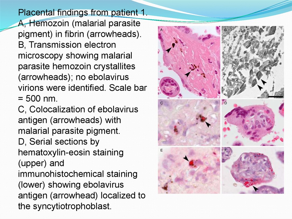
Pathologic FindingsThe placenta had mild subchorionitis and a moderate amount of malarial
parasite pigment (hemozoin) in fibrin and within macrophages embedded in
fibrin (Figure 1A). No parasitized erythrocytes or malarial intervillous
inflammatory infiltrates were present. By electron microscopy, hemozoin
crystallites were identified (Figure 1B), but no ebolavirus virions were seen.
The umbilical cord was normal.
Immunohistochemical analysis revealed ebolavirus antigen in the placenta,
primarily within areas of fibrin deposition, localized to embedded maternal
mononuclear cells, including malarial parasite pigment–laden macrophages
(Figure 1C). Focal immunostaining was seen within the syncytiotrophoblast
(Figure 1D). The decidua, fetal placental villous stroma, amnion, and umbilical
cord were negative by immunohistochemical analysis, and no tissue necrosis
or viral inclusions were noted.
Fetal tissues (lung, heart, liver, spleen, kidney, skin, and bone marrow) were
well preserved with minimal autolysis, were normal for gestational age, and had
no necrosis or viral inclusions. All fetal tissues were negative by
immunohistochemical analysis.
15.
Placental findings from patient 1.A, Hemozoin (malarial parasite
pigment) in fibrin (arrowheads).
B, Transmission electron
microscopy showing malarial
parasite hemozoin crystallites
(arrowheads); no ebolavirus
virions were identified. Scale bar
= 500 nm.
C, Colocalization of ebolavirus
antigen (arrowheads) with
malarial parasite pigment.
D, Serial sections by
hematoxylin-eosin staining
(upper) and
immunohistochemical staining
(lower) showing ebolavirus
antigen (arrowhead) localized to
the syncytiotrophoblast.
16.
RESULTS. Patient 2Patient 2, 29-year-old housewife, who was transferred from a health center
because of suspicion of EVD by a local clinician who was aware that her
relative died recently. She was admitted to the Ebola treatment center on day
4 of illness with fever, fatigue, headache, abdominal pain (with uterine
contractions), anorexia, dysphagia, vomiting, diarrhea, and muscle and joint
pain. Her last menstrual period date was unknown, but she was initially
estimated to be 7 months pregnant. Conjunctival injection was noted. Her
heart rate was 80 beats/minute, and her respiratory rate was 20
breaths/minute. Her cervix was 50% effaced with a 4-cm dilation, and fetal
movement was normal.
On day 5 of illness, her cervix was 100% effaced with an 8-cm dilation, and she
was treated with oxytocin. A malaria rapid diagnostic test was positive, and AL
was continued. That night (day 6 of illness), spontaneous vaginal delivery of a
live-born male infant occurred without assistance. The degree of vaginal
bleeding did not seem abnormal for a normal delivery, although she had had
an episode of black stool some hours later. She was treated with oxytocin,
ergometrine, intravenous fluids, and cefixime, and Plumpy′nut (Nutriset) was
provided. On day 7, the mother's condition rapidly deteriorated, with wheezing,
drowsiness, weakness, and a temperature of 38.5°C. Antibiotics were
switched to ceftriaxone. On day 8, she became comatose and died. A
postmortem skin sample was collected from the mother as part of the routine
17.
The infant appeared healthy at birth, with Apgar scores of8/10/10, and was clinically assessed to be at term on the basis
of examination of the nails and soles of the feet. Infant formula
was provided, although the baby may have briefly breastfed
immediately after delivery. A placental sample was collected to
evaluate for BDBV. Blood collected at 1 day of age (the second
day of life) was positive for BDBV by RT-PCR, with a cycle
threshold (Ct) of 29.2. Over the next few days, the baby was
noted to be quiet and inactive. He became febrile (temperature,
38.5°C) on day 4 of age, and repeat testing of the blood
revealed an RT-PCR Ct of 17.9 with negative IgM and IgG
ELISA results. Over the next few days, the baby had
hematemesis and bloody stools. He developed respiratory
distress and coma and died on the seventh day of age (eighth
day of life). No postmortem specimens were collected from the
infant.
18.
Pathologic FindingsIn the placenta, scattered atypical maternal macrophages were seen within the
intervillous space. These cells had degenerate-appearing nuclei, cytoplasmic
blebs, and small eosinophilic cytoplasmic granules, suggestive of viral inclusions
(Figure 2A). The placenta was otherwise normal, and the placental membranes
and umbilical cord were not sampled. No malarial parasite pigment or
parasitized erythrocytes were seen. No virions were seen by transmission
electron microscopy.
Ebolavirus antigen was seen by immunohistochemical analysis within the
circulating large atypical maternal mononuclear cells (Figure 2B). Antigen was
also present in multiple foci within the villous syncytiotrophoblast (Figure 2C),
frequently most intense at the basal aspect. Fetal stromal and endothelial cells
were negative by immunohistochemical analysis. In the basal plate,
immunostaining was prominent within the extravillous trophoblast (Figure
2D) ,with scattered additional cell types likely representing decidual and
maternal mononuclear cells. Focally, the lining cells of the maternal vessels of
the basal plate (likely endovascular trophoblasts) were positive. Within the
placenta, fetal stromal tissue, including villous blood vessels, was negative by
immunohistochemical analysis. The postmortem maternal skin specimen was
morphologically normal and immunohistochemically negative.
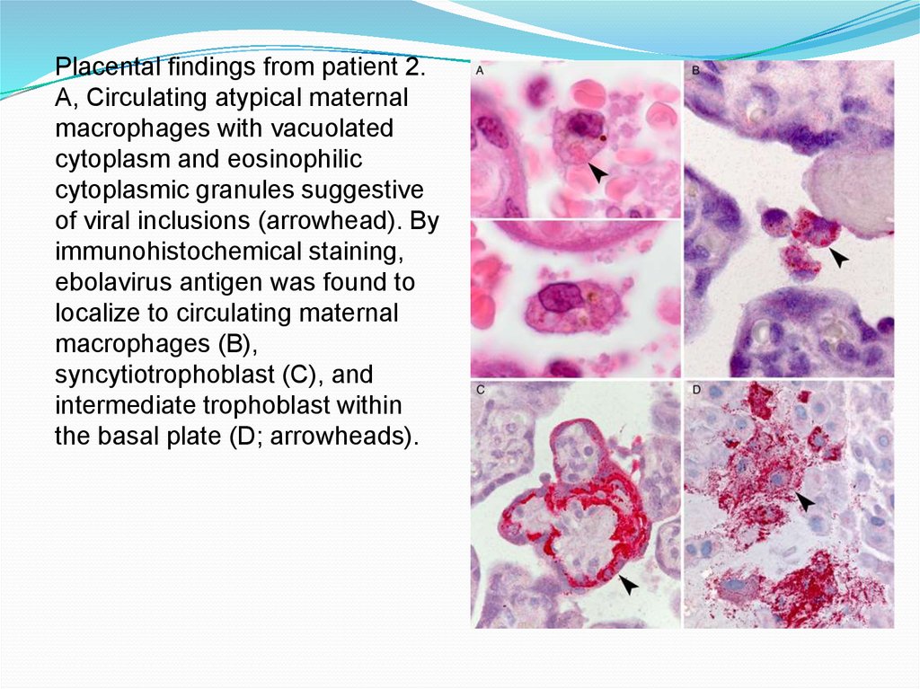
19.
Placental findings from patient 2.A, Circulating atypical maternal
macrophages with vacuolated
cytoplasm and eosinophilic
cytoplasmic granules suggestive
of viral inclusions (arrowhead). By
immunohistochemical staining,
ebolavirus antigen was found to
localize to circulating maternal
macrophages (B),
syncytiotrophoblast (C), and
intermediate trophoblast within
the basal plate (D; arrowheads).
20.
DISCUSSIONVertical transmission of pathogens can be by transplacental, transvaginal, or
by breastfeeding routes. Placenta sampling provides the opportunity to study
disease processes in living patients and gain insights regarding the mode and
mechanism of vertical transmission. In this study, SUDV or BDBV antigen was
noted in fetal trophoblast cells, suggesting that these viruses can infect and
potentially cross the placental epithelial barrier, resulting in transplacental
infection of the fetus. Transplacental infection of the fetus by EBOV has been
previously documented in stillbirths by PCR analysis of amniotic fluid, fetal
blood, and fetal swab specimens [7, 8]. The immunoprotective role of the
placenta may promote the persistence of virus observed in these cases even
after virus has been cleared from maternal blood [8, 9].
Several human pathogens can efficiently penetrate the placental barrier and
infect the fetus, including some herpesviruses, human immunodeficiency virus,
Zika virus, Treponema, and Toxoplasma. The trophoblast is the major cellular
barrier to fetal infection, and it comprises 2 major types: the villous trophoblast,
which is directly exposed to maternal blood, and the extravillous trophoblast,
which invades the maternal decidua and directly contacts maternal cells,
including lymphocytes and decidual stromal cells. In this study, both the
syncytiotrophoblast (in both patients) and the extravillous trophoblast (in the
patient from Isiro) demonstrated ebolavirus antigen by immunohistochemical
analysis.
21.
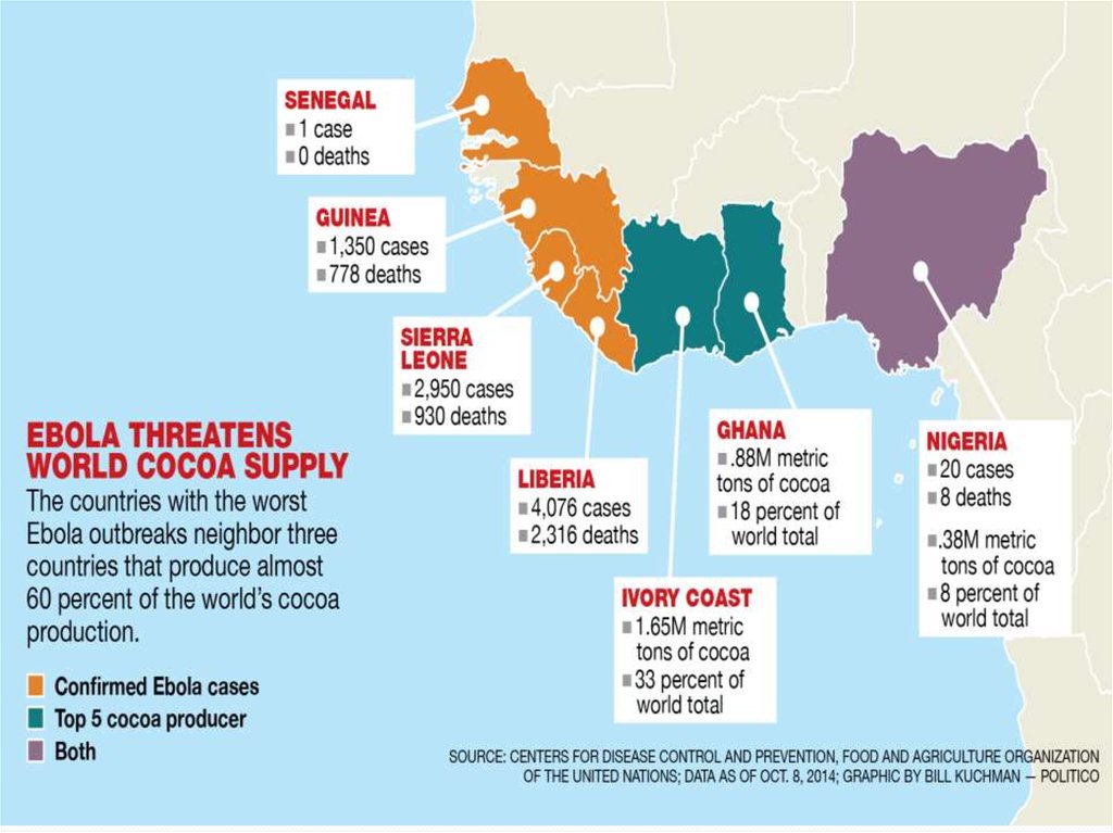
COUNTRIES AFFECTED WITH EVD22.
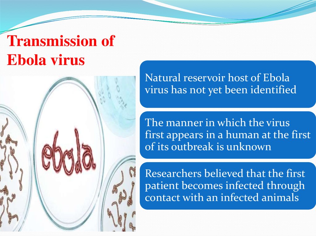
Transmission ofEbola virus
Natural reservoir host of Ebola
virus has not yet been identified
The manner in which the virus
first appears in a human at the first
of its outbreak is unknown
Researchers believed that the first
patient becomes infected through
contact with an infected animals
23.
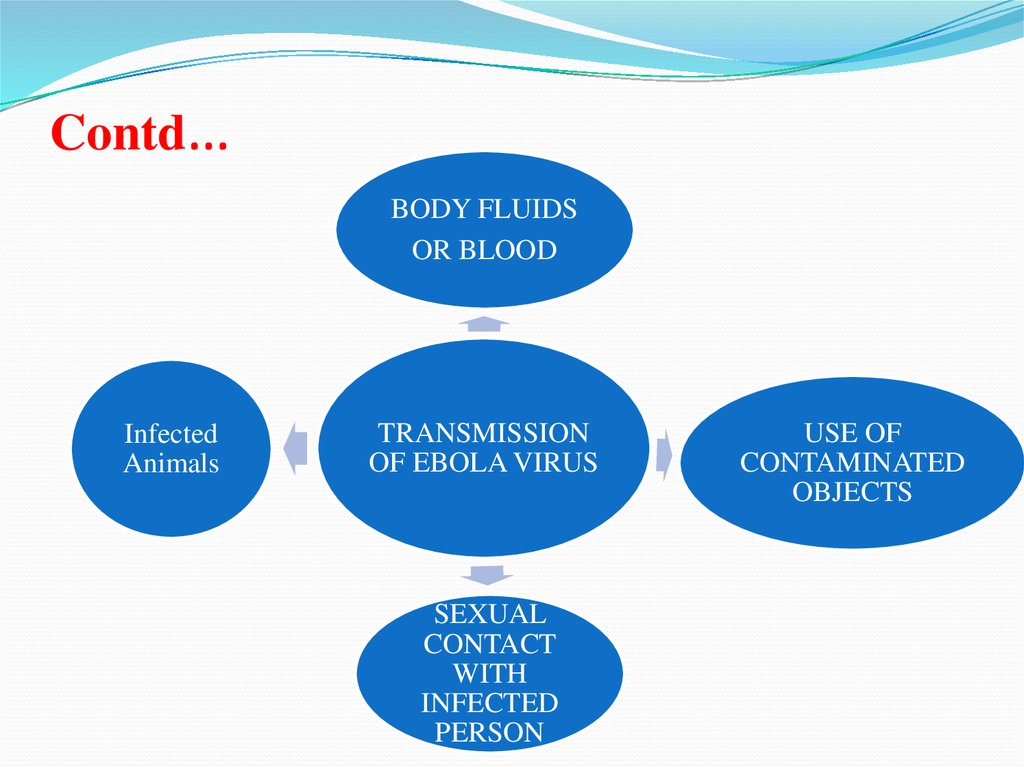
Contd…BODY FLUIDS
OR BLOOD
Infected
Animals
TRANSMISSION
OF EBOLA VIRUS
SEXUAL
CONTACT
WITH
INFECTED
PERSON
USE OF
CONTAMINATED
OBJECTS
24.
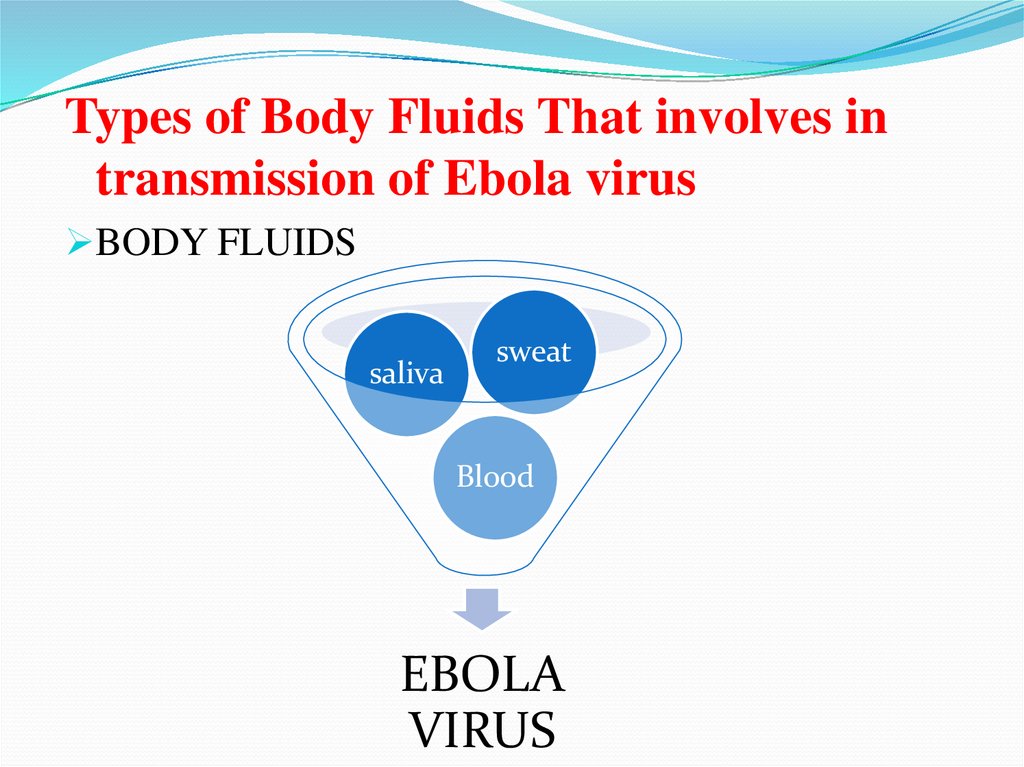
Types of Body Fluids That involves intransmission of Ebola virus
BODY FLUIDS
saliva
sweat
Blood
EBOLA
VIRUS
25.
Thanks for Attention!26.
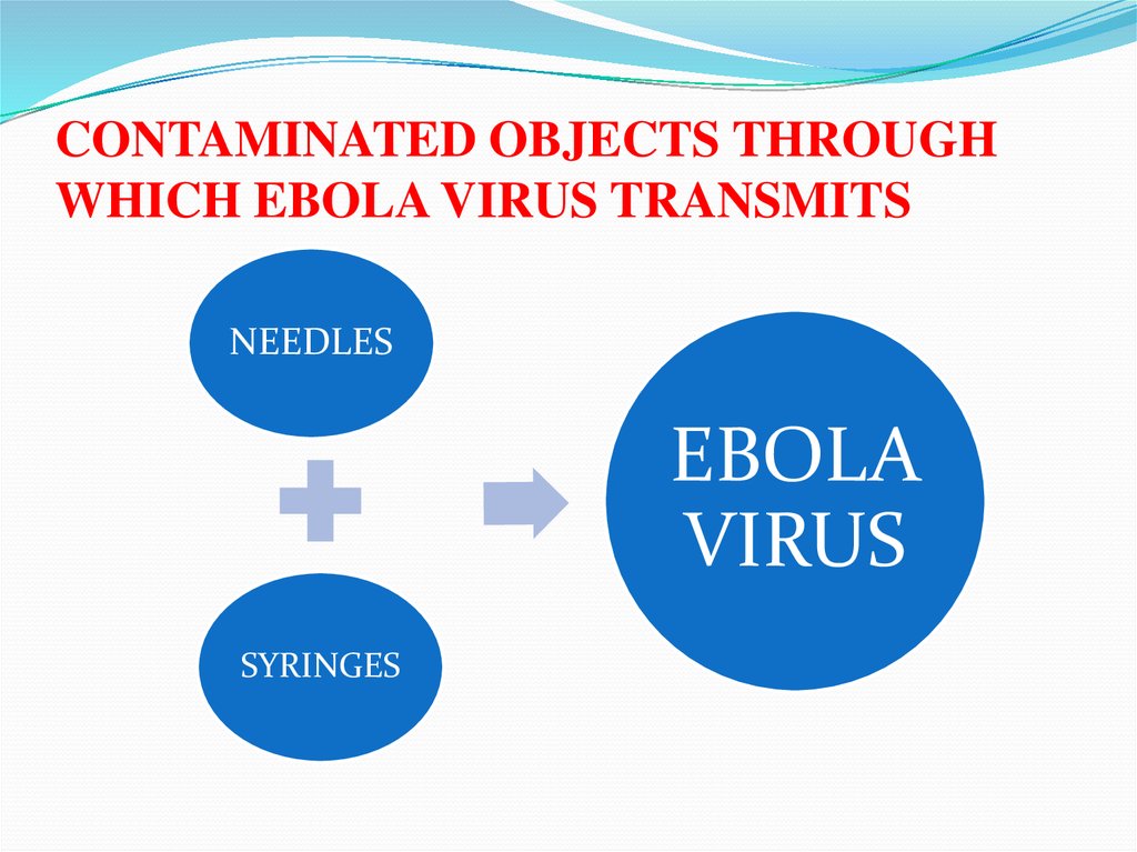
CONTAMINATED OBJECTS THROUGHWHICH EBOLA VIRUS TRANSMITS
NEEDLES
EBOLA
VIRUS
SYRINGES
27.
28.
TRADITIONALAFRICAN RITUALS
PLAYED ROLE IN
TRANSMISSION OF
VIRUS
29.
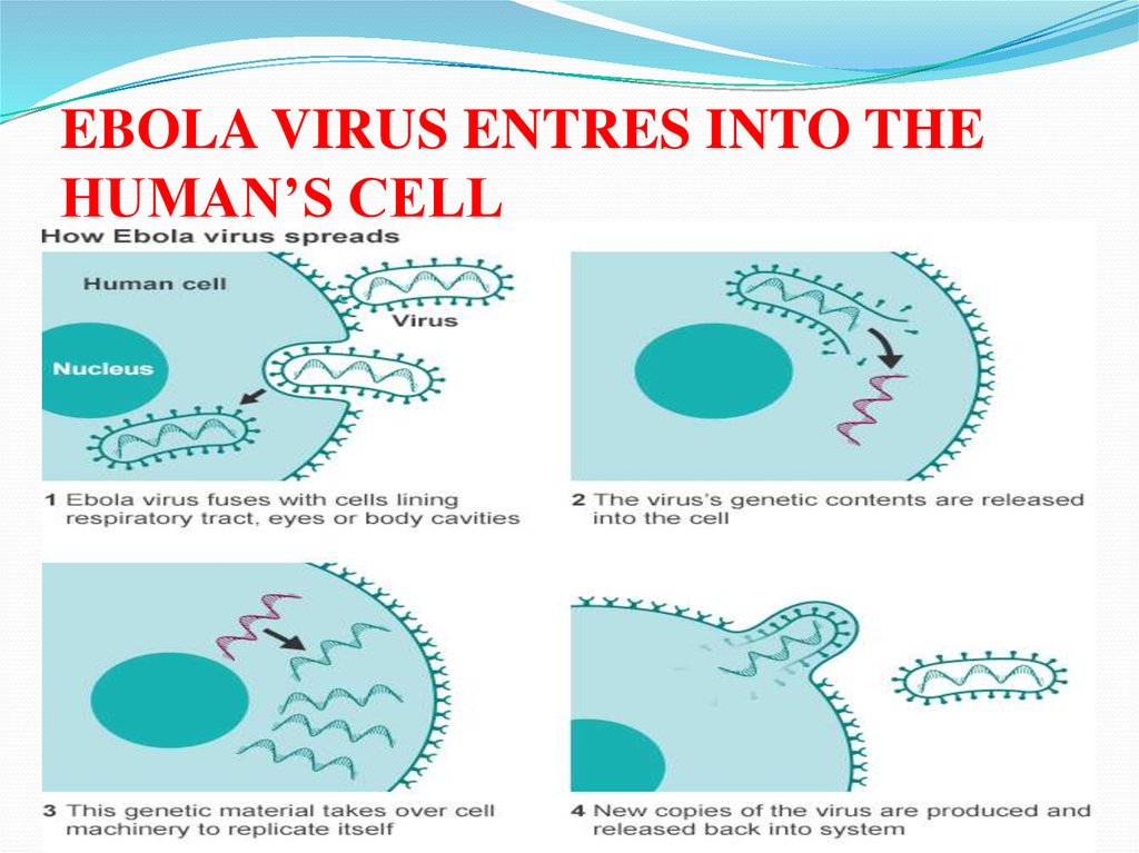
EBOLA VIRUS ENTRES INTO THEHUMAN’S CELL
30.
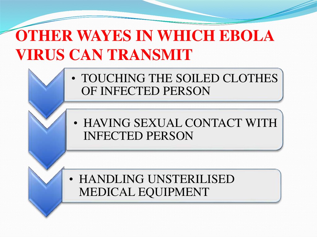
OTHER WAYES IN WHICH EBOLAVIRUS CAN TRANSMIT
• TOUCHING THE SOILED CLOTHES
OF INFECTED PERSON
• HAVING SEXUAL CONTACT WITH
INFECTED PERSON
• HANDLING UNSTERILISED
MEDICAL EQUIPMENT
31.
MASS CREMATION HAVE BEENSANCTIONED BY THE GOVERNMENT IN
LIBERIA IN BID TO HELP TO HALT THE
DEADLY VIRUS
32.
THESE SHOCKING PICTURES SHOW THEBODIES OF EBOLA VICTIMS BEING
BURNED ON HUGE FUNERAL PYRE
33.
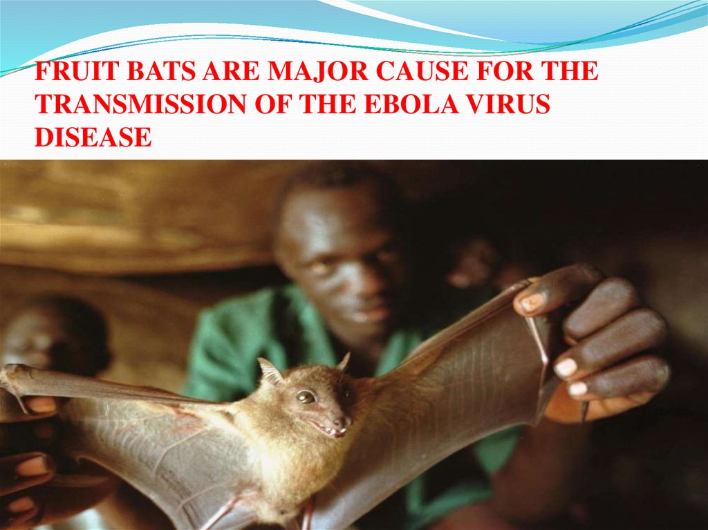
FRUIT BATS ARE MAJOR CAUSE FOR THETRANSMISSION OF THE EBOLA VIRUS
DISEASE
34.
UNHYGIENIC ENVIRONMENT MAY ALSO BE ACAUSE OF TRANSMISSION OF EBOLA VIRUS IN
WEST AFRICA
35.
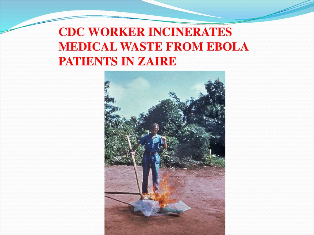
CDC WORKER INCINERATESMEDICAL WASTE FROM EBOLA
PATIENTS IN ZAIRE
36.
37.
38.
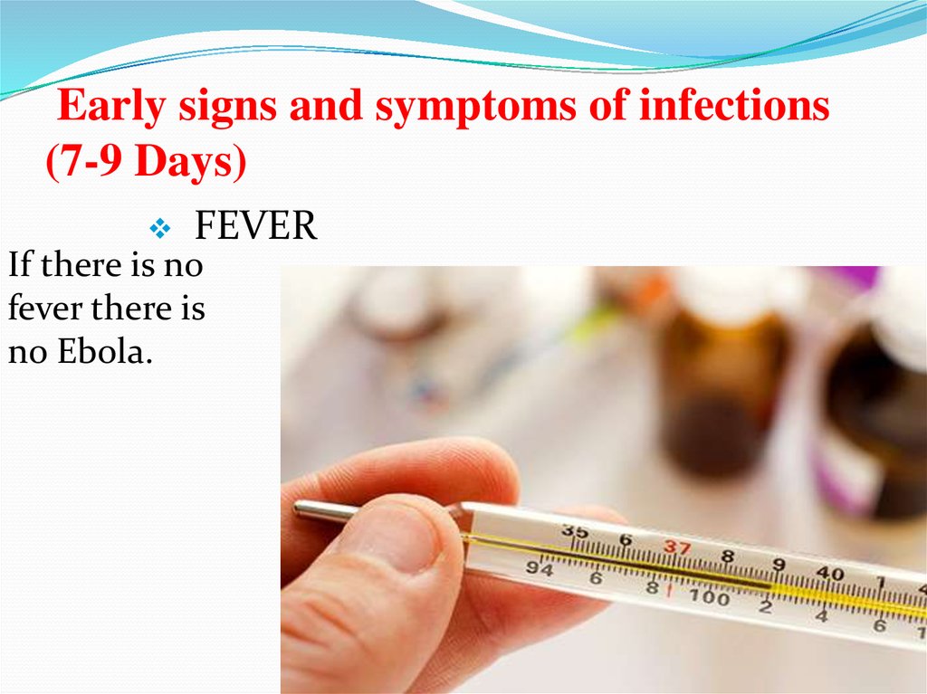
Early signs and symptoms of infections(7-9 Days)
FEVER
If there is no
fever there is
no Ebola.
39.
HEADACHESevere headaches start developing
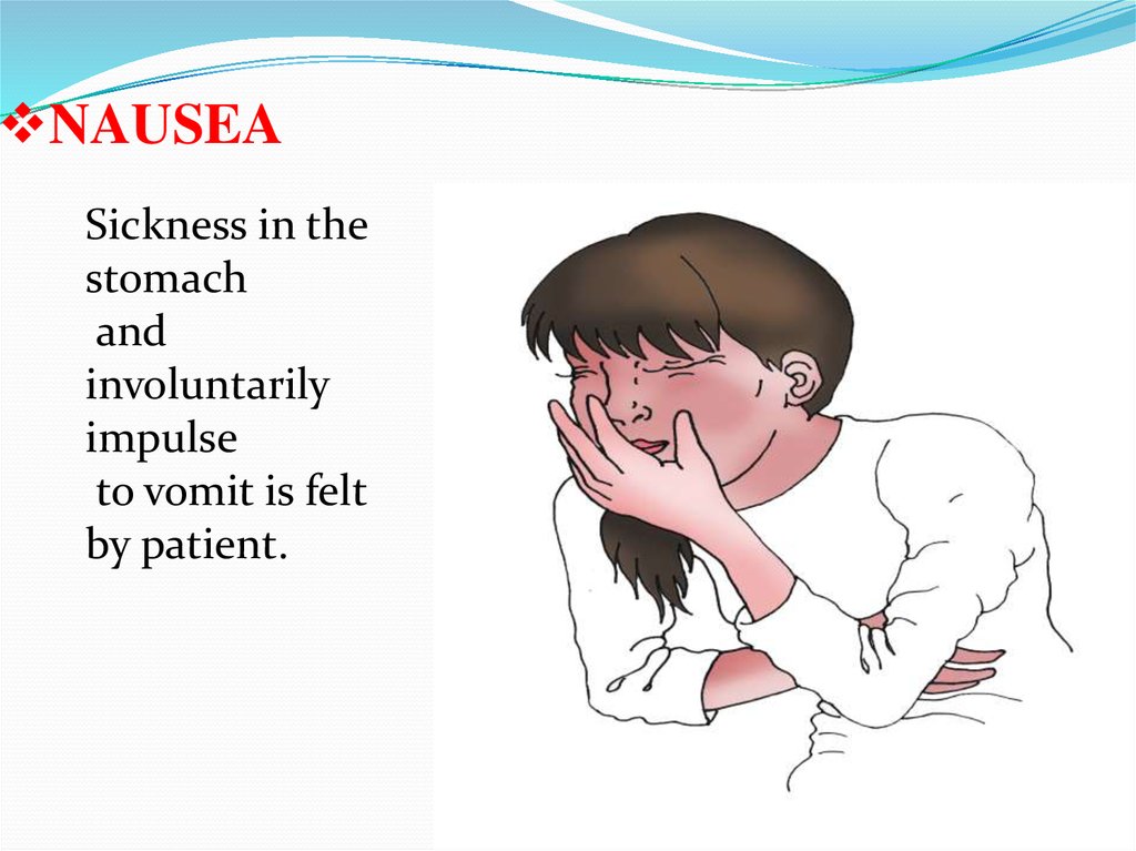
40.
NAUSEASickness in the
stomach
and
involuntarily
impulse
to vomit is felt
by patient.
41.
MUSCULARPAIN
Joint and
muscle pain
leads to
intense
weakness
throughout
the body of
the person.
42.
TIREDNESS43.
44.
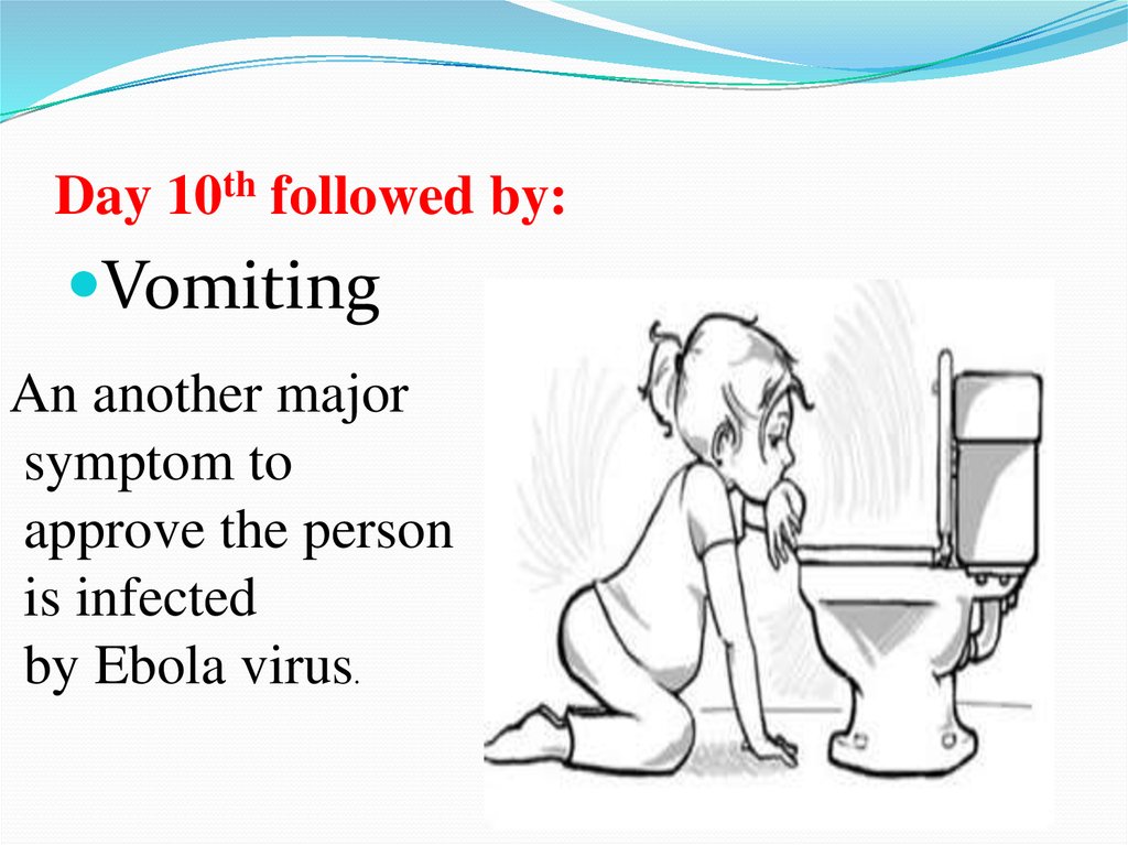
Day 10th followed by:Vomiting
An another major
symptom to
approve the person
is infected
by Ebola virus.
45.
Diarrhea46.
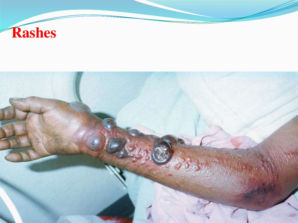
Rashes47.
Condition worsens on day 11thBRAIN DAMAGE
Loss of
consciousness ,
Seizures,
Massive internal
bleeding
leads to brain
damage.
48.
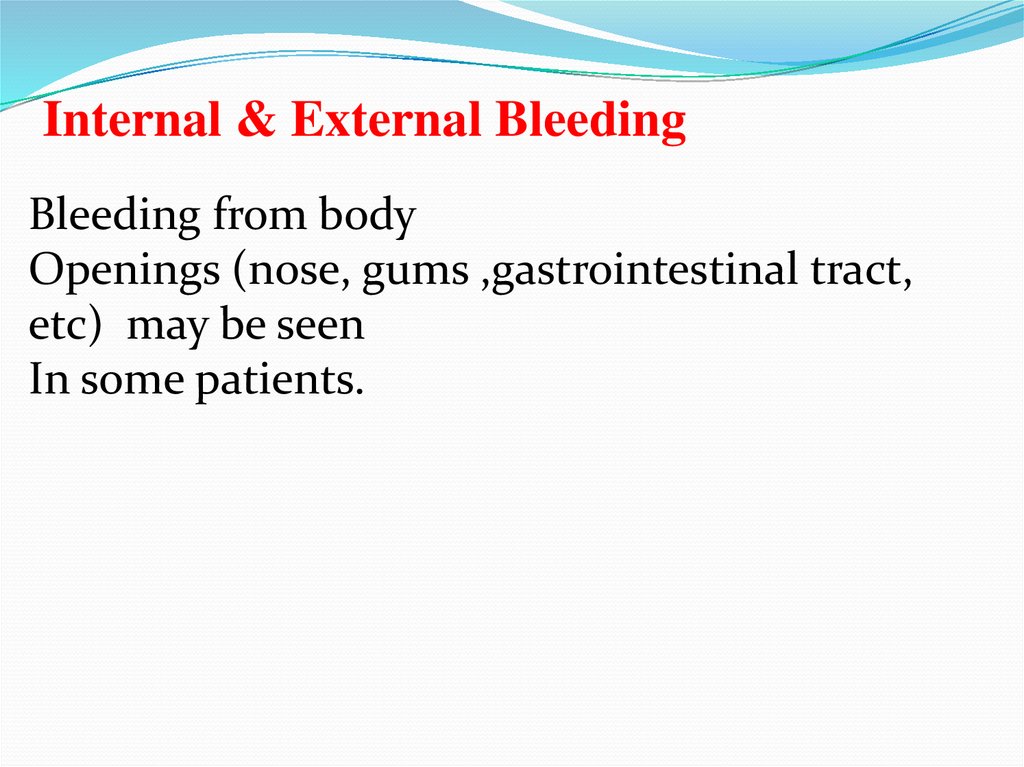
Internal & External BleedingBleeding from body
Openings (nose, gums ,gastrointestinal tract,
etc) may be seen
In some patients.
49.
How it is diagnosed?50.
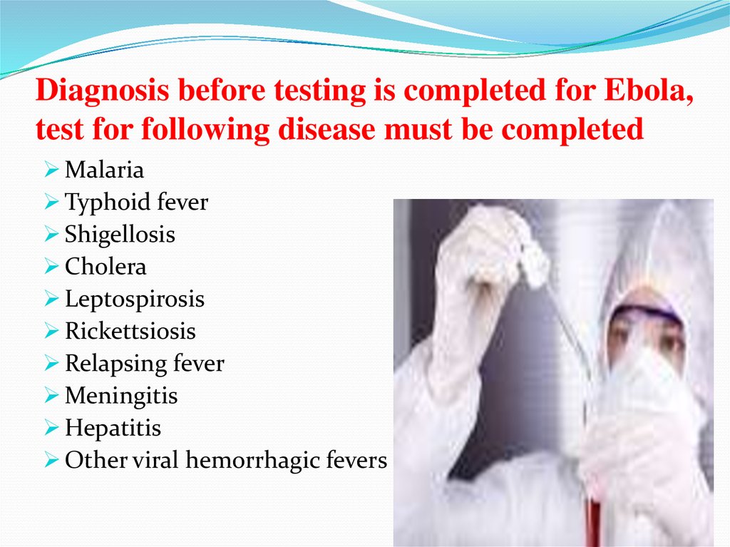
Diagnosis before testing is completed for Ebola,test for following disease must be completed
Malaria
Typhoid fever
Shigellosis
Cholera
Leptospirosis
Rickettsiosis
Relapsing fever
Meningitis
Hepatitis
Other viral hemorrhagic fevers
51.
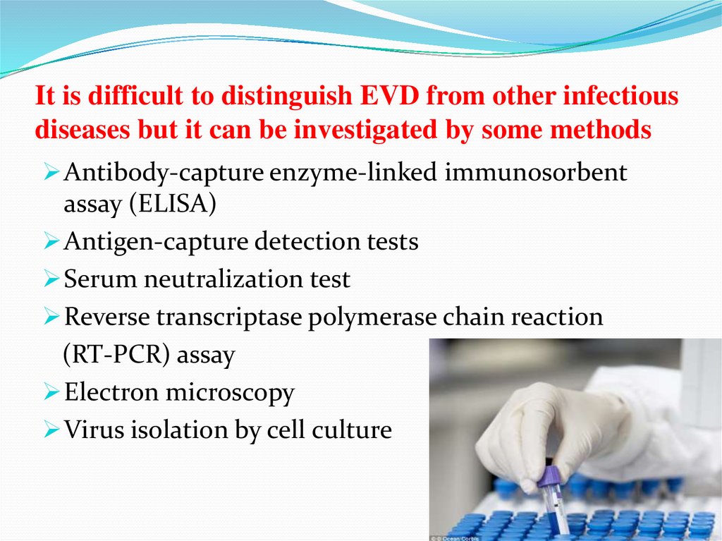
It is difficult to distinguish EVD from other infectiousdiseases but it can be investigated by some methods
Antibody-capture enzyme-linked immunosorbent
assay (ELISA)
Antigen-capture detection tests
Serum neutralization test
Reverse transcriptase polymerase chain reaction
(RT-PCR) assay
Electron microscopy
Virus isolation by cell culture
52.
Diagnostic Considerationsno
Although there is
approved specific
therapy for Ebola virus.
Clinical Findings - include fever of greater than 36.8 C (101.5 F) and
additional signs or symptoms like severe headache, muscle pain, vomiting,
diarrhea, abdominal pain
Risk factors
Those who have had contact with blood, body fluids or human remains of a
patient known to have or suspected to have Ebola virus disease.
Residence in or travel to an area where Ebola virus transmission is active.
Direct handling of bats, rodents or primates from endemic areas.
53.
A high risk exposure includes any of thefollowing
Percutaneous or mucous membrane exposure to blood
or body fluids of a person with Ebola virus disease.
Direct skin contact with patient having Ebola virus
disease without appropriate personal protective
equipment.
Direct contact with dead body without personal safety
equipments in a country where an Ebola virus disease
outbreak is occurring.
54.
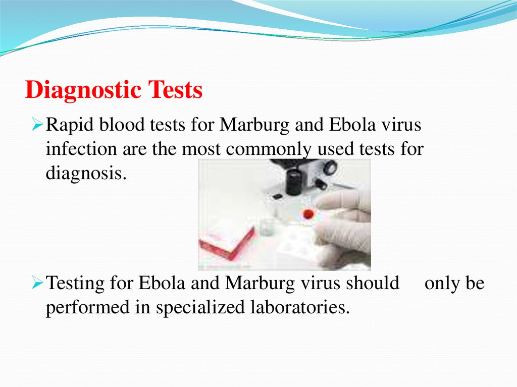
Diagnostic TestsRapid blood tests for Marburg and Ebola virus
infection are the most commonly used tests for
diagnosis.
Testing for Ebola and Marburg virus should
performed in specialized laboratories.
only be
55.
Other TestsAntigen detection may be used as a confirmatory test
for immediate diagnosis.
For individuals, who are recovering from Ebola virus
disease, PCR testing is also used to determine when a
patient can be discharged from hospital setting.
In some cases, testing for IgM or IgG antibodies to
Ebola virus may also be useful to monitor the immune
response over time and/or evaluate for past infection.
56.
Stages of symptoms of Ebola virusStage 1
Headache, sore throat, fever, muscle soreness
Stage 2
High fever, Vomiting, Passive Behaviour
Stage 3
Bruising, Bleeding from nose, mouth, eyes;
Blood in stool, Impaired liver function
Stage 4
Loss of consciousness, Seizures, Internal bleeding
leading to death
57.
Hospital Protocol for Ebola hitHandling Personal Protective Equipment (PPE)
Removal
Isolation
Fluid Control
Disinfecting
No Needles
58.
A new drug target for Ebola virusResearchers have recently developed a new drug
target in the Ebola virus that could be used against it
to fight the disease
University of Utah chemists have produced a
molecule known as peptide mimic that displays a
functionally critical region of the virus that is
universally conserved in all known species of Ebola
59.
Coffee, Fermented Soy, homeopathic Spider Venom, AndVitamin C, May All Hold Promise As Anti Ebola Virus
Therapies.
60.
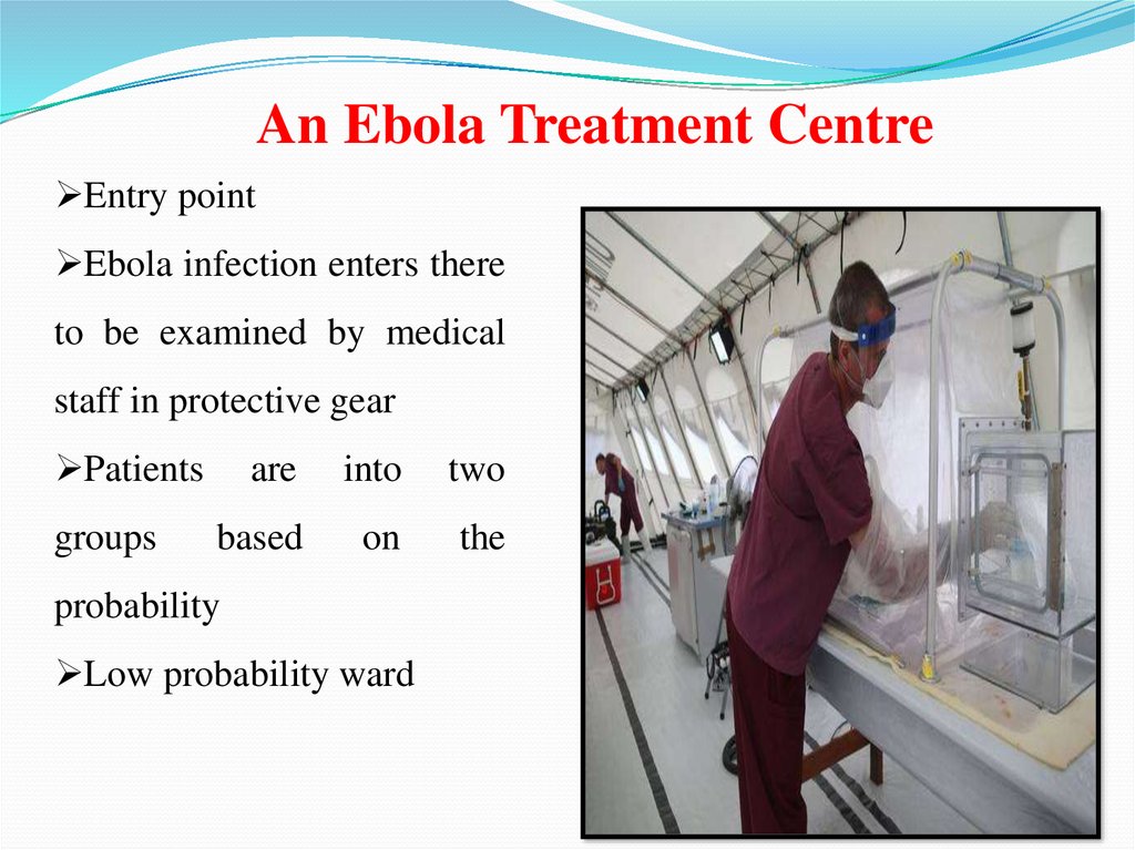
An Ebola Treatment CentreEntry point
Ebola infection enters there
to be examined by medical
staff in protective gear
Patients
groups
are
into
two
based
on
the
probability
Low probability ward
61.
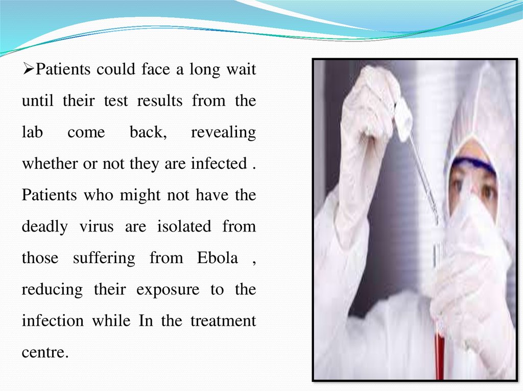
Patients could face a long waituntil their test results from the
lab
come
back,
revealing
whether or not they are infected .
Patients who might not have the
deadly virus are isolated from
those suffering from Ebola ,
reducing their exposure to the
infection while In the treatment
centre.
62.
High Probability WardPatients suspected of having Ebola based on the initial medical
examination remain here until official confirmation arrives that they
have the virus . Only once the Ebola diagnosis is confirmed they
transferred to another ward
63.
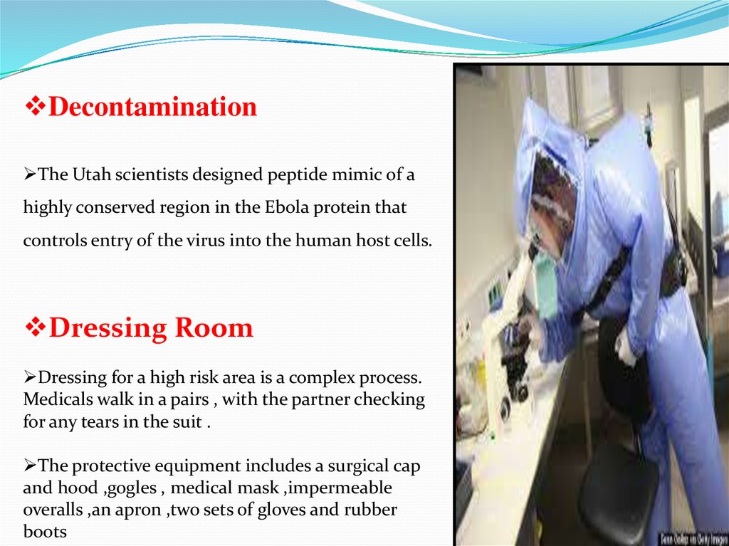
DecontaminationThe Utah scientists designed peptide mimic of a
highly conserved region in the Ebola protein that
controls entry of the virus into the human host cells.
Dressing Room
Dressing for a high risk area is a complex process.
Medicals walk in a pairs , with the partner checking
for any tears in the suit .
The protective equipment includes a surgical cap
and hood ,gogles , medical mask ,impermeable
overalls ,an apron ,two sets of gloves and rubber
boots
64.
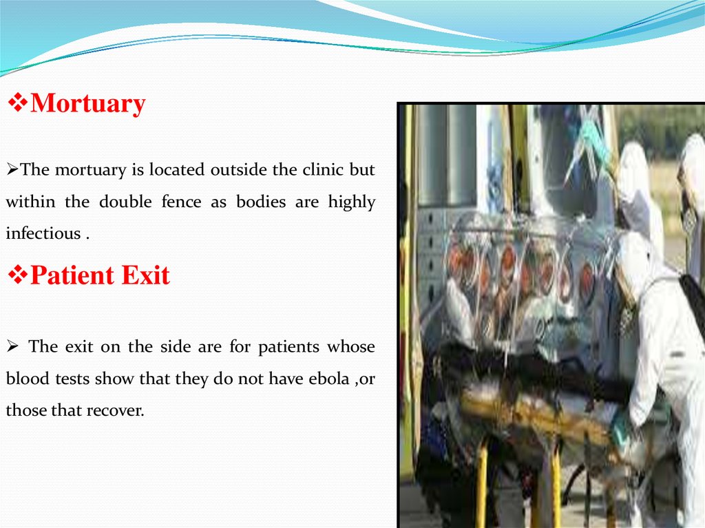
MortuaryThe mortuary is located outside the clinic but
within the double fence as bodies are highly
infectious .
Patient Exit
The exit on the side are for patients whose
blood tests show that they do not have ebola ,or
those that recover.
65.
Could Statins Treat EbolaStatins should be considered as a
possible treatment for Ebola
Statins also have been suggested as a
treatment for patient with sepsis, a
condition that involves an out of control
immune response similar to that seen in
Ebola patients.
66.
Canadian – Made Ebola Vaccine StartingClinical Trials In Humans
Experimental Canadian
made Ebola vaccine is
beginning clinical trials in
healthy humans .
The results are expected
in December
Clinical trails are now
starting
for
an
experimental made in
Canada Ebola vaccine
amid growing global
concern over the disease
that’s left more than
4,000 people dead.
67.
68.
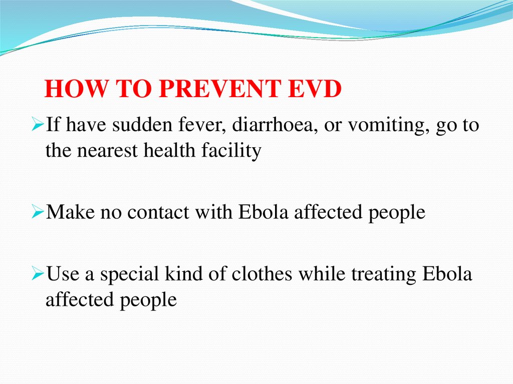
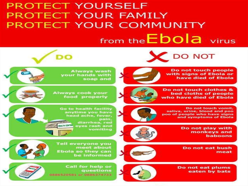
HOW TO PREVENT EVDIf have sudden fever, diarrhoea, or vomiting, go to
the nearest health facility
Make no contact with Ebola affected people
Use a special kind of clothes while treating Ebola
affected people
69.
70.
QUICK ACCESS TO APPROPRIATELABORATORY SERVICES
71.
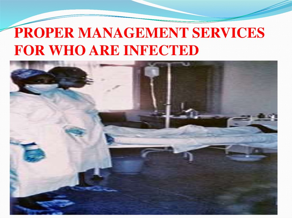
PROPER MANAGEMENT SERVICESFOR WHO ARE INFECTED
72.
PROPER DISPOSAL OF DEADTHROUGH CREMATION OR BURIAL
73.
People who go early to the health centrehave a better chance of survival.
CALL 117 WITH YOUR QUESTIONS