


 english
Similar presentations:


PCR: application in diagnostics
1. PCR: application in diagnostics
Done by: Naizabayeva D.2.
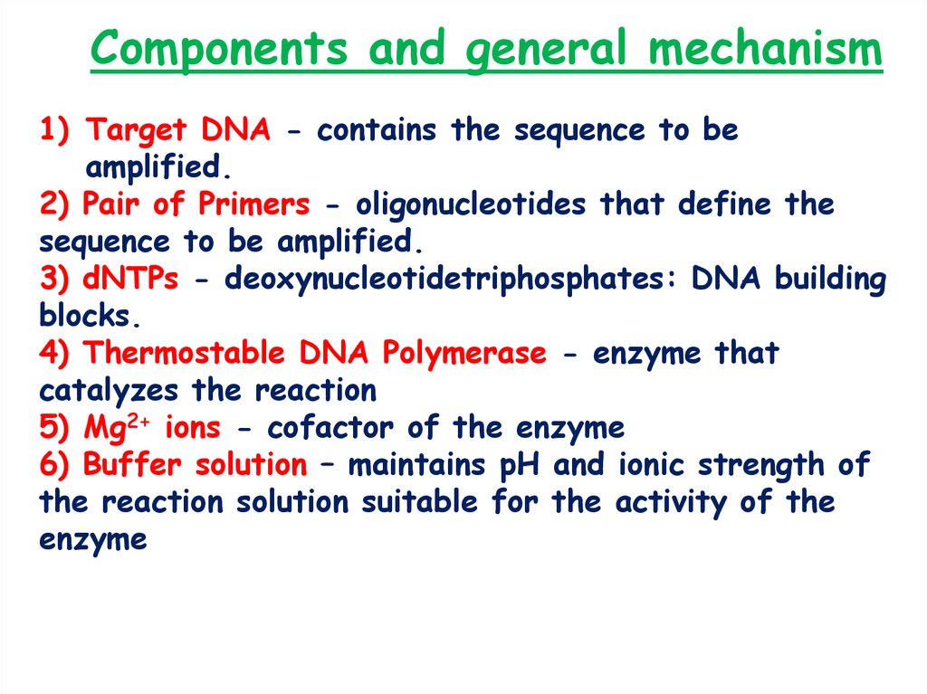
Components and general mechanism1) Target DNA - contains the sequence to be
amplified.
2) Pair of Primers - oligonucleotides that define the
sequence to be amplified.
3) dNTPs - deoxynucleotidetriphosphates: DNA building
blocks.
4) Thermostable DNA Polymerase - enzyme that
catalyzes the reaction
5) Mg2+ ions - cofactor of the enzyme
6) Buffer solution – maintains pH and ionic strength of
the reaction solution suitable for the activity of the
enzyme
3.
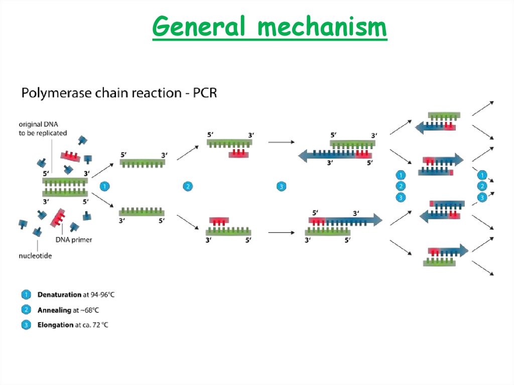
General mechanism4.
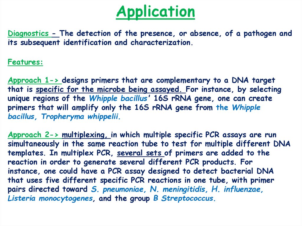
ApplicationDiagnostics - The detection of the presence, or absence, of a pathogen and
its subsequent identification and characterization.
Features:
Approach 1-> designs primers that are complementary to a DNA target
that is specific for the microbe being assayed. For instance, by selecting
unique regions of the Whipple bacillus' 16S rRNA gene, one can create
primers that will amplify only the 16S rRNA gene from the Whipple
bacillus, Tropheryma whippelii.
Approach 2-> multiplexing, in which multiple specific PCR assays are run
simultaneously in the same reaction tube to test for multiple different DNA
templates. In multiplex PCR, several sets of primers are added to the
reaction in order to generate several different PCR products. For
instance, one could have a PCR assay designed to detect bacterial DNA
that uses five different specific PCR reactions in one tube, with primer
pairs directed toward S. pneumoniae, N. meningitidis, H. influenzae,
Listeria monocytogenes, and the group B Streptococcus.
5.
Advantages/disadvantagesAdvantages- High sensitivity and specificity (specific
primer design), rapid test, ease of use, and
robustness, capability to detect pathogens which are
impossible to cultivate on media.
Disadvantages – Requirement of special conditions,
high cost equipment, expensive reagents.
6.
PCR in diagnosticsAssays are available for a variety of pathogens, including
HIV, HSV, hepatitis B virus, hepatitis C virus, cytomegalo virus,
ennterovirus, Chlamydia trachomatis, M. tuberculosis, T. whippelii,
and Neisseria gonorrhoeae, Brucella sp. For the detection of RNA
Viruses is applied RT-PCR method (Reverse Transcriptase PCR).
Reverse transcriptase is enzyme capable to synthesize DNA strand
from RNA template.
Generally the principle of detection is based on the
detection of pathogen’s specific DNA/RNA region, amplification of
that sequence and analyzing the presence or absence of detection
amplicons on electrophoretic agarose gel )
7.
The detection of Brucella sp. and strains (cause of brucellosis) using aPCR assay
Procedure
Analysis
of results
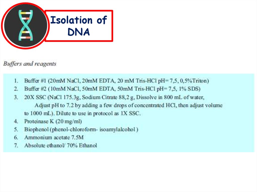
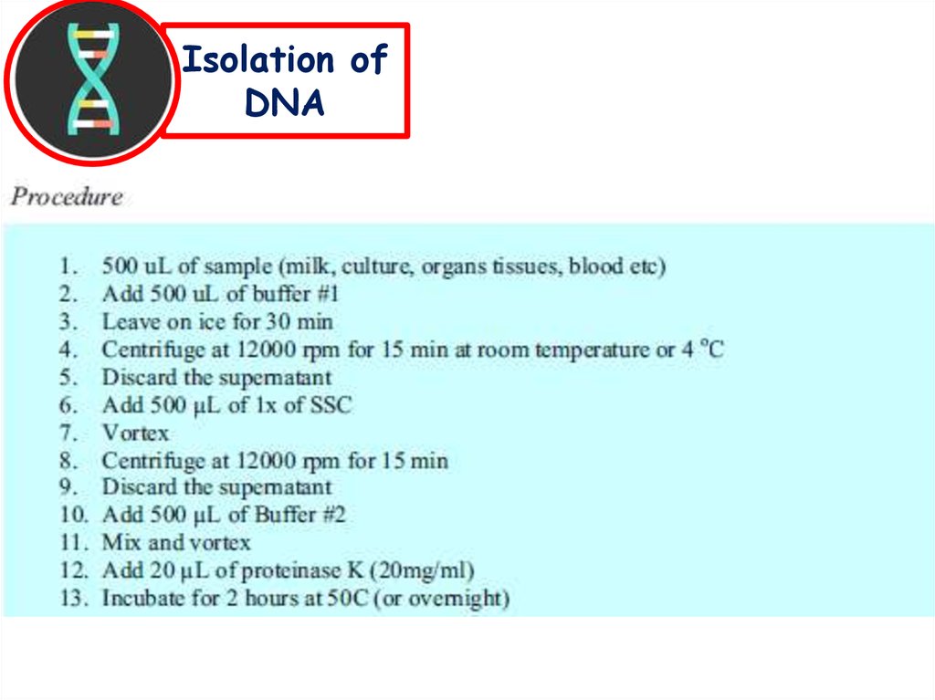
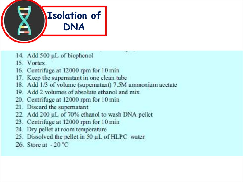
Isolation of
DNA
PCR
Gel
electrophoresis
8.
Isolation ofDNA
9.
Isolation ofDNA
10.
Isolation ofDNA
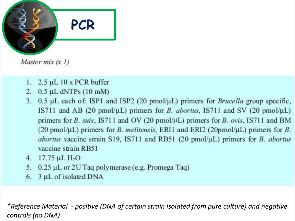
11.
PCR*Reference Material - positive (DNA of certain strain isolated from pure culture) and negative
controls (no DNA)
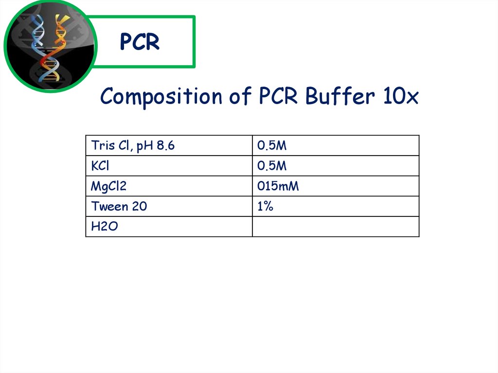
12.
PCRComposition of PCR Buffer 10x
Tris Cl, pH 8.6
0.5M
KCl
0.5M
MgCl2
015mM
Tween 20
1%
H2O
13.
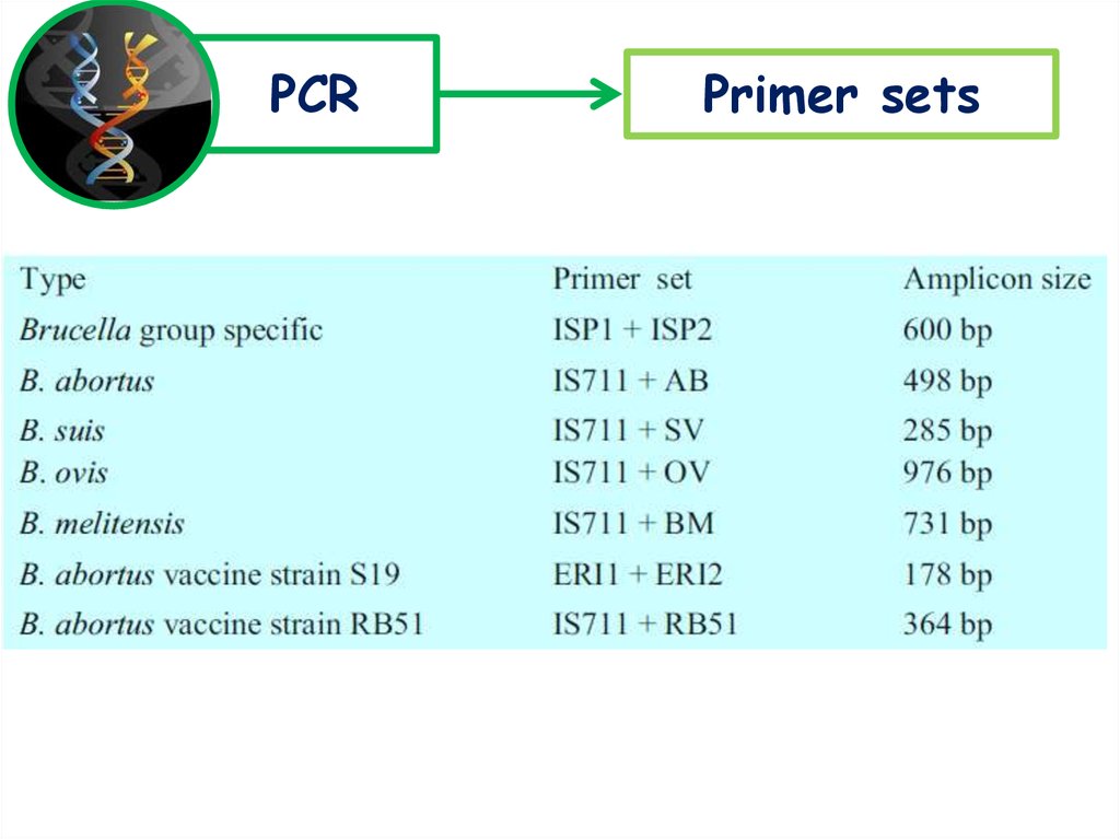
PCRPrimer sets
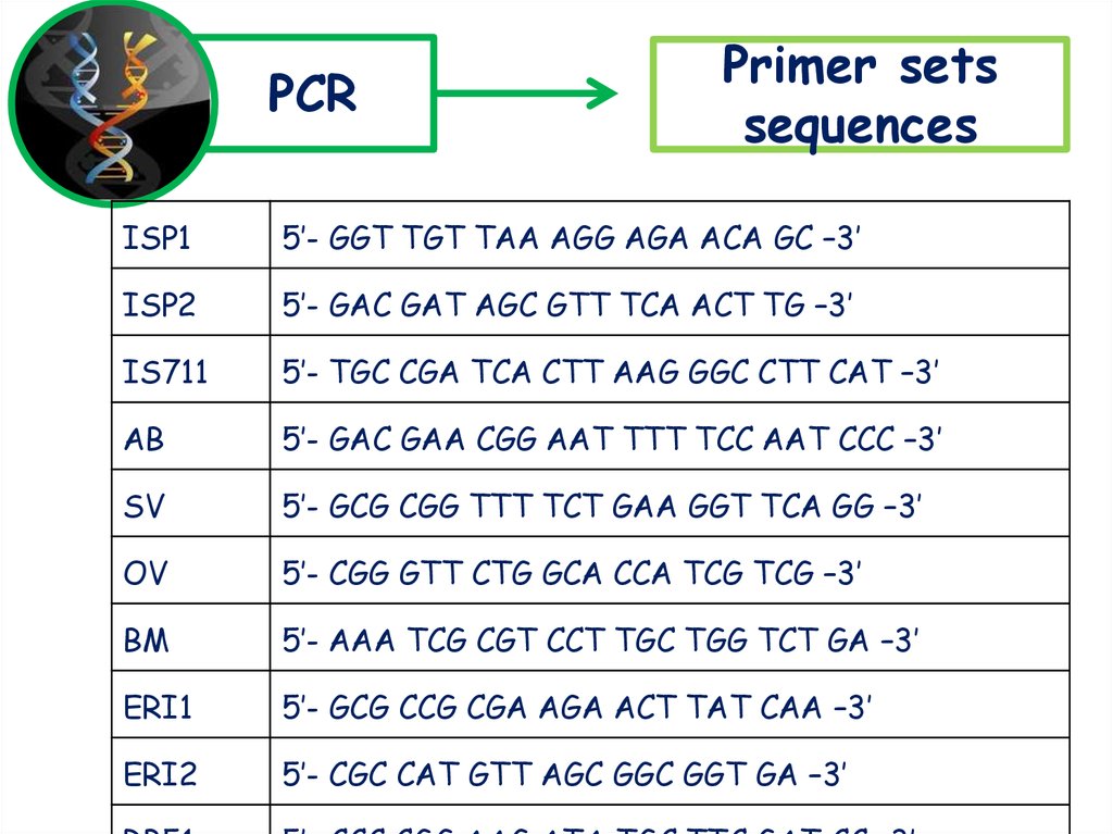
14.
PCRPrimer sets
sequences
ISP1
5’- GGT TGT TAA AGG AGA ACA GC –3’
ISP2
5’- GAC GAT AGC GTT TCA ACT TG –3’
IS711
5’- TGC CGA TCA CTT AAG GGC CTT CAT –3’
AB
5’- GAC GAA CGG AAT TTT TCC AAT CCC –3’
SV
5’- GCG CGG TTT TCT GAA GGT TCA GG –3’
OV
5’- CGG GTT CTG GCA CCA TCG TCG –3’
BM
5’- AAA TCG CGT CCT TGC TGG TCT GA –3’
ERI1
5’- GCG CCG CGA AGA ACT TAT CAA –3’
ERI2
5’- CGC CAT GTT AGC GGC GGT GA –3’
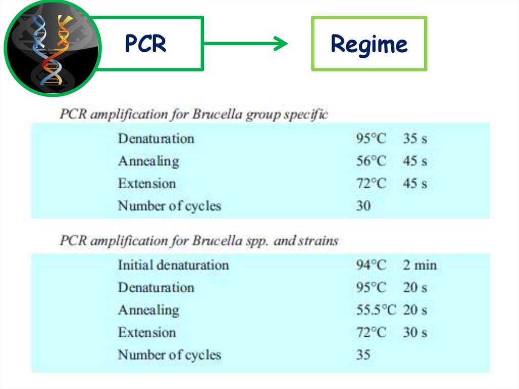
15.
PCRRegime
16.
Gelelectrophoresis
1. A 1,5% agarose gel stained with ethidium bromide is used
2. 10 μl of the product is loaded with 2 μl loading buffer
3. 2 μl of a 100 bp DNA molecular weight marker is loaded with 2 μl
loading buffer a single outside well
4. Gel electrophoresis is performed at 100 to 120V for 30 min
*The composition of LOADING buffer was not mentioned in manual, but on
practice it is possible to use loaders like bromphenol blue and xylene cyanol, or
cresol red.
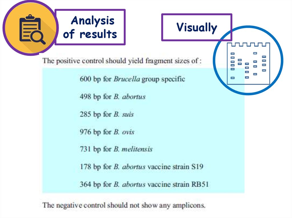
17.
Analysisof results
Visually
18.
Thanksfor
attention!