


 medicine
Similar presentations:


Tooth structure
1. Tooth structure Made by: Ismagambetov R. Dentistry 2-009 Karaganda 2016
Karaganda State Medical UniversityTOOTH STRUCTURE
MADE BY: ISMAGAMBETOV R.
DENTISTRY 2-009
KARAGANDA 2016
2. Tooth structure
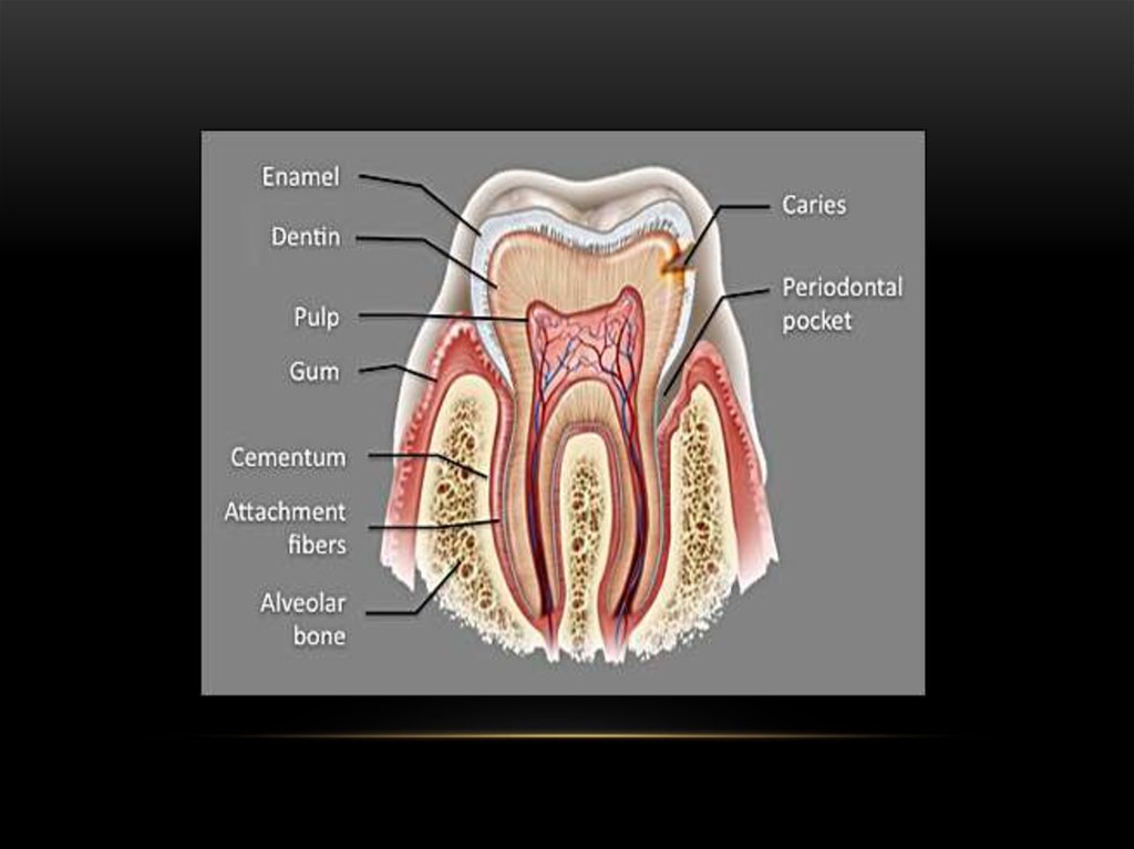
TOOTH STRUCTUREThe teeth of vertebrates represent the modified descendants of bony dermal (skin) plates
that armoured ancestral fishes. A tooth consists of a crown and one or more roots. The
crown is the functional part that is visible above the gum. The root is the unseen portion
that supports and fastens the tooth in the jawbone. The root is attached to the tooth bearing bone—the alveolar processes—of the jaws by a fibrous ligament called the
periodontal ligament or membrane. The “neck” of the root is embraced by the fleshy gum
tissue (a specialized area of connective tissue covered with mucous membrane that lines
the mouth cavity). The shape of the crown and root vary among different teeth.
All true teeth have the same general structure and consist of three layers. In mammals an
outer layer of enamel, which is wholly inorganic and is the hardest tissue in the body,
covers part or all of the crown of the tooth. The middle layer of the tooth is composed
of dentine, which is less hard than enamel and similar in composition to bone. The
dentine forms the main bulk, or core, of each tooth and extends almost the entire length of
the tooth, being covered by enamel on the crown portion and by cementum on the roots.
Dentine is nourished by the pulp, which is the innermost portion of the tooth. The pulp
consists of cells, tiny blood vessels, and a nerve and occupies a cavity located in the
center of the tooth.
3.
4. Enamel
ENAMELEnamel is the hardest and most highly mineralized substance of the body. It is one of the
four major tissues which make up the tooth, along with dentin, cementum, and dental
pulp.] It is normally visible and must be supported by underlying dentin. 96% of enamel
consists of mineral, with water and organic material comprising the rest. The normal color
of enamel varies from light yellow to grayish white. At the edges of teeth where there is no
dentin underlying the enamel, the color sometimes has a slightly blue tone. Since enamel
is semitranslucent, the color of dentin and any restorative dental material underneath the
enamel strongly affects the appearance of a tooth. Enamel varies in thickness over the
surface of the tooth and is often thickest at the cusp, up to 2.5mm, and thinnest at its
border, which is seen clinically as the CEJ. The wear rate of enamel, called attrition, is 8
micrometers a year from normal factors.
Enamel's primary mineral is hydroxyapatite, which is a crystalline calcium phosphate. The
large amount of minerals in enamel accounts not only for its strength but also for its
brittleness. Dentin, which is less mineralized and less brittle, compensates for enamel
and is necessary as a support. Unlike dentin and bone, enamel does not contain collagen.
Instead, it has two unique classes of proteins called amelogenins and enamelins. While
the role of these proteins is not fully understood, it is believed that they aid in the
development of enamel by serving as framework support among other functions.
5. Dentin
DENTINDentin is the substance between enamel or cementum and the pulp chamber. It is
secreted by the odontoblasts of the dental pulp. The formation of dentin is known
as dentinogenesis. The porous, yellow-hued material is made up of 70% inorganic
materials, 20% organic materials, and 10% water by weight. Because it is softer than
enamel, it decays more rapidly and is subject to severe cavities if not properly treated, but
dentin still acts as a protective layer and supports the crown of the tooth.
Dentin is a mineralized connective tissue with an organic matrix of collagenous proteins.
Dentin has microscopic channels, called dentinal tubules, which radiate outward through
the dentin from the pulp cavity to the exterior cementum or enamel border. The diameter
of these tubules range from 2.5 μm near the pulp, to 1.2 μm in the midportion, and 900 nm
near the dentino-enamel junction. Although they may have tiny side-branches, the tubules
do not intersect with each other. Their length is dictated by the radius of the tooth. The
three dimensional configuration of the dentinal tubules is genetically determined.
6. Cementum
CEMENTUMCementum is a specialized bone like substance covering the root of a tooth. It is
approximately 45% inorganic material (mainly hydroxyapatite), 33% organic material
(mainly collagen) and 22% water. Cementum is excreted by cementoblasts within the root
of the tooth and is thickest at the root apex. Its coloration is yellowish and it is softer than
either dentin or enamel. The principal role of cementum is to serve as a medium by which
the periodontal ligaments can attach to the tooth for stability. At the cementoenamel
junction, the cementum is acellular due to its lack of cellular components, and this
acellular type covers at least ⅔ of the root. The more permeable form of cementum,
cellular cementum, covers about ⅓ of the root apex.
7. Pulp
PULPThe dental pulp is the central part of the tooth filled with soft connective tissue . This tissue
contains blood vessels and nerves that enter the tooth from a hole at the apex of the
root. Along the border between the dentin and the pulp are odontoblasts, which initiate the
formation of dentin. Other cells in the pulp include fibroblasts,
preodontoblasts, macrophages and T lymphocytes. The pulp is commonly called "the
nerve" of the tooth.
8. Modal verbs
MODAL VERBSModal verbs (can, could, must, should, ought to, may, might, will, would, shall) are modal auxiliary
verbs that express ability, necessity, obligation, duty, request, permission, advice, desire,
probability, possibility, etc. Modal verbs express the speaker's attitude to the action indicated by
the main verb.
Modal verbs form questions without the help of the other auxiliary verbs. For example: Can you
do it? May I take it? Should I go there? Modal verbs also have quite a few peculiarities in the
formation of tenses.
Modal verbs do not have the future tense form. The future is expressed by the present tense
forms with the help of the context and adverbs of time referring to the future. (With the exception
of the modal verbs WILL, WOULD, of course, which express the future.)
Only two modal verbs can form the past by changing their forms directly. They are CAN, COULD
and WILL, WOULD (only in some of their meanings). The pair SHALL, SHOULD with the future
meaning can still work like that in British English. In American English, WILL is used for all
persons in the future (WOULD for the Future in the Past), and SHALL, SHOULD are used mostly
as separate modal verbs.
9. Examples
EXAMPLESHe should cure his teeth.
Dentist shouldn’t delete this tooth.
Can dentist save these teeth?
There might not be more gold fills