


 medicine
Similar presentations:

")


Parainfluenza
1. Parainfluenza
Turakhanov A. T.4-001 E)
2. Plan
Introduction
History
Etiology
Classification
Epidemiology
Pathogenesis
Clinical features
Diagnosis
Treatment
References
3. Introduction
• Parainfluenza refers to a group of viruses called human parainfluenzaviruses (HPIVs). There are four viruses in this group. Each one causes
different symptoms and illnesses. All forms of HPIV cause an
infection in either the upper or lower respiratory area of the body.
• Symptoms of HPIVs are like those of the common cold. When cases
are mild, the viruses are often misdiagnosed. Most healthy people
infected with an HPIV recover with no treatment. A person with a
weakened immune system is at risk for developing a life-threatening
infection.
4. History
• Parainfluenza first found in 1952 in Japan. It was look like influenza,this way renamed parainfluenza in 1959. Disease caused by
parainfluenza takes second place in diseases of ARD after influenza:
approximately 20 per cent of adults.
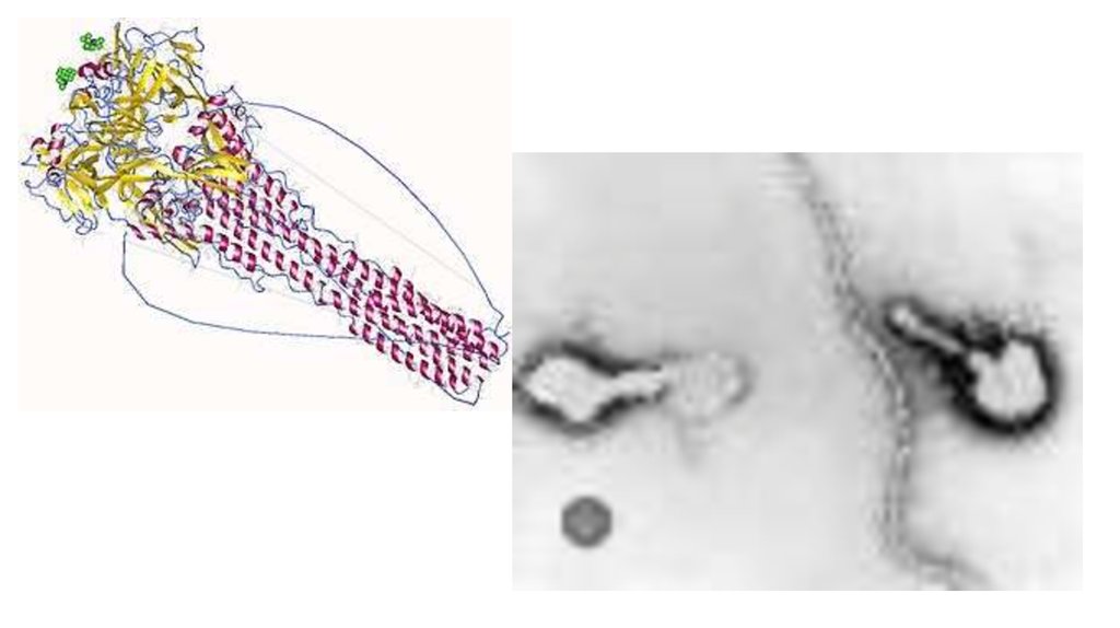
5. Etiology
• Order: Mononegavirales• Family: Paramyxoviridae
• Genus: Respirovirus & Rubulavirus
• Parainfluenza is RNA virus which includes Hemagglutinin (H) and
Neuraminidase (N). Four types of this virus calls disease in human.
Virions are approximately 150-250 nm in size
6.
7. Classification
• There are four different types of HPIV. They all cause a respiratory infection, but the typeof infection, symptoms, and location of the infection depend on the type of virus you
have. The four types of HPIV can infect anyone.
• HPIV-1 is the leading cause of croup in children. Croup is a respiratory illness that
manifests as swelling near the vocal cords and in other parts of the upper respiratory
system. HPIV-1 is responsible for outbreaks of croup in the autumn. In the United States,
the outbreaks tend to be more widespread in odd-numbered years.
• HPIV-2 causes croup in children, but doctors detect it much less often than HPIV-1. It’s
seen mostly in the autumn but to a lesser degree than HPIV-1.
• An HPIV-3 infection is mostly associated with pneumonia and bronchiolitis, which is
swelling from inflammation in the smallest airways in the lungs. It often causes infections
in the spring and early summer, but it appears in people throughout the year.
• HPIV-4 is rarer than the other types. Unlike the other strains of HPIV, there are no known
seasonal patterns of HPIV-4.
8. Epidemiology
• There are several ways you can become infected by an HPIV. An HPIVcan survive on a hard surface for up to 10 hours. If you touch a
contaminated surface with your hands and then touch your nose or
mouth, you can become infected.
• The viruses can also infect you through close contact with an infected
person. It usually takes between two and seven days after infection
for symptoms to occur.
9. Pathogenesis
• Viral replication is initiated only after successful entry into a cell by attachmentand fusion between the virus and the host cell lipid membrane. Viral RNA is
initially associated with nucleoprotein (NP), phosphoprotein (P) and the large
protein (L). The hemagglutinin–neuraminidase (HN) is involved with viral
attachment and thus hemadsorption and hemagglutination. Furthermore, the
fusion (F) protein is important in aiding the fusion of the host and viral cellular
membranes, eventually forming syncytia.
• Initially the F protein is in an inactive form (F0) but can be cleaved
by proteolysis to form its active form, F1 and F2, linked by di-sulphide bonds. Once
complete, this is followed by the HPIV nucleocapsid entering the cytoplasm of the
cell. Subsequently, genomic transcription occurs using the viruses own 'viral RNAdependant RNA polymerase' (L protein). The cells own ribosomes are then tasked
with translation, forming the viral proteins from the viral mRNA.
10. Pathogenesis
• Towards the end of the process, (after the formation of the viral proteins)the replication of the viral genome occurs. Initially this occurs with the
formation of a positive-sense RNA (intermediate step necessary for
producing progeny) and finally negative-sense RNA is formed which is then
associated with the nucleoprotein. This may then be either packaged and
released from the cell by budding or it can be used for subsequent rounds
of transcription and replication.
• The observable and morphological changes which can be seen in infected
cells includes the enlargement of the cytoplasm, decreased mitotic activity
and 'focal rounding,' with the potential formation of multi-nucleate cells.
• The pathogenicity of HPIVs is mutually dependent on the viruses having the
correct accessory proteins which are able to elicit antiinterferon properties. This is a major factor in the clinical significance of
disease.
11.
12. Clinical features
• Clinical picture. The incubation period lasts 3-4 days. The disease in mostcases begins gradually. Patients complain of malaise, mild headache,
mainly in the frontal region, less often in the temporal areas or eyeballs.
Sometimes there is a slight cognition, minor muscle pains. In a typical
parainfluenza, body temperature is subfebrile or normal, occasionally with
abrupt short-term elevations. From the 1st day
• Disease the leading symptom is a rough "barking" cough with hoarseness
or hoarseness of voice. There is a stuffy nose, followed by a rhinorrhea.
• Upon examination, the nasal mucosa is hyperemic and swollen. The soft
palate and the posterior wall of the pharynx are dimly hyperemic. In some
patients there is a fine granularity of the soft palate and a slight swelling of
the pharyngeal mucosa. There is an increase in the pulse, corresponding to
an increase in body temperature, with a serious course of the disease muffling heart sounds.
13. Clinical features
• The blood reveals normocytosis or moderate leukopenia. In the period ofconvalescence, monocytosis is possible; ESR within normal limits.
• Duration of the disease 1-3 weeks.
• In persons with chronic diseases of the respiratory system with
parainfluenza, the process quickly spreads to the lower respiratory tract. In
the early days of the disease, the phenomena of bronchitis are often
observed.
• Complications. The most frequent complications of parainfluenza are
pneumonia caused by a secondary bacterial flora and, as a rule, has a focal
character. In children in the first years of life, sometimes cereal occurs as a
result of edema and inflammatory infiltration of the laryngeal mucosa,
accumulation of secretion in its lumen and reflex muscle spasm.
• Parainfluenza leads to an exacerbation of chronic diseases.
14. Diagnosis
• Diagnosis can be made in several ways, encompassing a range of multifaceted techniques:Isolation and detection of the virus in cell culture.
Detection of viral antigens directly within bodily respiratory tract secretions
using immunofluorescence, enzyme immunoassays or fluroimmunoassays.
Polymerase chain reaction (PCR).
Analysis of specific IgG antibodies showing a subsequent rise in titre following
infection (using paired serum specimens).
Because of the similarity in terms of the antigenic profile between the
viruses, hemagglutination assay (HA) or hemadsorption inhibition (HAdI)
processes are often used. Both complement fixation, neutralisation
and enzyme linked immunosorbent assays – ELISA, can also be used to aid in
the process of distinguishing between viral serotypes.
15. Treatment
• Ribavirin is one medication which has shown good potential for thetreatment of HPIV-3 given recent in-vitro tests. Ribavirin is a
broadscale anti-viral and is currently being administered to those who
are severely immuno-compromised, despite the lack of conclusive
evidence for its use. Protein inhibitors and novel forms of medication
have also been proposed to relieve the symptoms of infection.
• Furthermore, antibiotics may be used if a secondary bacterial
infection develops. Corticosteroid treatment and nebulizers are also a
first line choice against croup if breathing difficulties ensue.
16. References
• Moscona A. ”Entry of parainfluenza virus into cells as a target forinterrupting childhood respiratory disease” 2015
• Chambers R, Takimoto T. ’’Parainfluenza Viruses’’ 2011
• Ющук Н. Д., Венгеров Ю. Я. “инфекционные болезни” 2003