Similar presentations:
Types of Pacemakers
1. CorePace – Module #2 Basic Pacing Concepts
2. Disclosures
This presentation is provided for general educationalpurposes only and should not be considered the exclusive
source for this type of information. At all times, it is the
professional responsibility of the practitioner to exercise
independent clinical judgment in a particular situation.
The device functionality and programming described in this
module are based on Medtronic products and can be
referenced in the device manuals.
Updated: April 2012
3. Objectives
• Explain the different types of pacemakers and the NBG Code• Identify the components of a pacemaker circuit
• Describe the relationship between voltage, current, and
resistance
• Describe the clinical significance of alterations in voltage,
current, and resistance
• Recognize low and high impedance conditions and possible
causes
• Identify a capture threshold and calculate safety margins
• Understand sensing and sensitivity in a pacemaker
4. Types of Pacemakers
TYPES OF PACEMAKERS5. Single Chamber System
• One lead– Atrium
– Ventricle (most common)
• May be used for patients in chronic AF
(VVI pacemaker) or patients with sinus
node dysfunction and no history of AV
block (AAI pacemaker)
VVI Pacemaker
AAI Pacemaker
6. Dual Chamber System
• Two leads– One lead implanted in the atrium
– One lead implanted in the ventricle
• Provides AV synchrony and pacing support
in both atrium and ventricle if needed
DDD Pacemaker
7. Dual Chamber Pacemaker
RA Lead in AppendageRV Lead at the Apex
8. Triple Chamber System
• Three Leads:– Right Atrium
– Right Ventricle
– Left Ventricle (via the Coronary Sinus vein)
• Most commonly called a Bi-Ventricular Pacemaker but also called
Cardiac Resynchronization Therapy (CRT–P)
• Paces both ventricles together to “resynchronize” the beat
DDD BiV Pacemaker
9. NBG Code – The Usual Pacing Modes
III
III
IV
Chamber(s)
Paced
Chamber(s)
Sensed
Response to
Sensing
Rate
Modulatio
n
O = None
A = Atrium
V = Ventricle
D = Dual (A + V)
S = Single (A or V)
O = None
A = Atrium
V = Ventricle
D = Dual (A + V)
S = Single (A or V)
O = None
T = Triggered
I = Inhibited
D = Dual (T + I)
O = None
R = Rate
V
Multisite
Pacing
O = None
A = Atrium
modulation V = Ventricle
D = Dual (A + V)
Examples of pacing modes which are typically programmed:
DDD
DDDR
VVI
DDIR
VVIR
AAIR
10. Knowledge Checkpoint
What type of pacemaker is this?11. Knowledge Checkpoint
What does VVIR mode mean?12. Key Learning Points
• There are three types of pacemakers– Important to identify which one the patient has and why
• The mode explains how the pacemaker should work
– Very important to understanding the basic function of the device
13. Components of the Pacemaker System
COMPONENTS OF THEPACEMAKER SYSTEM
14. Implantable Pacemaker Circuit
• Implantable pulse generator (IPG):Leads
– Battery
– Circuitry
– Connector(s)
IPG
• Leads or wires
– Cathode (negative electrode)
– Anode (positive electrode)
• Body tissue
Anode
Cathode
15. The Pulse Generator
• Lithium-Iiodine Battery– 2.8 V BOL
• Longevity
– Dependent on impedance
and output
– Ranges from 6-12 years
Circuitry
Battery
16. Leads are Insulated Wires
• Deliver electrical impulses from the pulsegenerator to the heart
• Sense cardiac depolarization
Lead
17. Lead Polarity
• Unipolar leads– May have a smaller diameter
lead body than bipolar leads
– May exhibit larger pacing
artifacts on the surface ECG
– May cause pectoral muscle
stimulation
Unipolar lead
• Bipolar leads
– Usually less susceptible to
oversensing of non-cardiac
signals (i.e., myopotentials,
EMI, etc.)
To tip (cathode)
From ring (anode)
Bipolar coaxial lead
Al-Ahmad, Amin, et. al. (2010). Pacemakers and Implantable Cardioverter Defibrillators: An Expert's Manual
Minneapolis: Cardiotext Publishing. (pg. 20-21).
18. Unipolar Pacing System
• The lead has only one electrode(the cathode) at the tip
• The pacemaker can is the anode
• When pacing, the impulse:
– Flows through the tip electrode
(cathode)
– Stimulates the heart
– Returns through body fluid and
tissue to the IPG can (anode)
• Why might this be important to
know during a procedure?
Anode
+
Cathode
-
19. Bipolar Pacing System
• The lead has both ananode and cathode
• The pacing impulse:
– Flows through the tip
electrode located at the end
of the lead wire
– Stimulates the heart
– Returns to the ring electrode,
the anode, above the lead tip
Anode +
Anode
Cathode -
Cathode
20. Transvenous Leads
Passive fixation (tined)Active fixation (screw-in)
The tines become lodged in the
trabeculae of the apex or the
pectinate of the appendage
which are fibrous meshworks of
heart tissue
The helix, or screw, extends
into the endocardial tissue
– Allows for lead positioning
anywhere in the heart’s
chamber
– The helix is extended using
an included tool
21. Epicardial Leads
• Leads applied directly tothe surface of the heart
– Utilized in pediatric patients
and patients contraindicated
for transvenous leads
– Fixation mechanisms include:
• Epicardial stab-in
• Myocardial screw-in
• Suture-on
– Applied via sternotomy,
thoroscopy, or limited
thoracotomy
22. Lead Insulators
Silicone insulated leadsInert
Biocompatible
Biostable
Repairable with medical adhesive
Historically very reliable
Polyurethane
Silicone
Polyurethane insulated
leads
Biocompatible
High tear strength
Low friction coefficient
Smaller lead diameter
Newer bipolar lead insulation
Hayes, David L., et. al. (2008). Cardiac pacing, defibrillation and resynchronization: a clinical
approach. New Jersey: Wiley-Blackwell Publishing. (pg. 127).
23. Knowledge Checkpoint
Where is the anode located in bipolar pacing?A. Tip Electrode
B. Ring Electrode
C
C. Device
D. Body Tissue
B
D
A
24. Key Learning Points
• The pacemaker circuit consists of the leads, device, andtissue
• Modern leads are usually bipolar, endocardial, and active
fixation but all types of leads are available
– Important to know what type of lead is implanted because it can be
helpful for diagnosing a problem and determining solutions
25. Electrical Concepts in Pacemakers
ELECTRICAL CONCEPTS INPACEMAKERS
26. Voltage
• Voltage is the force, or “push,” that causes electrons tomove through a circuit
• In a pacing system, voltage is:
–
–
–
–
Measured in volts (V)
Represented by the letter “V”
Provided by the pacemaker battery
Often referred to as amplitude or pulse amplitude
Note: The terms “amplitude” and “voltage” are often used interchangeably in pacing.
27. Initial Interrogation Report
Note: All clinic, physician, and patient names and data in this document are fictitious28. Voltage
29. Current
• The flow of electrons through a completed circuit• In a pacing system, current is:
– Measured in milliamps (mA)
– Represented by the letter “I”
– Determined by the amount of electrons that move through a circuit
Note: One ampere is a unit of electrical current produced by 1 volt acting through a
resistance of 1 ohm. 1 Ampere = 1000 milliamps
30. Current
31. Impedance
• The opposition to current flow• In a pacing system, impedance is:
– Measured in ohms (W)
– Represented by the letter “R”
– The sum of all resistances to the flow of current
• Lead conductor resistance
• The resistance to current flow from the electrode to the myocardium
• Polarization impedance (the accumulation of charges of opposite
polarity in the myocardium at the electrode-tissue interface)
32. Initial Interrogation Report
Note: All clinic, physician, and patient names and data in this document are fictitious33. Impedance
34. Summary Voltage, Current, and Impedance
• Voltage: The force moving the current (V)– In pacemakers it is a function of the battery chemistry
• Current: The actual continuing volume of flow of electricity (I)
– This flow of electrons causes the myocardial cells to depolarize (to
“beat”)
• Impedance: The sum of all resistance to current flow (R)
– Impedance is a function of the characteristics of the conductor (wire),
the electrode (tip), and the myocardium (tissue).
35. Ohm’s Law
• Describes the relationshipbetween voltage, current,
and resistance
(impedance)
V
• V=IXR
V
=
I X R
• I=V/R
V
I
I
R
• R=V/I
= R
V
I =
R
36. Ohm’s law tells us:
1. If the impedance (R) remains constant, and the voltagedecreases, the current decreases
2. If the voltage is constant, and the impedance decreases,
the current increases
V=IxR
Why is this important to clinical management of pacemakers?
The relationship between voltage, current, and impedance provides the rationale for
decisions we make during evaluation of pacing systems and reprogramming. Proper
management of electrical characteristics is important for patient safety and device longevity.
37. Knowledge Checkpoint
What is the delivered current from the Atrial Lead?38. Key Learning Points
• Know where to find the voltage and impedance on theprogrammer and report
• Ohm’s law and the relationship between voltage, current,
and impedance
– Knowing how these factors relate to each other can help you
understand how the pacemaker paces the heart
39. Testing the Pacemaker Circuit
TESTING THEPACEMAKER CIRCUIT
40. Typical Lead Impedance Range
• Most important that lead impedance is stable over thelifetime of the device.
• Generally, a 30% change or abrupt change is something
to be concerned about.
Typical Impedance range = 200 to 1,000 Ohms.*
*Impedance is higher for specially designed high impedance leads.
Hayes, David L., et. al. (2000). Cardiac pacing and defibrillation: a clinical approach. New York: Blackwell
Publishing. (pg. 398).
41. Lead Impedance Values Electrical Analogies
• Normal resistance – friction caused by the hose and nozzle• Low resistance – leaks in the hose reduce the resistance
Similar to a pacemaker lead with an insulation breach which
results in low resistance and high current drain; may cause
premature battery depletion.
• High resistance – a knot results in low total current flow
Similar to a pacemaker lead with a lead conductor break - impedance will
be high with little or no current reaching the myocardium.
42. Knowledge Checkpoint
What would you expect to happen if a lead wasfractured?
A. Impedance would drop
B. Current would decrease
C. Impedance would rise
D. Both B and C
43. High Impedance Conditions A Fractured Conductor
• A fractured wire can causeImpedance values to rise
– Current flow from the battery
may be too low to be
effective
• Impedance values may
exceed 3,000 W
Lead wire fracture
Increased resistance
Other reason for high impedance: Lead not seated properly
in pacemaker header (usually an acute problem).
44. Case Study: Clinic Visit
85 year old male with h/o pacemaker implant in 1996.Generator change in 2005. Follow up visits in clinic have
been normal. He now comes into your office complaining of
light-headedness and fatigue.
• You interrogate his pacemaker and find the ventricular
lead impedance is 1,867 ohms and it was usually trending
around 700 ohms.
45. Chest X Ray
Can you identify a problem?1st Rib-Clavicle Crush (lead fracture)
46. Lead Crush
Lead FractureNow that you know what the problem is,
How do you fix it?
47. Solutions for Lead Crush
• Unipolar configuration if the inner conductor is still intact• Lead replacement
48. Knowledge Checkpoint
What would you expect to happen if a lead has aninsulation break? Check all that apply.
Impedance would drop
Potential loss of capture
Current would increase
Battery longevity improves
49. Low Impedance Conditions An Insulation Break
• Insulation breaks can causeimpedance values to fall
– Current drain is high and can
lead to more rapid battery
depletion
– Current can drain through the
insulation break into the body or
other lead wire, not through
myocardium
• Impedance values may be less
than 300 W
Current will follow the path of
LEAST resistance
50. Case Study: Routine Follow Up
• A patient comes in for routine follow up and you notice thison the initial interrogation report:
51. Look at the EGM
Lead IIMarker
Channel
V EGM
• What do you suspect?
52. Insulation Break
• A low impedance usually means an insulation break• Oversensing can be a result of an insulation break and the
EGM shows abnormal electrical signals
Now that we know what the problem is,
how do you fix it?
53. Polarity Switch
• The automatic “Polarity Switch” of the pacemaker canautomatically notice an issue with the lead impedance and
switch to unipolar
54. Replace the Lead
• Since the lead is still oversensing and has a lowimpedance in the unipolar configuration, a lead
replacement still should be performed.
• The lead can be capped and a new ventricular pacing lead
can be placed at least 1 cm away to prevent lead-lead
noise.
55. Capture Threshold
• The minimum electrical stimulus needed to consistently capture theheart outside of the heart’s own refractory period
Capture
Ventricular pacemaker 60 ppm
Non-Capture
56. Effect of Lead Design on Capture
• Lead maturation– Fibrotic “capsule” develops around the electrode following lead
implantation
– May gradually raise threshold
– Usually no measurable effect on impedance
Ellenbogen, Kenneth A. and Mark A. Wood. (2005). Cardiac Pacing and ICDs. Massachusetts: Blackwell
Publishing. (pg. 342).
57. Steroid Eluting Leads
• Steroid eluting leadsreduce the inflammatory
process
– Exhibit little to no acute
stimulation threshold peaking
– Leads maintain low chronic
thresholds
Porous, platinized tip
for steroid elution
Silicone rubber plug
containing steroid
Tines for
stable
fixation
58. Effect of Steroid on Stimulation Thresholds
5Volts
4
Smooth Metal Electrode
3
Textured Metal Electrode
2
1
Steroid-Eluting Electrode
0
0
1
2
3
4
5
6
7
8
9
10 11 12
Implant Time (Weeks)
Pulse Width = 0.5 msec
References: Pacing Reference Guide, Bakken Education Center, 1995, UC199601047aEN. Cardiac Pacing,
2nd Edition, Edited by Kenneth A. Ellenbogen. 1996.
59. Factors That Can Affect Thresholds
– Pacemaker circuit (lead) integrity• Insulation break
• Wire fracture
–
–
–
–
–
The characteristics of the electrode
Electrode placement within the heart
Drugs
Electrolytes
Sleeping/Eating
Hayes, Cardiac Pacing and Defibrillation, 2010
60. Myocardial Capture
• Capture is a function of:– Amplitude—the strength of the impulse expressed in volts
• The amplitude of the impulse must be large enough to cause
depolarization (i.e., to “capture” the heart)
• The amplitude of the impulse must be sufficient to provide an
appropriate pacing safety margin
– Pulse width—the duration of the current flow expressed in
milliseconds
• The pulse width must be long enough for depolarization to disperse to
the surrounding tissue
61. Comparison
5.0 Volt Amplitude at Different Pulse WidthsAmplitude
5.0 V
0.5 ms
0.25 ms
1.0 ms
62. Strength-Duration Curve
• Strength-duration curveshows relationship of
amplitude and pulse width
• Adequate safety margins are
important because thresholds
can fluctuate slightly
Stimulation Threshold (Volts)
Strength-Duration Curve
2.0
X
Programmed Output
1.5
Capture
1.0
.50
.25
No Capture
0.5
1.0
Duration
Pulse Width (ms)
1.5
63. Strength Duration Curve Example
Safety Margin =2 x Amplitude Threshold
OR
3 x Pulse Width Threshold
64. Programming Outputs
• Primary goal: Ensure patient safety and appropriatedevice performance
• Secondary goal: Extend the service life of the battery
– Typically program amplitude to < 2.5 V, but always maintain
adequate safety margins
– Amplitude values greater than the cell capacity of the pacemaker
battery (usually about 2.8 V) require a voltage multiplier, resulting
in markedly decreased battery longevity
65. Knowledge Checkpoint
What is the threshold?1.25 V
A. 0.05 V
B. 0.75 V
C. 1.00 V
D. 1.25 V
1.00 V
0.75 V
0.05 V
66.
Case Study: ER VisitA patient presented to the ER with the complaint that he felt
just the way he did when he first received his pacemaker.
What is your interpretation?
SETUP: Unknown
67. Order a Chest X-ray
The chest x-ray revealed a dislodged lead68. Twiddler’s Syndrome
69. Sensing
• Sensing is the ability of the pacemaker to “see” when anatural (intrinsic) depolarization is occurring
– Pacemakers sense cardiac depolarization by measuring changes
in electrical potential of myocardial cells between the anode and
cathode
0.5 mV signal
2.0 mV signal
70. Sensing
Acceptable Sensing Values (mV)11Curtis,
Acute
Chronic
Atrium
>1.5
>1.0
Ventricle
>7.0
>5.0
Anne B. (2010). Fundamentals of Cardiac Pacing. Massachusetts: Jones and Bartlett
Publishers. (pg. 98).
71. Sensitivity
Amplitude (mV)5.0
2.5
1.25
Time
72. Less Sensitive = High Sensitivity Number
Amplitude (mV)5.0
2.5
1.25
Time
73. More Sensitive = Low Sensitivity Number
Amplitude (mV)5.0
2.5
1.25
Time
74. Adequate Sensitivity
Amplitude (mV)5.0
2.5
1.25
Time
75. Sensing Amplifiers/Filters
Vectors and GradientsSense
2.5 mV
The wave of depolarization produced by
normal conduction creates a gradient
across the cathode and anode. This
changing polarity creates the signal.
Once this signal exceeds the
programmed sensitivity – it is
sensed by the device.
76. Vectors and Gradients
Changing the VectorSense
2.5 mV
A PVC occurs, which is conducted
abnormally. Since the vector relative
to the lead has changed, what effect
might this have on sensing?
In this case, the wave of
depolarization strikes the anode
and cathode almost simultaneously.
This will create a smaller gradient
and thus, a smaller signal.
77. Changing the Vector
Sensing Accuracy• Affected by:
– Pacemaker circuit (lead) integrity
• Insulation break
• Wire fracture
–
–
–
–
–
–
The characteristics of the electrode
Electrode placement within the heart
The sensing amplifiers of the pacemaker
Lead polarity (unipolar vs. bipolar)
The electrophysiological properties of the myocardium
EMI – Electromagnetic Interference
78. Sensing Accuracy
Undersensing . . .Overpacing• Pacemaker does not “see” the intrinsic beat, and therefore
does not respond appropriately
Intrinsic beat
not sensed
Scheduled pace
delivered
VVI / 60
79. Undersensing . . .Overpacing
Oversensing …Underpacing• An electrical signal other than the intended P or R wave
is detected
Marker channel
shows intrinsic
activity...
...though no
activity is present
VVI / 60
80. Oversensing …Underpacing
Knowledge CheckpointWhich of these pacemakers is more sensitive?
Pacemaker
A
Programmed
Sensitivity 0.5 mV
OR
Pacemaker
B
Programmed
Sensitivity 2.5 mV
81. Knowledge Checkpoint
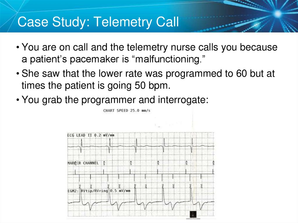
Case Study: Telemetry Call• You are on call and the telemetry nurse calls you because
a patient’s pacemaker is “malfunctioning.”
• She saw that the lower rate was programmed to 60 but at
times the patient is going 50 bpm.
• You grab the programmer and interrogate:
82. Case Study: Telemetry Call
SolutionNow that we know what the problem is,
How do we fix it?
• Measure the size of the R waves
• Make the ventricular lead less sensitive by increasing the
ventricular sensitivity
83. Solution
Key Learning Points• The NBG code indicates the pacing mode and whether the
pacemaker is pacing, sensing, and inhibiting in either the atrium
and ventricle.
• There is a mathematical relationship between voltage, current,
and resistance. These variables should be considered for
patient safety (to ensure capture) and device longevity.
• Lead impedance is a key measure of lead integrity. Low or high
impedance may indicate a faulty lead.
• Appropriate safety margins should be applied to the capture
threshold to ensure patient safety.
• Proper sensing is vital to the operation of the pacemaker.
84. Key Learning Points
Brief Statement: IPGs and ICDsIndications
• Implantable Pulse Generators (IPGs) are indicated for rate adaptive pacing in patients who
may benefit from increased pacing rates concurrent with increases in activity and increases in
activity and/or minute ventilation. Pacemakers are also indicated for dual chamber and atrial
tracking modes in patients who may benefit from maintenance of AV synchrony. Dual chamber
modes are specifically indicated for treatment of conduction disorders that require restoration of
both rate and AV synchrony, which include various degrees of AV block to maintain the atrial
contribution to cardiac output and VVI intolerance (e.g. pacemaker syndrome) in the presence of
persistent sinus rhythm.
• Implantable cardioverter defibrillators (ICDs) are indicated for ventricular antitachycardia
pacing and ventricular defibrillation for automated treatment of life-threatening ventricular
arrhythmias.
• Cardiac Resynchronization Therapy (CRT) ICDs are indicated for ventricular antitachycardia
pacing and ventricular defibrillation for automated treatment of life-threatening ventricular
arrhythmias and for the reduction of the symptoms of moderate to severe heart failure (NYHA
Functional Class III or IV) in those patients who remain symptomatic despite stable, optimal
medical therapy and have a left ventricular ejection fraction less than or equal to 35% and a
prolonged QRS duration.
• CRT IPGs are indicated for the reduction of the symptoms of moderate to severe heart failure
(NYHA Functional Class III or IV) in those patients who remain symptomatic despite stable,
optimal medical therapy, and have a left ventricular ejection fraction less than or equal to 35%
and a prolonged QRS duration.
85. Brief Statement: IPGs and ICDs
Contraindications• IPGs and CRT IPGs are contraindicated for dual chamber atrial pacing in patients with chronic
refractory atrial tachyarrhythmias; asynchronous pacing in the presence (or likelihood) of
competitive paced and intrinsic rhythms; unipolar pacing for patients with an implanted
cardioverter defibrillator because it may cause unwanted delivery or inhibition of ICD therapy;
and certain IPGs are contraindicated for use with epicardial leads and with abdominal
implantation.
• ICDs and CRT ICDs are contraindicated in patients whose ventricular tachyarrhythmias may
have transient or reversible causes, patients with incessant VT or VF, and for patients who have
a unipolar pacemaker.
Warnings/Precautions
• Changes in a patient’s disease and/or medications may alter the efficacy of the device’s
programmed parameters. Patients should avoid sources of magnetic and electromagnetic
radiation to avoid possible underdetection, inappropriate sensing and/or therapy delivery, tissue
damage, induction of an arrhythmia, device electrical reset or device damage. Do not place
transthoracic defibrillation paddles directly over the device. Additionally, for CRT ICDs and CRT
IPGs, certain programming and device operations may not provide cardiac resynchronization.
Also for CRT IPGs, Elective Replacement Indicator (ERI) results in the device switching to VVI
pacing at 65 ppm. In this mode, patients may experience loss of cardiac resynchronization
therapy and / or loss of AV synchrony. For this reason, the device should be replaced prior to
ERI being set.
86. Brief Statement: IPGs and ICDs
Potential Complications• Potential complications include, but are not limited to, rejection phenomena, erosion through the
skin, muscle or nerve stimulation, oversensing, failure to detect and/or terminate arrhythmia
episodes, and surgical complications such as hematoma, infection, inflammation, and
thrombosis. An additional complication for ICDs and CRT ICDs is the acceleration of ventricular
tachycardia.
See the device manual for detailed information regarding the implant
procedure, indications, contraindications, warnings, precautions, and
potential complications/adverse events. For further information, please call
Medtronic at 1-800-328-2518 and/or consult Medtronic’s website at
www.medtronic.com.
Caution: Federal law (USA) restricts these devices to sale
by or on the order of a physician.
87. Brief Statement: IPGs and ICDs
Brief Statement: LeadsIndications
• Medtronic leads are used as part of a cardiac rhythm disease management system. Leads are
intended for pacing and sensing and/or defibrillation. Defibrillation leads have application for
patients for whom implantable cardioverter defibrillation is indicated. The Attain Leads have
application as part of a Medtronic biventricular pacing system.
Contraindications
Medtronic leads are contraindicated for the following:
• Ventricular use in patients with tricuspid valvular disease or a tricuspid mechanical heart valve.
• Patients for whom a single dose of 1.0 mg of dexamethasone sodium phosphate or
dexamethasone acetate may be contraindicated. (includes all leads which contain these
steroids).
• Epicardial leads should not be used on patients with a heavily infarcted or fibrotic myocardium.
The SelectSecure Model 3830 Lead is also contraindicated for the following:
• Patients for whom a single dose of 40.µg of beclomethasone dipropionate may be
contraindicated.
• Patients with obstructed or inadequate vasculature for intravenous catheterization.
The Attain leads are contraindicated for patients with coronary venous vasculature that is
inadequate for lead placement, as indicated by venogram. For the Model 4193 and 4194 leads,
do not use steroid eluting leads in patients for whom a single dose of 1.0 mg dexamethasone
sodium phosphate may be contraindicated
88. Brief Statement: Leads
Warnings/Precautions• People with metal implants such as pacemakers, implantable cardioverter defibrillators (ICDs),
and accompanying leads should not receive diathermy treatment. The interaction between the
implant and diathermy can cause tissue damage, fibrillation, or damage to the device
components, which could result in serious injury, loss of therapy, or the need to reprogram or
replace the device.
• For the SelectSecure Model 3830 lead, total patient exposure to beclomethasone 17,21dipropionate should be considered when implanting multiple leads. No drug interactions with
inhaled beclomethasone 17,21-dipropionate have been described. Drug interactions of
beclomethasone 17,21-dipropionate with the Model 3830 lead have not been studied.
• Attain leads, stylets, and guidewires should be handled with great care at all times. When using
a Model 4193 or 4194 lead, only use compatible stylets (stylets with downsized knobs and are 3
cm shorter than the lead length). Output pulses, especially from unipolar leads, may adversely
affect device sensing capabilities. Back-up pacing should be readily available during implant.
Use of leads may cause heart block. For the Model 4193 and 4194 leads, it has not been
determined if the warnings, precautions, or complications usually associated with injectable
dexamethasone sodium phosphate apply to the use of this highly localized, controlled-release
device. For a list of potential adverse effects, refer to the Physician’s Desk Reference. Patients
should avoid diathermy. Previously implanted pulse generators, implantable cardioverterdefibrillators, and leads should generally be explanted.
89. Brief Statement: Leads
Potential Complications• Potential complications related to the use of leads include, but are not limited to the following
patient- related conditions: cardiac dissection, cardiac perforation, cardiac tamponade, coronary
sinus dissection, death, endocarditis, erosion through the skin, extracardiac muscle or nerve
stimulation, fibrillation or other arrhythmias, heart block, heart wall or vein wall rupture,
hemoatoma/seroma, infection, myocardial irritability, myopotential sensing, pericardial effusion,
epicardial or pericardial rub, pneumothorax, rejection phenomena, threshold elevation,
thrombosis, thrombotic or air embolism, and valve damage. Other potential complications
related to the lead may include lead dislodgement, lead conductor fracture, insulation failure,
threshold elevation or exit block.
• See the specific device manual for detailed information regarding the implant procedure,
indications, contraindications, warnings, precautions, and potential complications/adverse
events. For further information, please call Medtronic at 1-800-328-2518 and/or consult
Medtronic’s website at www.medtronic.com.
• Caution: Federal law (USA) restricts these devices to sale by or on the order of a physician.
90. Brief Statement: Leads
Brief Statement: 2090 ProgrammerIntended Use
The Medtronic CareLink programmer system is comprised of prescription devices indicated for use in
the interrogation and programming of implantable medical devices. Prior to use, refer to the
Programmer Reference Guide as well as the appropriate programmer software and implantable device
technical manuals for more information related to specific implantable device models. Programming
should be attempted only by appropriately trained personnel after careful study of the technical manual
for the implantable device and after careful determination of appropriate parameter values based on the
patient's condition and pacing system used. The Medtronic CareLink programmer must be used only for
programming implantable devices manufactured by Medtronic or Vitatron.
See the device manual for detailed information regarding the instructions for use, indications,
contraindications, warnings, precautions, and potential adverse events. For further information, please
call Medtronic at 1-800-328-2518 and/or consult Medtronic’s website at www.medtronic.com.
Caution: Federal law (USA) restricts this device to sale by or on the order of a physician.
91. Brief Statement: 2090 Programmer
World Headquarters Contact InformationWorld Headquarters
Medtronic, Inc.
710 Medtronic Parkway
Minneapolis, MN 55432-5604
USA
Tel: (763) 514-4000
Fax: (763) 514-4879
www.medtronic.com
Medtronic USA, Inc.
Toll-free: 1(800) 328-2518
(24-hour technical support for
physicians and medical
professionals)
©Medtronic, Inc. 2012
Minneapolis, MN
http://www.medtronic.com
All Rights Reserved
April 2012





 medicine


