


 medicine
Similar presentations:


")


Аутоиммунные заболевания ЦНС
1.
Санкт-Петербургский государственный университетБиологический факультет
НЕЙРОИММУНОЛОГИЯ
Лекция 7
Аутоиммунные заболевания ЦНС
Санкт-Петербург
2013
2.
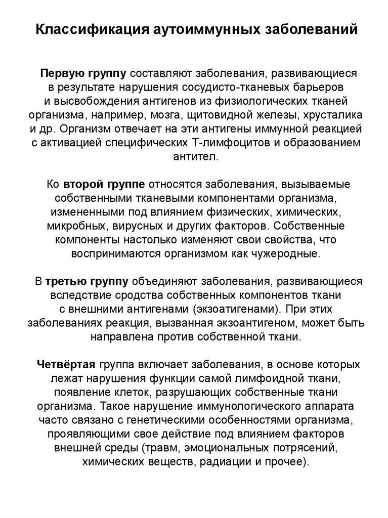
Классификация аутоиммунных заболеванийПервую группу составляют заболевания, развивающиеся
в результате нарушения сосудисто-тканевых барьеров
и высвобождения антигенов из физиологических тканей
организма, например, мозга, щитовидной железы, хрусталика
и др. Организм отвечает на эти антигены иммунной реакцией
с активацией специфических Т-лимфоцитов и образованием
антител.
Ко второй группе относятся заболевания, вызываемые
собственными тканевыми компонентами организма,
измененными под влиянием физических, химических,
микробных, вирусных и других факторов. Собственные
компоненты настолько изменяют свои свойства, что
воспринимаются организмом как чужеродные.
В третью группу объединяют заболевания, развивающиеся
вследствие сродства собственных компонентов ткани
с внешними антигенами (экзоатигенами). При этих
заболеваниях реакция, вызванная экзоантигеном, может быть
направлена против собственной ткани.
Четвёртая группа включает заболевания, в основе которых
лежат нарушения функции самой лимфоидной ткани,
появление клеток, разрушающих собственные ткани
организма. Такое нарушение иммунологического аппарата
часто связано с генетическими особенностями организма,
проявляющими свое действие под влиянием факторов
внешней среды (травм, эмоциональных потрясений,
химических веществ, радиации и прочее).
3.
Аутоиммунные заболевания нервной системы.В 2000 г. D. Karussis предложил классификацию
аутоиммунных заболеваний нервной системы, согласно
которой выделяют три основных группы (Пономарёв В.В.
Аутоиммунные заболевания в неврологии. – Минск:
Беларуская навука, 2010. – 259с. )
1. Идиопатические аутоиммунные неврологические
заболевания:
а) с первичным поражением ЦНС и ПНС; б) системные
заболевания с вторичным поражением ЦНС и ПНС;
К идиопатическим заболеваниям с первичным поражением
нервной системы относятся рассеянный склероз,
миастению Гравис, синдром Гийена – Барре, хроническую
воспалительную демиелинизирующюю полинейропатию, а
также более редкие болезни: – мультифокальная моторная
нейропатия, изолированные церебральные васкулиты,
синдром «ригидного человека», синдром Исаакса, энцефалит
Расмуссена.
Идиопатические воспалительные (аутоиммунные)
демиелинизирующие заболевания центральной нервной
системы представляют собой широкий спектр расстройств,
различающихся клинически и иммунологически. Наибольшую
долю среди них занимает рассеянный склероз с
классическими вариантами течения: ремитирующим,
ремитирующе – прогрессирующим, вторично и
первично прогрессирующим. Кроме этого различают
острые (фульминантные) формы: болезнь Марбурга, болезнь
Шильдера, концентрический склероз Бало, острый
рассеянный энцефаломиелит. Первые три рассматриваются
сегодня как атипичные варианты рассеянного склероза.
4.
Аутоиммунные заболевания нервной системы.(продолжение)
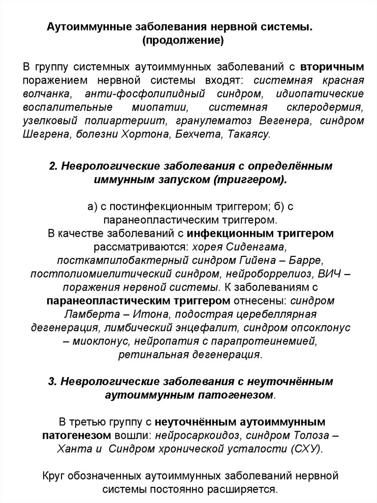
В группу системных аутоиммунных заболеваний с вторичным
поражением нервной системы входят: системная красная
волчанка, анти-фосфолипидный синдром, идиопатические
воспалительные
миопатии,
системная
склеродермия,
узелковый полиартериит, гранулематоз Вегенера, синдром
Шегрена, болезни Хортона, Бехчета, Такаясу.
2. Неврологические заболевания с определённым
иммунным запуском (триггером).
а) с постинфекционным триггером; б) с
паранеопластическим триггером.
В качестве заболеваний с инфекционным триггером
рассматриваются: хорея Сиденгама,
посткампилобактерный синдром Гийена – Барре,
постполиомиелитический синдром, нейроборрелиоз, ВИЧ –
поражения нервной системы. К заболеваниям с
паранеопластическим триггером отнесены: синдром
Ламберта – Итона, подострая церебеллярная
дегенерация, лимбический энцефалит, синдром опсоклонус
– миоклонус, нейропатия с парапротеинемией,
ретинальная дегенерация.
3. Неврологические заболевания с неуточнённым
аутоиммунным патогенезом.
В третью группу с неуточнённым аутоиммунным
патогенезом вошли: нейросаркоидоз, синдром Толоза –
Ханта и Синдром хронической усталости (СХУ).
Круг обозначенных аутоиммунных заболеваний нервной
системы постоянно расширяется.
5.
РАССЕЯННЫЙ СКЛЕРОЗ6.
7.
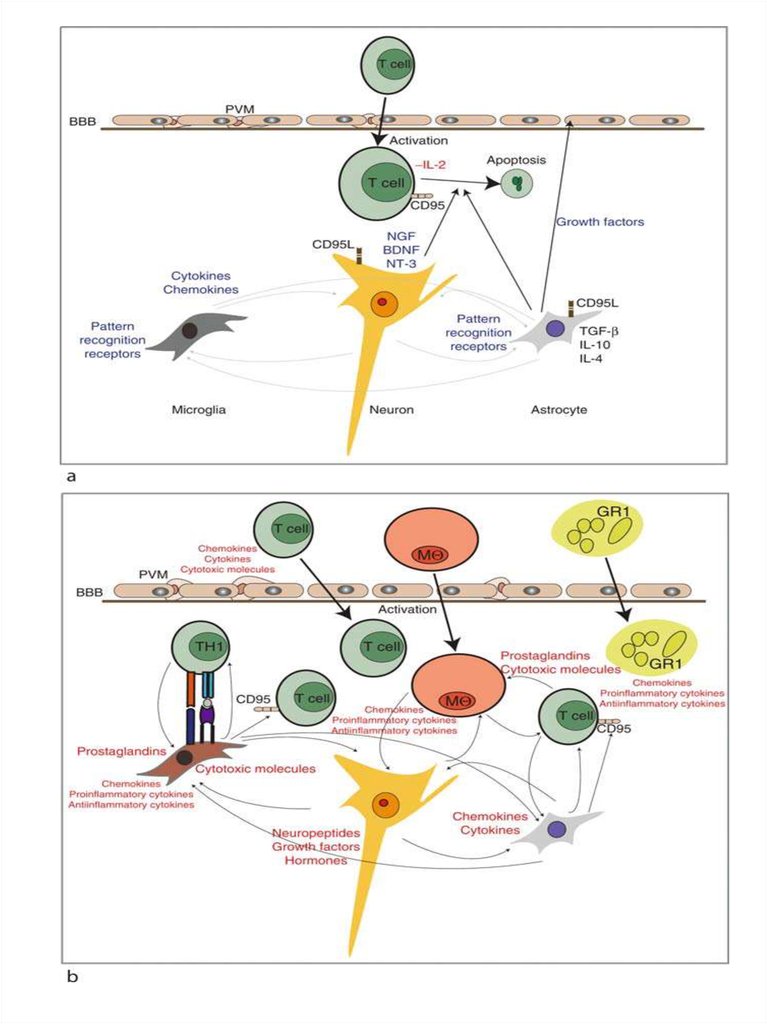
The glioneuronal micromilieu regulates immunologicalmechanisms within the brain.
The upper panel illustrates the physiologic situation (i.e.,
without an inflammatory stimulus), which is characterized by
secretion of immunosuppressive factors/mediators in order to
maintain an immunologically downregulated state. Activated T
cells may cross the blood–brain barrier (BBB), but regularly
undergo apoptosis under normal conditions, because they do
not recognize antigen. Astrocytes produce immunosuppressive
TGF‐b, IL‐4, and IL‐10.
Neurons produce neurotrophins such as brain‐derived
neurotrophic factor (BDNF), nerve growth factor (NGF), and
neurotrophin‐3 (NT‐3), which have been shown to prevent
expression of proinflammatory cytokines on astrocytes and
microglial cells. Neurons and astrocytes express fas‐ligand
(CD95L). Both astrocytes and microglial cells constitutively
express PRR. Perivascular macrophages are not activated and
secrete low or no inflammatory mediators.
The lower panel shows inflammation which activates neurons,
astrocytes, and microglial cells as well as endothelial cells and
perivascular macrophages to produce chemokines, cytokines,
growth factors, and cytotoxic mediators as well as
prostaglandins. A chemokine gradient is thought to attract
inflammatory leukocytes (T cells), granulocytes (GRA),
macrophages(mf), and microglial cells to the lesion site. Specific
antigen recognition stimulates T cells locally and leads to
enhanced production of cytotoxic mediators and proinflammatory
cytokines. Antigen presentation to invading T cells is assured by
activated microglial cells, which have upregulated their MHC
production
8.
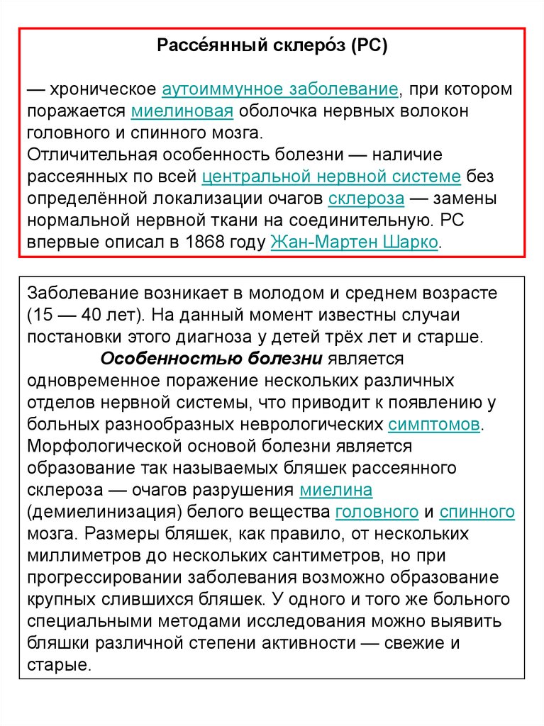
Рассе́янный склеро́з (РС)— хроническое аутоиммунное заболевание, при котором
поражается миелиновая оболочка нервных волокон
головного и спинного мозга.
Отличительная особенность болезни — наличие
рассеянных по всей центральной нервной системе без
определённой локализации очагов склероза — замены
нормальной нервной ткани на соединительную. РС
впервые описал в 1868 году Жан-Мартен Шарко.
Заболевание возникает в молодом и среднем возрасте
(15 — 40 лет). На данный момент известны случаи
постановки этого диагноза у детей трёх лет и старше.
Особенностью болезни является
одновременное поражение нескольких различных
отделов нервной системы, что приводит к появлению у
больных разнообразных неврологических симптомов.
Морфологической основой болезни является
образование так называемых бляшек рассеянного
склероза — очагов разрушения миелина
(демиелинизация) белого вещества головного и спинного
мозга. Размеры бляшек, как правило, от нескольких
миллиметров до нескольких сантиметров, но при
прогрессировании заболевания возможно образование
крупных слившихся бляшек. У одного и того же больного
специальными методами исследования можно выявить
бляшки различной степени активности — свежие и
старые.
9.
Возникновение рассеянного склероза связано сослучайным индивидуальным сочетанием
неблагоприятных эндогенных и экзогенных
факторов риска.
К эндогенным факторам прежде всего следует отнести
мутации в комплексе локусов генов HLA II класса и,
возможно, генов, кодирующих ФНО-a,
обусловливающих генетическую несостоятельность
иммунорегуляции.
Среди внешних факторов могут иметь значение: зона
проживания в детском возрасте, особенности питания,
частота вирусных и бактериальных инфекций и др.
Следует подчеркнуть, что ни один взятый изолированно
фактор не может иметь значение в возникновении
рассеянного склероза, только определённое сочетание
ряда факторов. В организме, имеющем генетически
обусловленную несостоятельность регуляторных
систем иммунитета, происходит активация иммунной
системы одним из неспецифических провоцирующих
факторов, например, вирусной инфекцией, травмой,
стрессовой ситуацией.
При этом антиген-стимулированные макрофаги и
активированные Т-хелперы фиксируются на клетках
эндотелия гемато-энцефалического барьера (ГЭБ).
Цитокины, выделяемые фиксированными клетками,
экспрессируют на поверхности ГЭБ антигены основного
комплекса гистосовместимости I и II класса (для
представления антигена), а также молекулы клеточной
адгезии.
10.
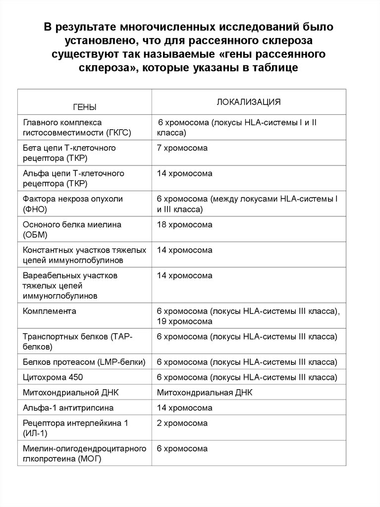
В результате многочисленных исследований былоустановлено, что для рассеянного склероза
существуют так называемые «гены рассеянного
склероза», которые указаны в таблице
ЛОКАЛИЗАЦИЯ
ГЕНЫ
Главного комплекса
гистосовместимости (ГКГС)
6 хромосома (локусы HLA-системы I и II
класса)
Бета цепи Т-клеточного
рецептора (ТКР)
7 хромосома
Альфа цепи Т-клеточного
рецептора (ТКР)
14 хромосома
Фактора некроза опухоли
(ФНО)
6 хромосома (между локусами HLA-системы I
и III класса)
Осноного белка миелина
(ОБМ)
18 хромосома
Константных участков тяжелых
цепей иммуноглобулинов
14 хромосома
Вареабельных участков
тяжелых цепей
иммуноглобулинов
14 хромосома
Комплемента
6 хромосома (локусы HLA-системы III класса),
19 хромосома
Транспортных белков (ТАРбелков)
6 хромосома (локусы HLA-системы III класса)
Белков протеасом (LMP-белки)
6 хромосома (локусы HLA-системы III класса)
Цитохрома 450
6 хромосома (локусы HLA-системы III класса)
Митохондриальной ДНК
Митохондриальная ДНК
Альфа-1 антитрипсина
14 хромосома
Рецептора интерлейкина 1
(ИЛ-1)
2 хромосома
Миелин-олигодендроцитарного
глкопротеина (МОГ)
6 хромосома
11.
12.
13.
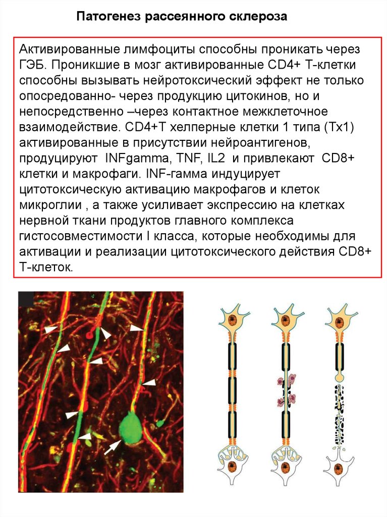
Патогенез рассеянного склерозаАктивированные лимфоциты способны проникать через
ГЭБ. Проникшие в мозг активированные CD4+ Т-клетки
способны вызывать нейротоксический эффект не только
опосредованно- через продукцию цитокинов, но и
непосредственно –через контактное межклеточное
взаимодействие. CD4+Т хелперные клетки 1 типа (Тх1)
активированные в присутствии нейроантигенов,
продуцируют INFgamma, TNF, IL2 и привлекают CD8+
клетки и макрофаги. INF-гамма индуцирует
цитотоксическую активацию макрофагов и клеток
микроглии , а также усиливает экспрессию на клетках
нервной ткани продуктов главного комплекса
гистосовместимости I класса, которые необходимы для
активации и реализации цитотоксического действия CD8+
Т-клеток.
14.
B-Cell functions in autoimmunity:(a) Antibody-producing cells–plasma cells.
(b) (b) Antigen-presenting cells (autoreactive T cells with a
specific antigen; regulatory with low levels of nonspecific
antigen).
(c) (c) Cytokineproducing cells; regulatory (B-cell activation
with isolated CD40 stimulation), polarizing (B-cell
activation with dual stimulation of BCR and CD40),
lymphoneogenesis (memory B cells, primarily produce
proinflammatory cytokines, TNFa/LT following dual
stimulation of BCR and CD40).
(d) (d) Development of tertiary/ectopic germinal centers
15.
The immunopathogenesis of MSA limited number of resting T cells may enter the CNS after
crossing the intact blood-brain barrier (BBB). Autoreactive CD4+
T cells may be activated in the periphery by antigens that are
structurally similar to myelin antigens (molecular mimicry). When
activated T lymphocytes interact with endothelial cells (EC) of
CNS venules, cell trafficking across the BBB increases.
Reactivation of myelin antigen-specific T cells by local antigenpresenting cells (APC) may initiate myelin damage by several
mechanisms: CD4+ T cell production of proinflammatory
cytokines and chemokines that further recruit effector cells
through an inflamed BBB; CD8+ T inducing cytolysis and
chemokine production; activated macrophages producing oxygen
radicals and NO (nitric oxide); deposition of myelin antigenspecific antibodies activating complement and facilitating myelin
damage by activated macrophages. Resolution of inflammatory
damage may be modulated by regulatory cells, that produce antiinflammatory cytokines, and by elimination of effector cells by
apoptosis.
16.
Models of demyelination in multiple sclerosis.a.
b.
c.
Primary autoimmune attack. Autoreactive Th lymphocytes initiate the
autoimmune attack when they gain entry across the blood–brain barrier. This
leads to recruitment of microglia and macrophages and the release
ofinflammatory mediators, leading to the destruction of myelin, death of
oligodendrocytes, and phagocytic clearance of the debris.
Primary oligodendrogliopathy. Oligodendrocyte apoptosis is the primary event,
leading to activation of microglia, which are overwhelmed by the amount of
myelin debris. Circulating autoreactive T lymphocytes are then activated by
the myelin debris and initiate the immune attack on myelin.
Axonal injury. Axonal injury is the primary event, leading into microglial, and in
turn, T‐cell activation. The stereotypical autoimmune destruction of myelin
ensues as a secondary event
17.
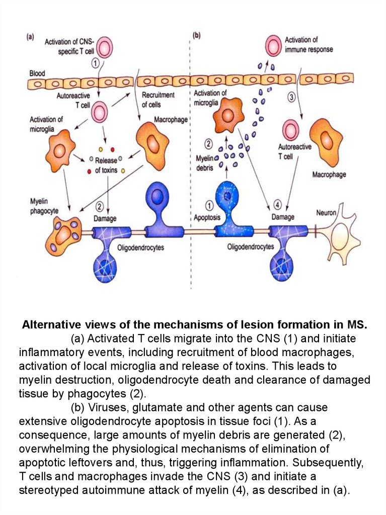
Alternative views of the mechanisms of lesion formation in MS.(a) Activated T cells migrate into the CNS (1) and initiate
inflammatory events, including recruitment of blood macrophages,
activation of local microglia and release of toxins. This leads to
myelin destruction, oligodendrocyte death and clearance of damaged
tissue by phagocytes (2).
(b) Viruses, glutamate and other agents can cause
extensive oligodendrocyte apoptosis in tissue foci (1). As a
consequence, large amounts of myelin debris are generated (2),
overwhelming the physiological mechanisms of elimination of
apoptotic leftovers and, thus, triggering inflammation. Subsequently,
T cells and macrophages invade the CNS (3) and initiate a
stereotyped autoimmune attack of myelin (4), as described in (a).
18.
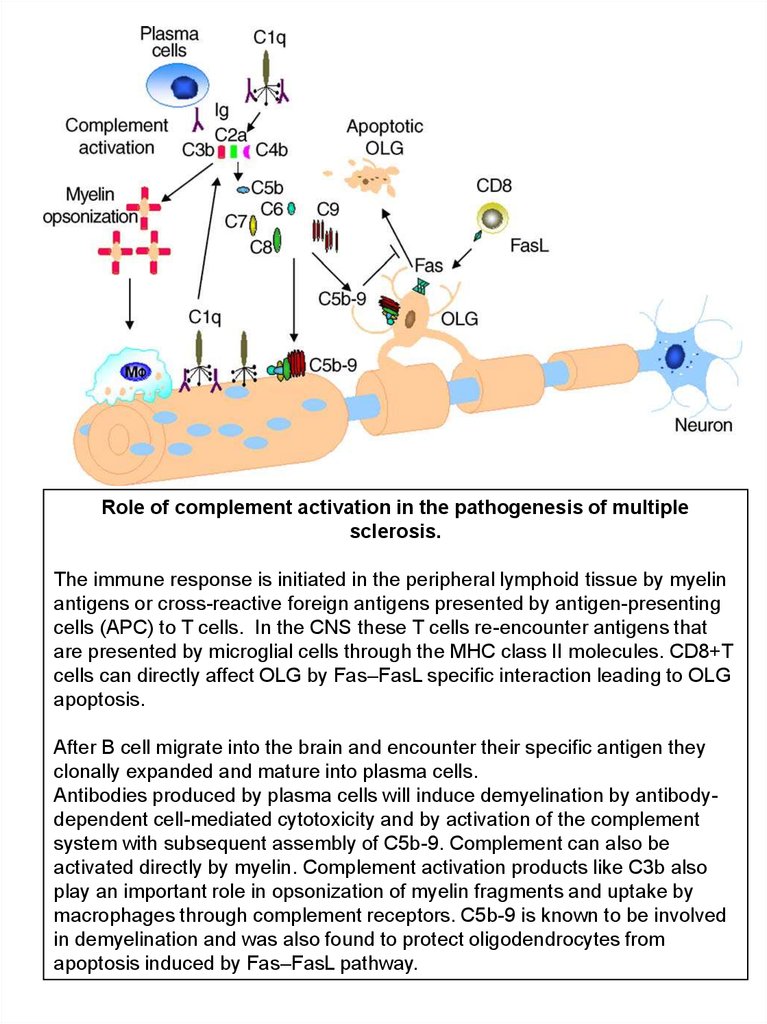
Role of complement activation in the pathogenesis of multiplesclerosis.
The immune response is initiated in the peripheral lymphoid tissue by myelin
antigens or cross-reactive foreign antigens presented by antigen-presenting
cells (APC) to T cells. In the CNS these T cells re-encounter antigens that
are presented by microglial cells through the MHC class II molecules. CD8+T
cells can directly affect OLG by Fas–FasL specific interaction leading to OLG
apoptosis.
After B cell migrate into the brain and encounter their specific antigen they
clonally expanded and mature into plasma cells.
Antibodies produced by plasma cells will induce demyelination by antibodydependent cell-mediated cytotoxicity and by activation of the complement
system with subsequent assembly of C5b-9. Complement can also be
activated directly by myelin. Complement activation products like C3b also
play an important role in opsonization of myelin fragments and uptake by
macrophages through complement receptors. C5b-9 is known to be involved
in demyelination and was also found to protect oligodendrocytes from
apoptosis induced by Fas–FasL pathway.
19.
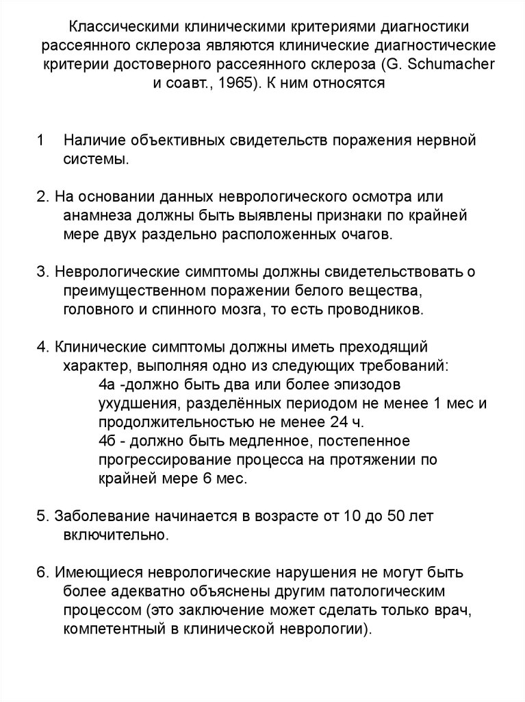
Классическими клиническими критериями диагностикирассеянного склероза являются клинические диагностические
критерии достоверного рассеянного склероза (G. Schumacher
и соавт., 1965). К ним относятся
1
Наличие объективных свидетельств поражения нервной
системы.
2. На основании данных неврологического осмотра или
анамнеза должны быть выявлены признаки по крайней
мере двух раздельно расположенных очагов.
3. Неврологические симптомы должны свидетельствовать о
преимущественном поражении белого вещества,
головного и спинного мозга, то есть проводников.
4. Клинические симптомы должны иметь преходящий
характер, выполняя одно из следующих требований:
4а -должно быть два или более эпизодов
ухудшения, разделённых периодом не менее 1 мес и
продолжительностью не менее 24 ч.
4б - должно быть медленное, постепенное
прогрессирование процесса на протяжении по
крайней мере 6 мес.
5. Заболевание начинается в возрасте от 10 до 50 лет
включительно.
6. Имеющиеся неврологические нарушения не могут быть
более адекватно объяснены другим патологическим
процессом (это заключение может сделать только врач,
компетентный в клинической неврологии).
20.
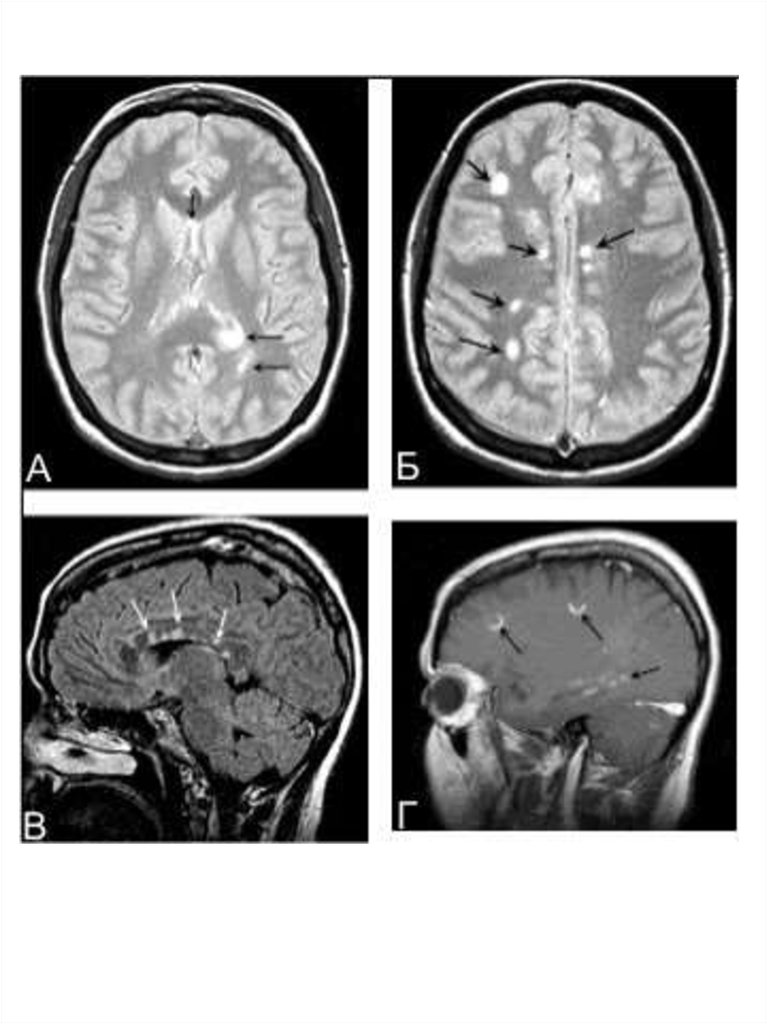
МРТ в диагностике рассеянного склерозаМРТ — метод диагностики, основанный на том, что из-за
особенностей белково-липидного строения мембран миелина
вода располагается в определённом порядке между слоями
миелиновой оболочки. Этот порядок нарушается при
демиелинизации, когда высвобождается часть воды. А так как
вода имеет более длительное время релаксации в магнитном
поле, участки демиелинизации выявляются как участки
пониженной плотности в режиме Т1 и как участки повышенной
плотности на Т2-взвешенных изображениях. При рассеянном
склерозе выявляются множественные разнообразных
размеров и форм очаги в различных отделах вещества
головного и спинного мозга.
Существуют разные диагностические критерии данных МРТ.
Критерии Фазекас (F.Fazekas и соавт., 1988): для
рассеянного склероза характерно не менее трёх областей с
повышенной интенсивностью сигнала, две из них должны быть
в перивентрикулярном пространстве и по крайней мере один
— супратенториально; размеры очагов должны быть не более
5 мм в диаметре.
Критерии Пати (D.Paty и соавт., 1988): должно быть не
менее четырёх очагов гиперинтенсивности на Т2
изображениях, размерами более 3 мм, или три очага, один из
которых расположен перивентрикулярно.
21.
MRI –Т1 ( left ),MWF – Т2
(middle ) and
Luxol fast blue
myelin stain of
post-mortem
tissue
( right )
It is also possible to assess myelin in vivo using positron
emission tomography (PET) and tracers such as 1,4-bis( p
-aminostyryl)-2-methoxy benzene (BMB)
22.
23.
24.
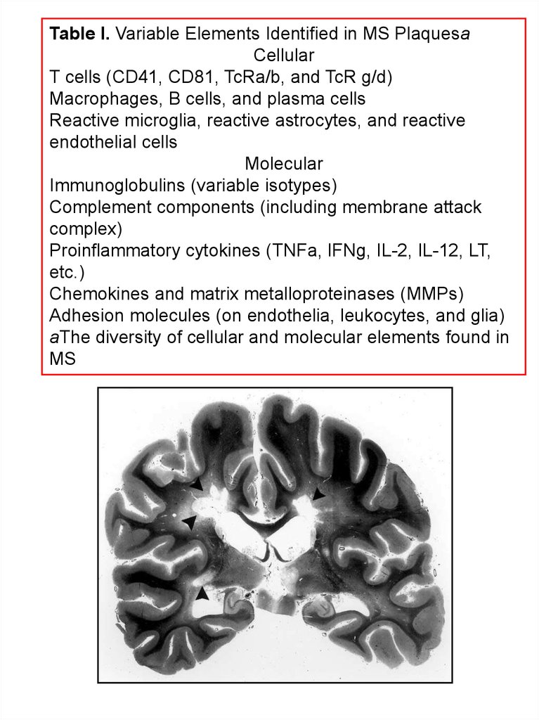
очаги демиелинизациикора
Склеротические бляшки в коре мозга при рассеянном
склерозе.
Специфическим, необходимым патоморфологическим
маркером РС является
бляшка рассеянного склероза - очаг хронической
воспалительной демиелинизации.
Располагаются очаги в любом отделе белого вещества
головного и спинного мозга. При длительно текущем
рассеянном склерозе и выраженном разрушении
миелина может происходить вторичная дегенерация
осевых цилиндров нервных волокон, в последующем —
нервных клеток и олигодендроцитов. Это приводит к
атрофии головного и спинного мозга, расширению
желудочков мозга.
25.
In this acute plaque, the central blood vessel is almostobliterated by a lymphocytic infiltrate, which extended
through the wall to lie perivascularly. Inflammatory cells also
lie in the tissue away from the vessels
Histology of a plaque silver stained for axons. There is a
major dropout of black axons, some of which are thickened
and swollen indicating damage
26.
Actively demyelinating MS lesion.Myelin basic protein (MBP) is a major constituent of
the myelin sheath of oligodendrocytes in the central nervous
system. Macrophages phagocytosing myelin debris can be
easily detected in actively demyelinating lesions
27.
Table I. Variable Elements Identified in MS PlaquesaCellular
T cells (CD41, CD81, TcRa/b, and TcR g/d)
Macrophages, B cells, and plasma cells
Reactive microglia, reactive astrocytes, and reactive
endothelial cells
Molecular
Immunoglobulins (variable isotypes)
Complement components (including membrane attack
complex)
Proinflammatory cytokines (TNFa, IFNg, IL-2, IL-12, LT,
etc.)
Chemokines and matrix metalloproteinases (MMPs)
Adhesion molecules (on endothelia, leukocytes, and glia)
aThe diversity of cellular and molecular elements found in
MS
28.
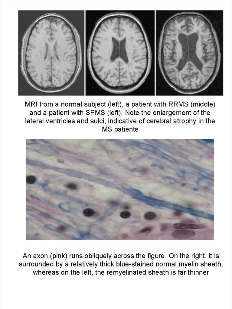
MRI from a normal subject (left), a patient with RRMS (middle)and a patient with SPMS (left). Note the enlargement of the
lateral ventricles and sulci, indicative of cerebral atrophy in the
MS patients
An axon (pink) runs obliquely across the figure. On the right, it is
surrounded by a relatively thick blue-stained normal myelin sheath,
whereas on the left, the remyelinated sheath is far thinner
29.
30.
Electron micrograph showing a field of demyelinatedaxons. On the right, two remyelinated axons, with thin
sheaths, are present
The white matter in the occipital lobes of this
brain is almost totally replaced with diffuse gray
plaque
31.
Разрушение нерва цитотоксическими Т-лимфоцитами32.
ba
1
2
3
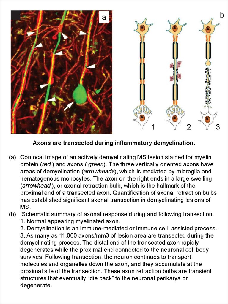
Axons are transected during inflammatory demyelination.
(a) Confocal image of an actively demyelinating MS lesion stained for myelin
protein (red ) and axons ( green). The three vertically oriented axons have
areas of demyelination (arrowheads), which is mediated by microglia and
hematogenous monocytes. The axon on the right ends in a large swelling
(arrowhead ), or axonal retraction bulb, which is the hallmark of the
proximal end of a transected axon. Quantification of axonal retraction bulbs
has established significant axonal transection in demyelinating lesions of
MS.
(b) Schematic summary of axonal response during and following transection.
1. Normal appearing myelinated axon.
2. Demyelination is an immune-mediated or immune cell–assisted process.
3. As many as 11,000 axons/mm3 of lesion area are transected during the
demyelinating process. The distal end of the transected axon rapidly
degenerates while the proximal end connected to the neuronal cell body
survives. Following transection, the neuron continues to transport
molecules and organelles down the axon, and they accumulate at the
proximal site of the transection. These axon retraction bulbs are transient
structures that eventually “die back” to the neuronal perikarya or
degenerate.
33.
Cortical demyelination and neuronal pathology.Three types of cortical lesions have been described in MS
brains (a–c, orange areas).
(a) Type I lesions affect both white and gray matter.
(b) (b) Type II lesions are small perivascular areas of
demyelination.
(c) (c) Type III lesions extend from the pial surface into the cortex
and often demyelinate multiple gyri.
(d) Cortical demyelination occurs without significant infiltration of
hematogenous leukocytes (ctx, cortex; wm, white matter).
(e) Axons and dendrites are transected (white arrowheads) during
cortical demyelination. ( f ) Apoptotic neurons (red arrows),
identified by tunnel staining, are increased in demyelinated
cortex
34.
Spinal cord demyelination.The spinal cord is a predilection site for MS lesions, and
inmany advanced cases, only islands of myelinated
tissue remain in both the white and gray matter.
Immunolabeled for MBP (основной белок миелина)
35.
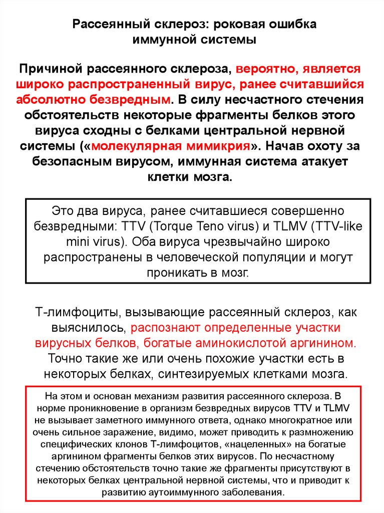
Рассеянный склероз: роковая ошибкаиммунной системы
Причиной рассеянного склероза, вероятно, является
широко распространенный вирус, ранее считавшийся
абсолютно безвредным. В силу несчастного стечения
обстоятельств некоторые фрагменты белков этого
вируса сходны с белками центральной нервной
системы («молекулярная мимикрия». Начав охоту за
безопасным вирусом, иммунная система атакует
клетки мозга.
Это два вируса, ранее считавшиеся совершенно
безвредными: TTV (Torque Teno virus) и TLMV (TTV-like
mini virus). Оба вируса чрезвычайно широко
распространены в человеческой популяции и могут
проникать в мозг.
Т-лимфоциты, вызывающие рассеянный склероз, как
выяснилось, распознают определенные участки
вирусных белков, богатые аминокислотой аргинином.
Точно такие же или очень похожие участки есть в
некоторых белках, синтезируемых клетками мозга.
На этом и основан механизм развития рассеянного склероза. В
норме проникновение в организм безвредных вирусов TTV и TLMV
не вызывает заметного иммунного ответа, однако многократное или
очень сильное заражение, видимо, может приводить к размножению
специфических клонов Т-лимфоцитов, «нацеленных» на богатые
аргинином фрагменты белков этих вирусов. По несчастному
стечению обстоятельств точно такие же фрагменты присутствуют в
некоторых белках центральной нервной системы, что и приводит к
развитию аутоиммунного заболевания.
36.
SUMMARY POINTS1. Multiple sclerosis is an immune-mediated demyelinating
disease of the human central nervous system.
2. Neurodegeneration is the major cause of permanent
neurological disability in MS patients.
3. The cerebral cortex of MS patients is demyelinated without
significant influx of immune cells.
4. Despite indirect evidence supporting an autoimmune
etiology of MS, it remains to be determined if inflammation
is primary or secondary to a degenerative process in the
brain.
FUTURE ISSUES
1. If new inflammatory demyelinating lesions can be
prevented inMSbrains, will the disease process be
halted?
2. Are relapsing-remittingMSand primary progressiveMSthe
same disease or two different diseases?
3. Is there a global cortical-based disease in MS patients?
4. Investigators need to develop brain imaging modalities that
can detect cortical demyelination.
Multiple Sclerosis: An Immune or Neurodegenerative Disorder? Bruce D. Trapp, and KlausArmin Nave AnnRevNeurosc., 2008
37.
Рассеянный склероз: иммунные клетки нервнойсистемы (жёлтые) нападают на олигодендроциты,
38.
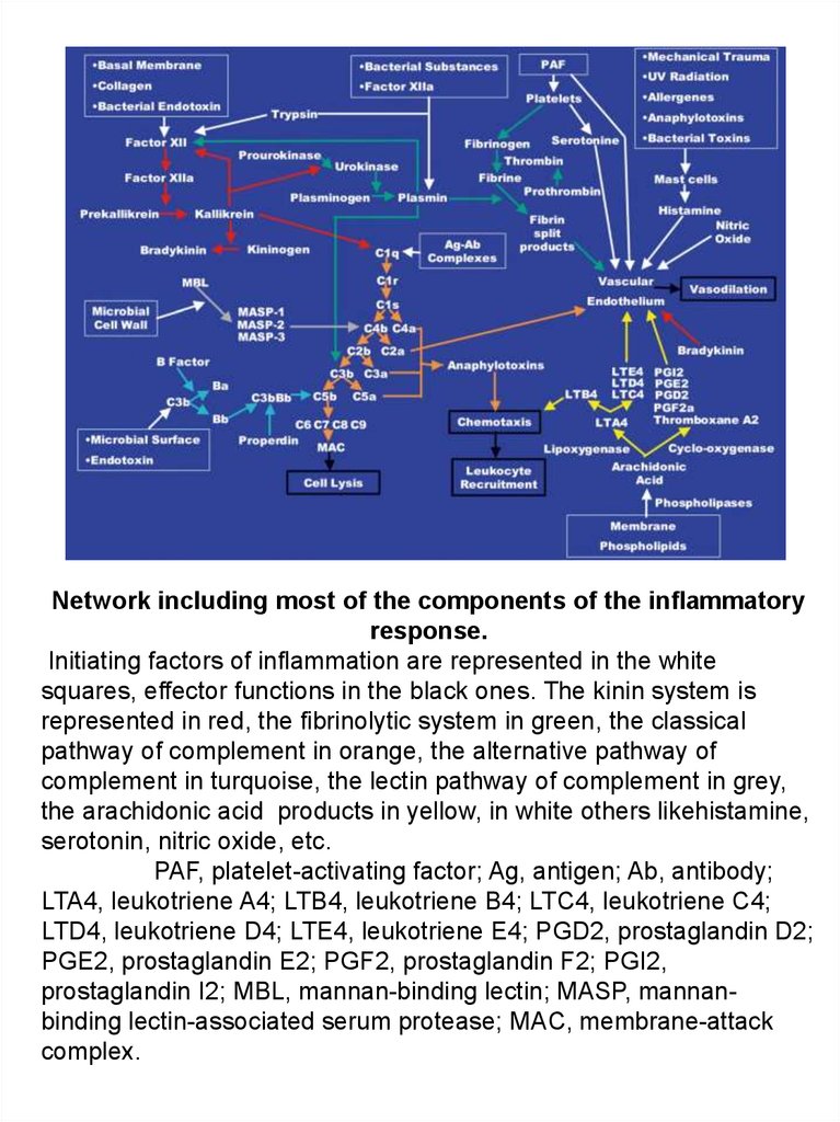
Network including most of the components of the inflammatoryresponse.
Initiating factors of inflammation are represented in the white
squares, effector functions in the black ones. The kinin system is
represented in red, the fibrinolytic system in green, the classical
pathway of complement in orange, the alternative pathway of
complement in turquoise, the lectin pathway of complement in grey,
the arachidonic acid products in yellow, in white others likehistamine,
serotonin, nitric oxide, etc.
PAF, platelet-activating factor; Ag, antigen; Ab, antibody;
LTA4, leukotriene A4; LTB4, leukotriene B4; LTC4, leukotriene C4;
LTD4, leukotriene D4; LTE4, leukotriene E4; PGD2, prostaglandin D2;
PGE2, prostaglandin E2; PGF2, prostaglandin F2; PGI2,
prostaglandin I2; MBL, mannan-binding lectin; MASP, mannanbinding lectin-associated serum protease; MAC, membrane-attack
complex.
39.
40.
41.
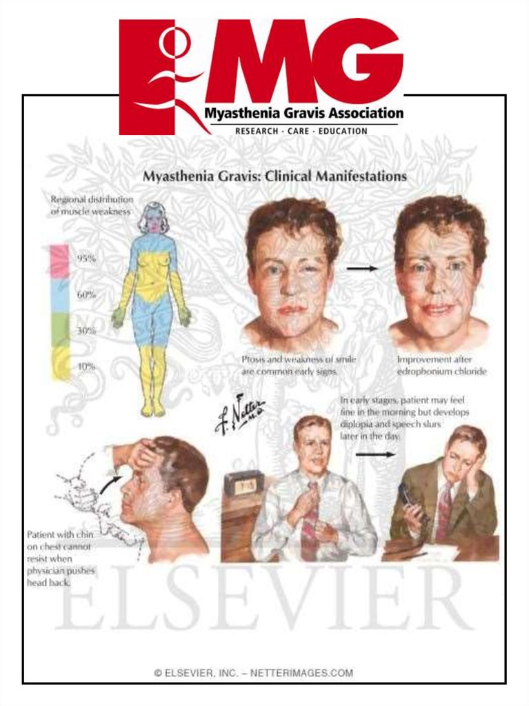
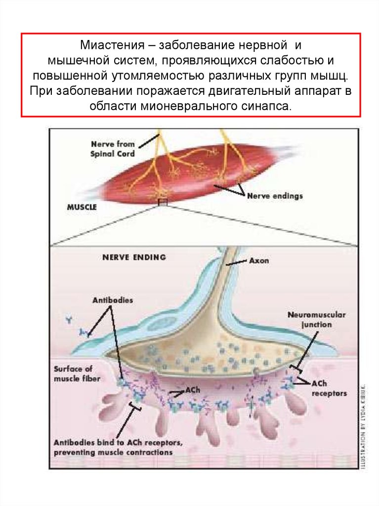
Миастения – заболевание нервной имышечной систем, проявляющихся слабостью и
повышенной утомляемостью различных групп мышц.
При заболевании поражается двигательный аппарат в
области мионеврального синапса.
42.
Myasthenia gravispseudoparalytica
Болезнь Эрба-Гольдфлама
1. Проявляется обычно в 3 декаде жизни, но может
встречаться в любом возрасте после первого года
жизни, наиболее часто с птозом иди диплопией. У
пациентов с генерализованным поражением
развивается безболезненная усталость, часто после
физических нагрузок, которая усиливается к концу дня
и провоцируется инфекцией или стрессом.
2. Симптомы миастении gravis. Самый важный
симптом — утомление, затрагивающее поперечнополосатую мускулатуру конечностей, связанное с
изменением выражения лица, движений глаз,
процессов жевания и речи.
а) периферические симптомы: слабость, особенно в
руках и проксимальных мышц ног. Долговременные
миопатические изменения могут быть при длительно
существующем заболевании;
б) фациальные симптомы: амимичность
(миопатическое лицо) и птоз;
в) бульбарные нарушения: трудности с глотанием
(дисфагия), при разговоре (дизартрия) и жевании;
г) дыхательные нарушения: редки, но всегда к этому
следует относиться серьезно.
43.
Cellular and molecular players in the multi-steprecruitment of T lymphocytes across the blood-CNS
barriers.
A For the superficial vessels of inflamed brain, Eand-P-selectin,PSGL-1 and α4-integrin are involved in
lymphocyte tethering and rolling. G protein-dependent
activation of LFA-1 on T lymphocytes, probably by
chemokines (yet to be identified), leads to their firm
adhesion on endothelial ICAM-1. Lymphocytes migrate
transcellularly through the BBB endothelium, leaving TJs
intact. In EAE, the inflammatory lymphocytes present in
brain (and spinal cord) parenchyma are Th1 effector
memory cells with a characteristic surface-marker
phenotype.
B In the spinal cord white matter, T lymphocytes
arrest immediately through α4-integrin. G protein-dependent
increase in α4-integrin avidity on the T lymphocytes is
required for firm adhesion to endothelial counter-receptors.
LFA-1 supports T lymphocyte diapedesis, adjacent to TJs.
C Molecular mechanisms in lymphocyte transit
across fenestrated CP microvessels, and subsequent
migration across the CPE into the CSF are relatively
unknown, although α4-integrin is required. Endothelial Pselectin mediates T lymphocyte recruitment into the CP
stroma. Pathways of T lymphocyte traversal across CP
endothelial and epithelial barriers are equally mysterious. T
lymphocytes in the CSF, both of healthy individuals and MS
patients, are predominantly central-memory CD4+ T
lymphocytes, about half of which express the recent
activation marker CD69
44.
Myasthenia gravispseudoparalytica
Болезнь Эрба-Гольдфлама
45.
Myasthenia gravis initially manifestptosis.
46.
Патология является результатом выработки антителпротив альфа-субединицы никотиновых
холинорецепторов.
Антитела к никотиновым рецепторам подобно кураре
затрудняют синаптическую передачу и приводят к
мышечной слабости.
Аутоантитела вытесняют ацетилхолин в
мионевральных соединениях, блокируют нейромышечную синаптическую передачу.
Заболевание спорадическое, однако описаны и
семейные случаи.
47.
Направления виды лечения миастении1 — антихолинестеразная терапия,
2 — иммуносупрессивная терапия (глюкокортикостероидная
и цитостатическая),
3 — тимэктомия и лучевая терапия,
4 — сорбционные методы
5 — иммуно-стимулирующая и иммуномодулирующая терапия
(развивается)
6 — неспецифическая терапия, усиливающая действие
патогенетических средств.
При отсутствии эффекта от АХЭ терапии проведение
иммуносупрессивной терапии, сорбционных методов лечения,
молодым пациентам — тимэктомии, а пожилым — курса лучевой
терапии на область вилочковой железы.
При правильно подобранном лечении, более 95 %
пациентов возвращаются к нормальному качеству жизни.
48.
49.
50.
51.
52.
аb
c
e
1
2
3
f
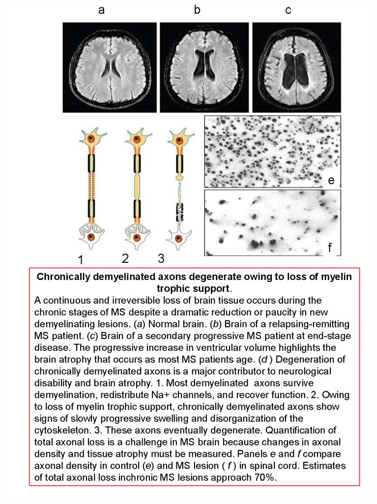
Chronically demyelinated axons degenerate owing to loss of myelin
trophic support.
A continuous and irreversible loss of brain tissue occurs during the
chronic stages of MS despite a dramatic reduction or paucity in new
demyelinating lesions. (a) Normal brain. (b) Brain of a relapsing-remitting
MS patient. (c) Brain of a secondary progressive MS patient at end-stage
disease. The progressive increase in ventricular volume highlights the
brain atrophy that occurs as most MS patients age. (d ) Degeneration of
chronically demyelinated axons is a major contributor to neurological
disability and brain atrophy. 1. Most demyelinated axons survive
demyelination, redistribute Na+ channels, and recover function. 2. Owing
to loss of myelin trophic support, chronically demyelinated axons show
signs of slowly progressive swelling and disorganization of the
cytoskeleton. 3. These axons eventually degenerate. Quantification of
total axonal loss is a challenge in MS brain because changes in axonal
density and tissue atrophy must be measured. Panels e and f compare
axonal density in control (e) and MS lesion ( f ) in spinal cord. Estimates
of total axonal loss inchronic MS lesions approach 70%.
53.
A model of MS immunopathogenesis. Myelin-autoreactive T cells are found inan enhanced state of activation in the circulation of MS patients (1) due to
genetic and environmental factors (including viral infection, bacteria, and
superantigens).
Endothelial adhesion molecules, such as ICAM-1 and VCAM-1, are
upregulated (2), which further facilitates the migration of T cells into the CNS.
Chemoattraction via chemokines (3) and elaboration of matrix
metalloproteinases, which may enhance migration by degrading extracellularmatrix proteins (4), results in invasion of activated autoreactive T cells across
the blood– brain barrier (BBB). Inside the central nervous system (CNS),
reactivation of T cells by local or infiltrating antigen presenting cells (APCs)
results in release of proinflammatory and cytotoxic mediators, leading to
tissue injury. The protective myelin sheath may be injured (6) via several
mechanisms: cytokine-mediated injury; digestion of surface myelin antigens
by macrophages, which may include the binding of antibodies; complementmediated injury; and direct injury by CD41 and CD81 T cells.
54.
Functions of microglia in immune and inflammatorymechanisms in the CNS.
Microglia produce a variety of neuroprotective and
neurotoxic factors. These factors play a role on the
development of demyelination, neuronal degeneration,
regeneration, and gliosis. Infiltrating T cells, especially
Th1 cells, activate microglia to function as effector cells
in neuroinflammation. Arrows indicate functions of
soluble factors from microglia, and dotted lines indicate
functions of Th1-derived factors. E: endothelial cells,
oligo: oligodendrocytes,
55.
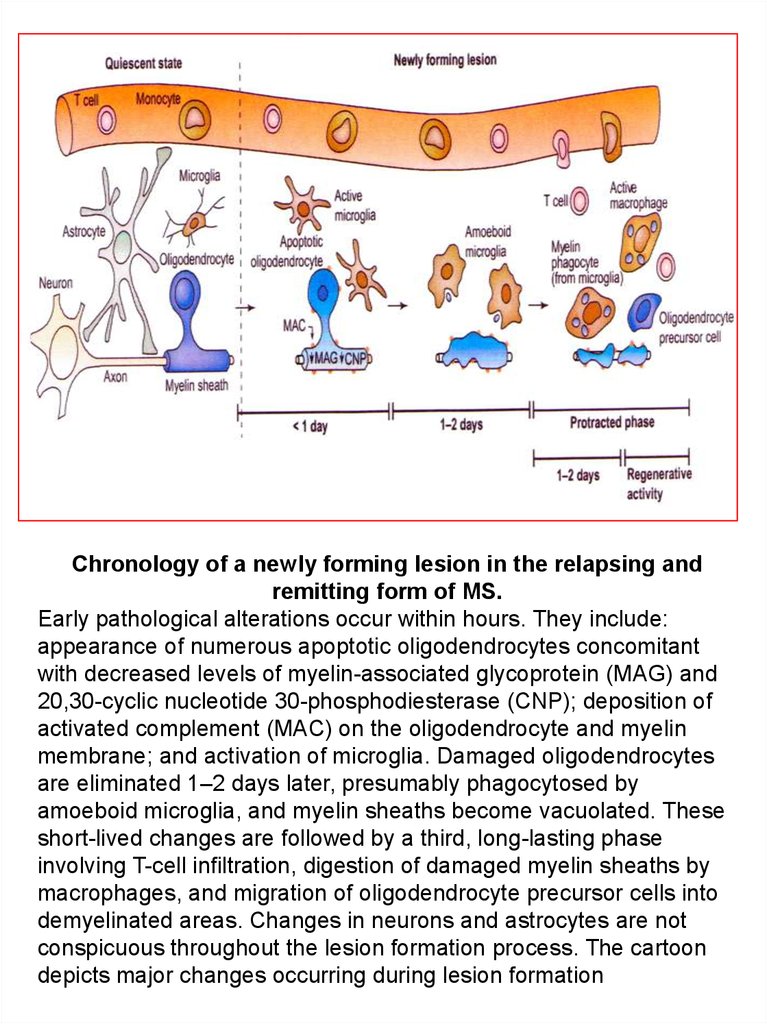
Chronology of a newly forming lesion in the relapsing andremitting form of MS.
Early pathological alterations occur within hours. They include:
appearance of numerous apoptotic oligodendrocytes concomitant
with decreased levels of myelin-associated glycoprotein (MAG) and
20,30-cyclic nucleotide 30-phosphodiesterase (CNP); deposition of
activated complement (MAC) on the oligodendrocyte and myelin
membrane; and activation of microglia. Damaged oligodendrocytes
are eliminated 1–2 days later, presumably phagocytosed by
amoeboid microglia, and myelin sheaths become vacuolated. These
short-lived changes are followed by a third, long-lasting phase
involving T-cell infiltration, digestion of damaged myelin sheaths by
macrophages, and migration of oligodendrocyte precursor cells into
demyelinated areas. Changes in neurons and astrocytes are not
conspicuous throughout the lesion formation process. The cartoon
depicts major changes occurring during lesion formation
56.
Pathogenesis of EAE.In the peripheral immune system, antigen‐presenting cells
(mainly DCs but also B cells and macrophages) produce
IL‐12 and IL‐23 which prime native CD4. T cells during
activation by interaction of the T‐cell receptor with cognate
antigen presented by MHC class II complexes.
Activated Th1 and Th17 cells cross the blood–brain barrier
into the CNS. Resident antigen‐presenting cells (APCs)
interact with activated myelin‐specific Th1 and Th17 cells
which become reactivated. Inflammatory cytokines and
chemokines are produced and an inflammatory cascade is
initiated. Infiltrating macrophages are activated and attack the
myelin sheath on neurons
57.
The role of lymphoid chemokines in CNS homeostatic andpathogenic states.
А. CCL19 and CCL21 are constitutively expressed in
cerebrovascular endothelium and the choroid plexus. The majority
of cells in the CSF are TCM cells that express CCR7 and may play
a role in immunosurveillance of the subarachnoid and perivascular
spaces, including in the clearance of local infections,
В. Conversely, aberrant production of CCL19, CCL21 and
CXCL13 may support CNS infiltration by leukemic T and B cells and
the establishment of hematopoetic tumors (inset) in the brain and
spinal cord,
С. CXCL13 is not expressed in the healthy CNS but is
upregulated during autoimmune inflammation where it may have a
role in the cognate interaction of encephalitogenic T cells and DC,
while later in disease CXCL13 has been associated with the
development of ectopic lymphoid structures (inset) in the meninges,
58.
Photograph of a brain with well-demarcated gray/pink MS plaquesaround the ventricles and extending into the white matter. Adjacent to the
plaques, there are areas of less well-defined white matter changes,
representing dirty-appearing white matter. Note the atrophy due to myelin
and axon loss in the corpus callosum on the left and the ventricular
dilatation
59.
Neurodegeneration and Neuroprotection – Innate Immune Response.Figure 1 Innate (natural) defensemechanisms in health and diseases of the CNS: (i): The
physical blood brain barrier composed ofendothelial cells linked by tight junctions and
surrounded by the end feet of astrocytes prevents the infiltrationby pathogens. (ii): Brain cells
such as microglia and astrocytes can also induce apoptosis of autoimmuneT lymphocytes
through the Fas/FasL pathway. (iii): In response to infection or tissue injury, resident cells
willbe activated, i.e., reactive gliosis with the activation and proliferation of astrocytes and
microglia. (iv):Remarkably, innate immune molecules will contribute to the selective
recognition and removal of pathogensand toxic cell debris (apoptotic corpses, amyloid fibrils)
while preserving self cells. Furthermore, several innateimmune molecules will initiate tissue
repair (neurogenesis). (v): This response will have to be kept under safeguard through the
expression of inhibitory/regulatory molecules for example by neurons to controlphagocytosis
by microglia. (vi): In sharp contrast, several innate immune molecules have been involved
inneuronal loss and oligodendrocyte damage in diseases such as Alzheimer’s disease and
multiple sclerosis,respectively. For example, host defense complement proteins are known to
induce adverse cytotoxic activitiesagainst myelin-forming cells and neurons. Hence, a fine
balance must exist and which is at the route of futuretherapeutic strategies.
60.
61.
Use of EAE in neuroimmunology research.Healthy mice are immunized with a subcutaneous injection of
strainspecific, immunogenic myelin peptide in complete Freud’s
adjuvant (CFA) and pertussis toxin is injected on days 0 and 2.
Disease onset, evident as progressive neurological disability,
occurs between days 9 and 14.
Tissues are collected and are either directly analyzed
histologically and by PCR or are cultured and later harvested for
phenotypic and molecular analysis. Results are analyzed,
interpreted, and used to evaluate the original hypothesis of the
experiment and plan further studies
62.
Schematic drawing representing signal transductionof interleukin-1 and tumor necrosis factor.
AA: arachidonic acid; AC: adenylate cyclase; cAMP:
cyclic adenosine monophosphate; CAPK: ceramide
activated protein kinase; DAG: diacylglycerol; MAPK:
mitogen activated protein kinase; NF: nuclear factor; PG:
prostaglandin; PKC: protein kinase C; PLC:phospholipase
C; SMase: sphingomyelinase.
63.
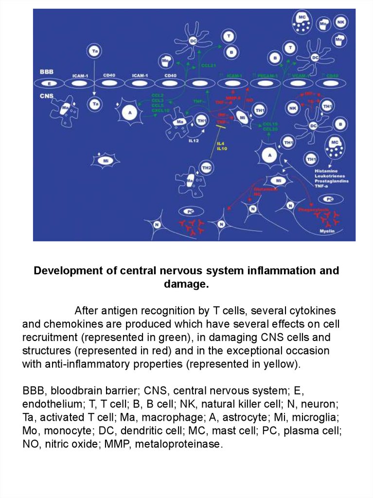
Development of central nervous system inflammation anddamage.
After antigen recognition by T cells, several cytokines
and chemokines are produced which have several effects on cell
recruitment (represented in green), in damaging CNS cells and
structures (represented in red) and in the exceptional occasion
with anti-inflammatory properties (represented in yellow).
BBB, bloodbrain barrier; CNS, central nervous system; E,
endothelium; T, T cell; B, B cell; NK, natural killer cell; N, neuron;
Ta, activated T cell; Ma, macrophage; A, astrocyte; Mi, microglia;
Mo, monocyte; DC, dendritic cell; MC, mast cell; PC, plasma cell;
NO, nitric oxide; MMP, metaloproteinase.
64.
65.
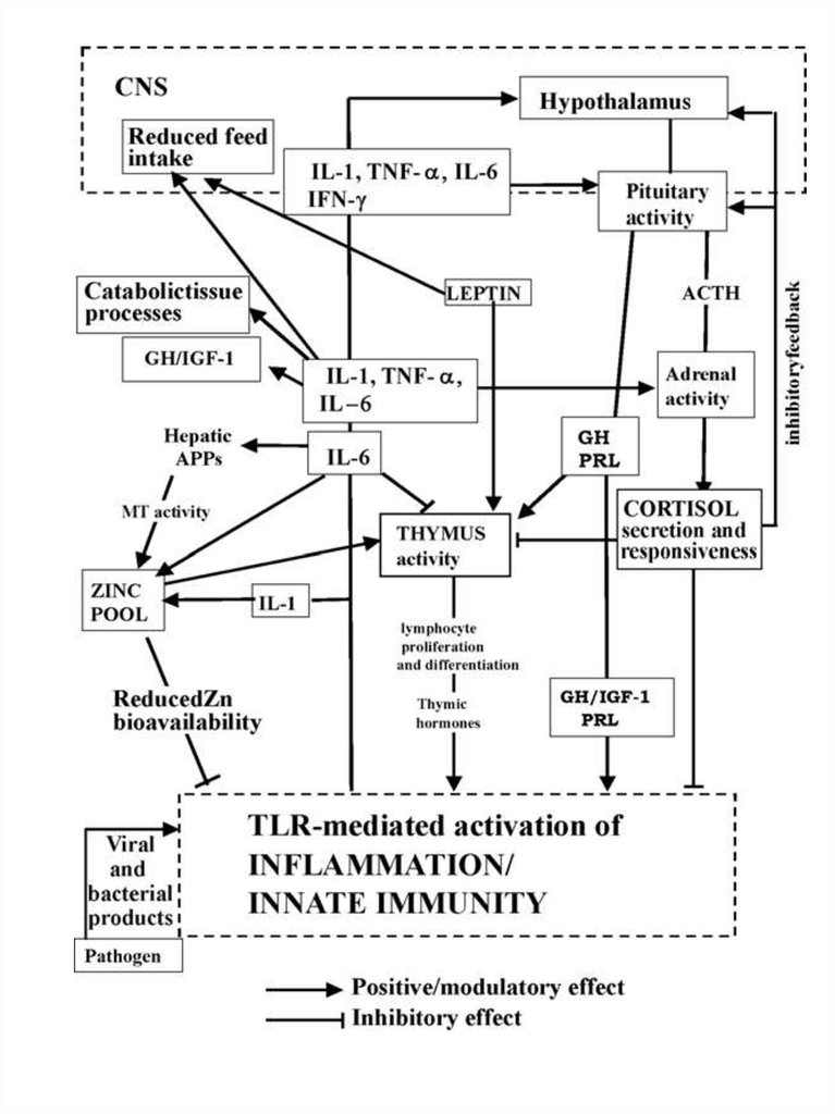
. Possible pathways involved in controlled immune response or inuncontrolled inflammation: the central role of pro-inflammatory
cytokine levels and glucocorticoids.
(a) IL-1, TNF-a and IL-6 production sustains an early and
efficient inflammatory/innate response which limits and destroys the
pathogen and primes acquired immunity; glucocorticoids (GCs) control the
local and systemic inflammatory response preventing damage by excessive
inflammation;
(b) uncoupled regulatory linkage between GH and IGF-1 during
infection: increased levels of GH could be considered as a compensatory
attempt to sustain innate immunity and counteract excessive GC activity;
(c) reduced GH and IGF-1 activity drives metabolic and nutritional
modifications towards supporting the immune function; cytokine production in
adipose and muscle tissue can favour energy partitioning; (d) uncontrolled
high levels of pro-inflammatory cytokines, IL-1, TNF-a particularly IL-6, cause
inflammatory tissue damage concomitantly with
(e) HPA axis dysregulation, altered levels
of ACTH and/or GCs and reduced GCs Receptor (GR) expression; (f)
persistent levels of IL-6 mediate chronic inflammation