


 medicine
Similar presentations:


Injections. Punctures
1. Injections. Punctures.
INJECTIONS.PUNCTURES.
A. A. KANTAY VS-316
2. Injections
INJECTIONS• An injection is an infusion method of putting fluid into the body, usually with a syringe
and a hollow needle which is pierced through the skin to a sufficient depth for the
material to be administered into the body. An injection follows a parenteral route of
administration; that is, administration via a route other than through the digestive tract.
3. methods of injection
METHODS OF INJECTION• There are several methods of injection or infusion used in animals, including intradermal,
subcutaneous, intramuscular, intravenous, intraosseous, intraperitoneal, intrathecal,
epidural, intracardiac, intraarticular, intracavernous, and intravitreal. Rodents used for
research are often administered intracerebral, intracerebroventricular, or intraportal
injections as well.
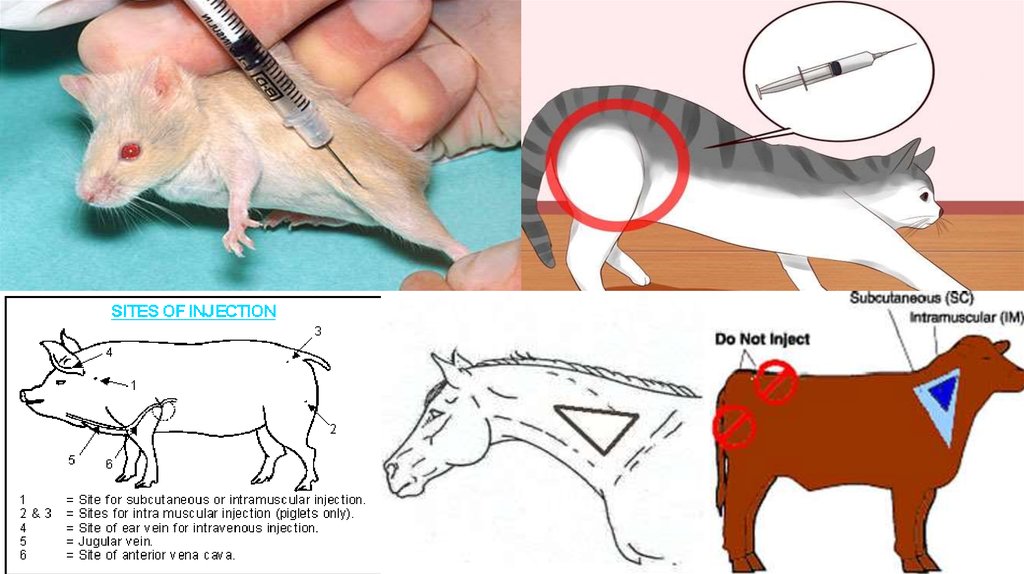
4. Intramuscular Injections
INTRAMUSCULAR INJECTIONS• Choose muscle tissue of lesser value to consumers for intramuscular injections. In cattle, for example,
intramuscular injections where possible, are often given in the neck area instead of the hip.
• Give intramuscular injections deep into a muscle. Use a needle long enough to penetrate skin,
subcutaneous tissue and fat to reach the muscle. The needle should enter the skin perpendicular to the
skin surface.
• Insert the needle into the animal, and then attach the syringe to the needle. Check that the needle is not
in a blood vessel by pulling back on the plunger and observing for blood flow in the tip of the syringe. If
blood appears, remove the needle and put it in a different location at least one inch away from the
original injection site.
5.
6. Subcutaneous Injections
SUBCUTANEOUS INJECTIONS• Give Subcutaneous injections half way up the neck in front of the shoulder, or over the
ribs well behind the shoulder.
• Use a 0.5 to 1 inch long needle.
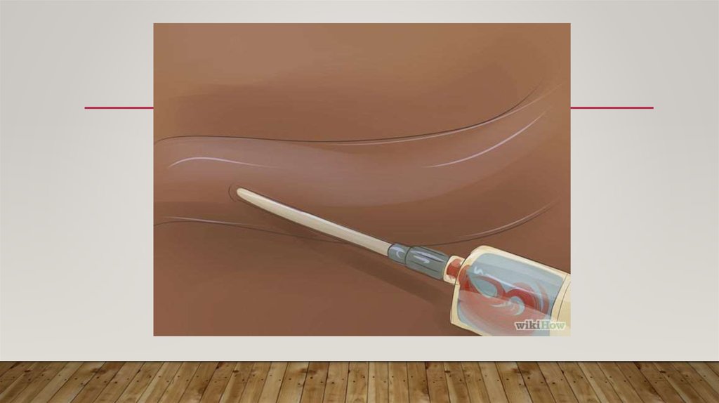
• To give Subcutaneous injections for cattle, lift a fold of skin to make a skin "tent". Insert
the needle through one side of the tent at an angle of 30 to 45 degrees relative to the
surface of the body. For swine, it won't be possible to make a "tent", so slide the needle
under the skin at an angle of about 30 degrees from parallel to the skin surface and
inject.
7.
8. Intravenous therapy
INTRAVENOUS THERAPY• Intravenous therapy is the infusion of liquid substances directly into a vein. Intravenous simply means
"within vein". Therapies administered intravenously are often included in the designation of specialty
drugs. It is commonly referred to as a drip because many systems of administration employ a drip
chamber, which prevents air from entering the blood stream (air embolism), and allows an estimation of
flow rate.
• Intravenous therapy may be used to correct electrolyte imbalances, to deliver medications, for blood
transfusion or as fluid replacement to correct, for example, dehydration. Intravenous therapy can also be
used for chemotherapy.
• Compared with other routes of administration, the intravenous route is the fastest way to deliver fluids
and medications throughout the body. The bioavailability of the medication is 100% in Intravenous
therapy.
9.
10. Consequences of Poor Injection Techniques
CONSEQUENCES OF POOR INJECTIONTECHNIQUES
• Treatment failure, if product absorption is delayed or blocked.
• Drug residues in meat or milk if the drug can not be absorbed and metabolized in a timely
manner.
• Animal suffering and incapacitation due to nerve damage and swelling from tissue reactions.
• Excessive trim at slaughter due to abscess, scarring, broken needles.
• Shock or death of the animal being treated, if medications unintentionally enter the
bloodstream.
• Accidental human injection.
11. Records of Treatment
RECORDS OF TREATMENT• All treatments given to food animals should be permanently recorded to ensure
withdrawal time requirements are met and to improve treatment decisions and success.
• Keep permanent written records of treatments administered to individuals or groups of
animals.
• Record the animal's identification, date(s) the treatment was given, product name, amount
given, the route, site and time when meat or milk will be ready for sale.
12. Cardiac puncture
CARDIAC PUNCTURE• Cardiac puncture is a suitable technique to obtain a single, large, good quality sample
from a euthanised mouse or a mouse under deep terminal anaesthesia if coagulation
parameters, a separate arterial or venous sample or cardiac histology are not required. It
is appropriate for all strains of mouse.
• Cardiac puncture should not be used if the peritoneum needs to be lavaged to harvest
cells, as this technique can cause blood to escape into the peritoneal cavity.
13. ARTHROCENTESIS
• Arthrocentesis is performed in cases of suspected inflammation or infection of a peripheraljoint. Cytologic and microbiologic analyses of joint fluid help differentiate septic and infectious
arthritis from immune-mediated or simple inflammatory arthritis. Aspiration of a joint may
also help confirm hemarthrosis following trauma or in association with clotting abnormalities.
One or more joints may be aspirated, depending upon the clinical presentation and history. In
cases of polyarthritis, multiple joints are often aspirated.
• Arthrocentesis is also performed for the purpose of insertion of radiologic contrast medium
when positive contrast arthrography is undertaken. Occasionally arthrocentesis is used to
administer therapeutic medications intrasynovially.
14. TECHNIQUE
• Sedation and local anesthesia - Many animals require mild sedation for the procedure. Ill and debilitated animalsmay only require manual restraint. A subcuticular infusion of 2% lidocaine provides local anesthesia and is
particularly helpful in animals with painful joints. Care must be taken to avoid infusing the lidocaine into the
joint space. Contrast arthrography is routinely performed under heavy sedation or general anesthesia.
• Positioning - The animal is placed in lateral recumbency when arthrocentesis of the shoulder, elbow, stifle or
hock joints is performed. The carpal joint may be aspirated in either sternal or lateral recumbency.
• Materials - A 3 or 6 ml syringe with a 22 g. needle is most commonly used. An EDTA tube should be available
for collection of fluid for cytologic analysis. A culturette in transport medium or thioglycolate broth is used
when samples are submitted for bacterial culture.
15.
• Procedure - The skin over the joint(s) is clipped and prepared using aseptictechnique. The joint capsule is usually distended and is carefully palpated to
identify the ideal point of insertion of the needle.
• When aspirating the stifle, the joint is flexed and the distal portion of the
patella is located. The proximal edge of the tibial tuberosity is also palpated
and the needle is inserted approximately 2/3 the way between the
tuberosity and the patella, just lateral to the patellar tendon. The needle is
directed towards the center of the joint, between the two femoral condyles.
16.
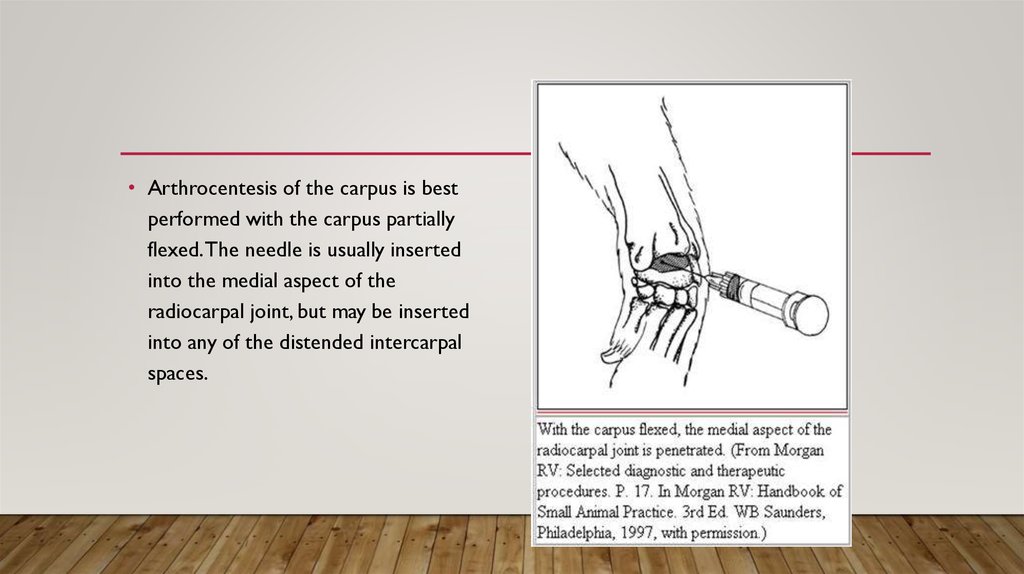
• Arthrocentesis of the carpus is bestperformed with the carpus partially
flexed. The needle is usually inserted
into the medial aspect of the
radiocarpal joint, but may be inserted
into any of the distended intercarpal
spaces.