


 biology
Similar presentations:
")


")
")
")
Pathologic Protozoa
1.
Pathologic Protozoa2.
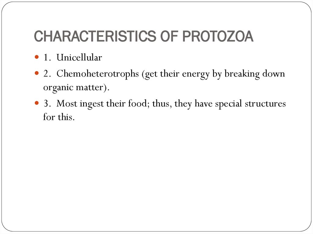
CHARACTERISTICS OF PROTOZOA1. Unicellular
2. Chemoheterotrophs (get their energy by breaking down
organic matter).
3. Most ingest their food; thus, they have special structures
for this.
3.
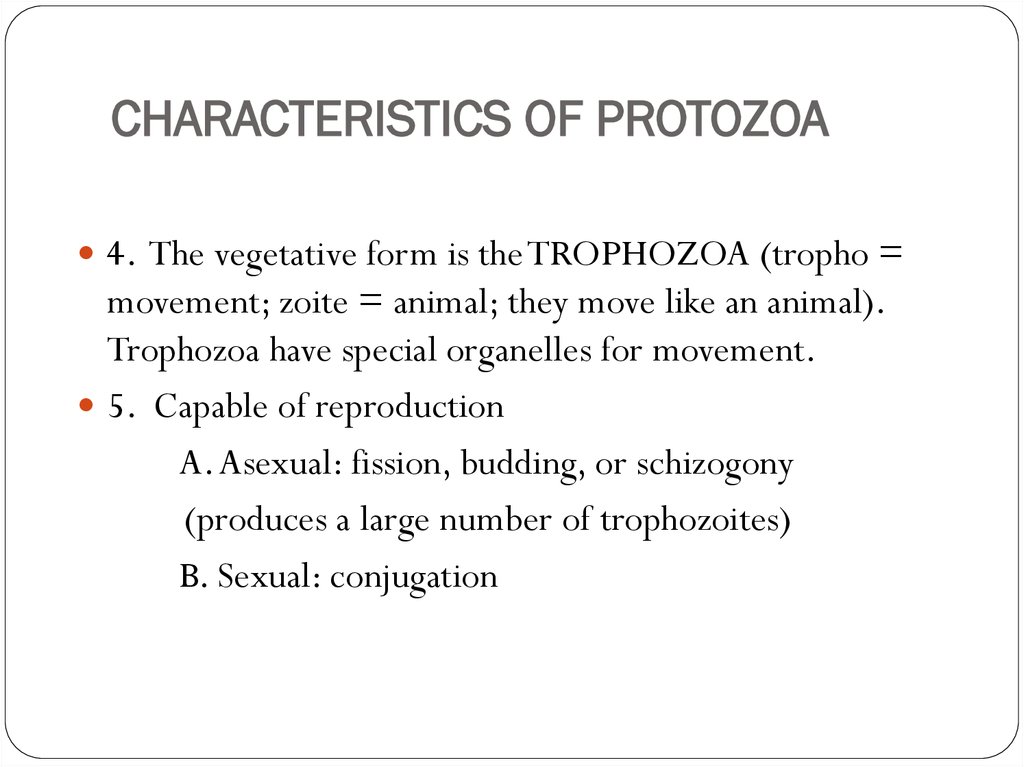
CHARACTERISTICS OF PROTOZOA4. The vegetative form is the TROPHOZOA (tropho =
movement; zoite = animal; they move like an animal).
Trophozoa have special organelles for movement.
5. Capable of reproduction
A. Asexual: fission, budding, or schizogony
(produces a large number of trophozoites)
B. Sexual: conjugation
4.
CHARACTERISTICS OF PROTOZOA6. Some produce cysts.
These are not tissue cysts like a human gets under their
skin; protozoa cysts are cellular.
They have a thick cell wall that allows for survival in
harsh environments better than the trophozoite form.
5.
PROTOZOA CYSTSCysts are not as resistant as a bacterial endospore.
You can kill cysts by boiling them.
They can live in the soil or water for months.
A cyst is no motile, so it is not trophozoic.
A cyst does not procure its nutrients or ingest food, but
it can absorb nutrients.
It has no organelles to ingest food.
6.
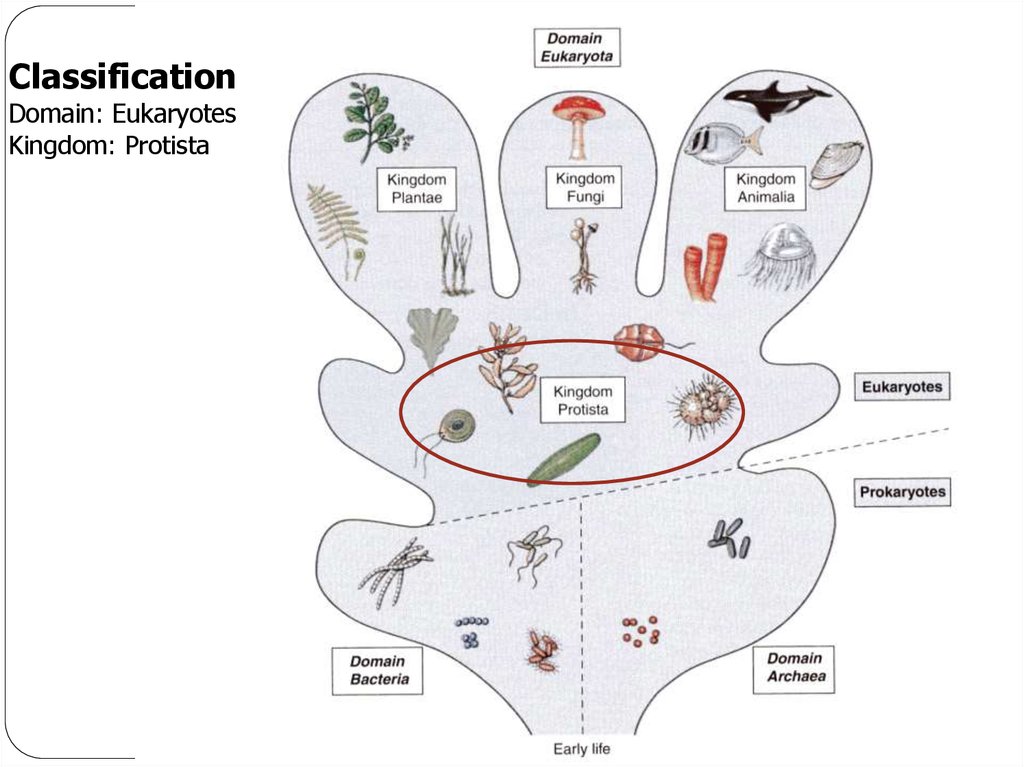
ClassificationDomain: Eukaryotes
Kingdom: Protista
7.
ClassificationTraditional classification of protozoa phylae was based on mode of
locomotion.
MASTIGOPHORA (flagella)
CILIOPHORA (cilia)
SARCODINA (amoebas)
SPOROZOA (spore-formers)
Apicomplexa (attachment organ)
8.
Modern ClassificationModern classification of protozoa is based on how they evolved and how
closely related they are (phylogenetic taxonomy), as determined by their
ribosomal RNA. The human pathogenic protozoa may be classified as
follows:
METAMONADA (multiple flagella with feeding grooves)
AMOEBOZOA (amoebas)
APICOMPLEXA (attachment organ)
CILIOPHORA (cilia)
EUGLENOZOA (flagella and disc-shaped cristae in mitochondria)
9.
EUGLENOZOAEUGLENOZOA (older classification = Mastigophora): has flagella
and its mitochondria have disc-shaped cristae
Organisms
Trypanosoma
Disease: Trypanosomiasis
Leishmania donovani
Disease: Leishmaniasis
10.
MASTIGOPHORA DISEASESTrypanosomiasis
Leishmaniasis
11.
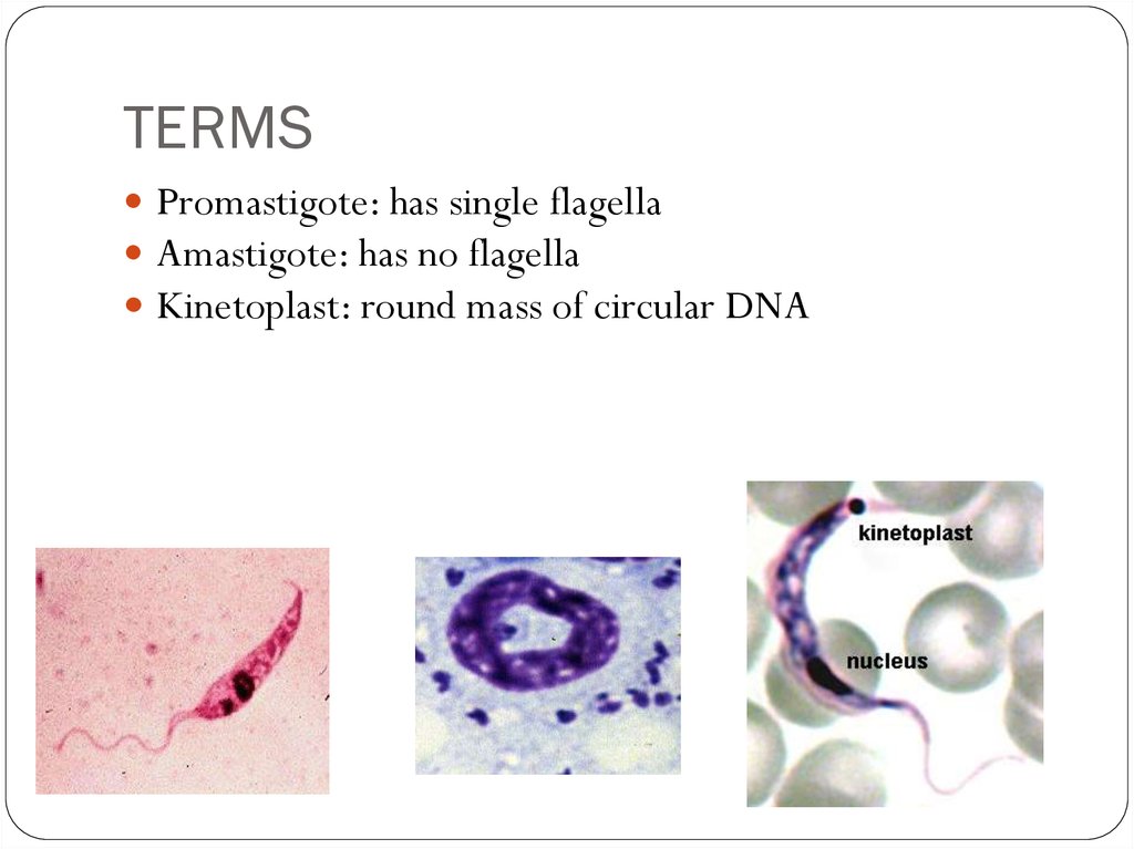
TERMSPromastigote: has single flagella
Amastigote: has no flagella
Kinetoplast: round mass of circular DNA
12.
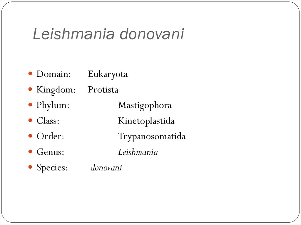
Leishmania donovaniDomain:
Kingdom:
Phylum:
Class:
Order:
Genus:
Species:
Eukaryota
Protista
Mastigophora
Kinetoplastida
Trypanosomatida
Leishmania
donovani
13.
Leishmania donovaniDisease: Leishmaniasis
Vector-borne disease transmitted by sandflies.
14.
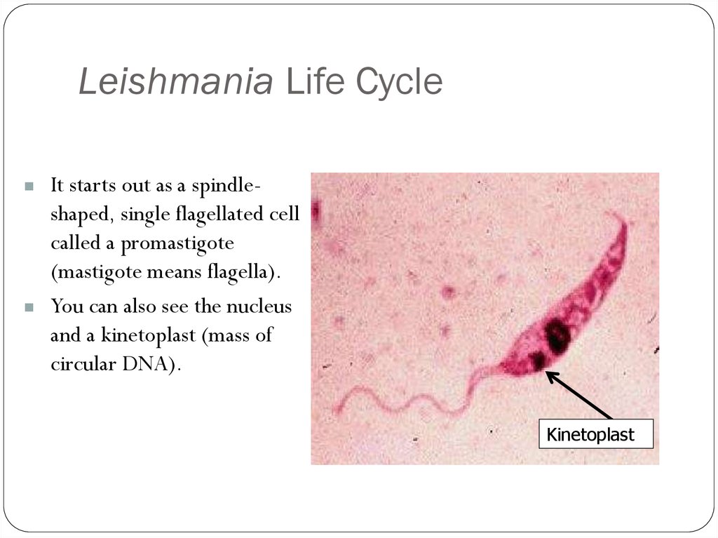
Leishmania Life CycleIt starts out as a spindleshaped, single flagellated cell
called a promastigote
(mastigote means flagella).
You can also see the nucleus
and a kinetoplast (mass of
circular DNA).
Kinetoplast
15.
Leishmania rosetteIn prepared slides you can see
promastigotes align their nose
in a circle, called a rosette.
16.
Leishmaniasis rosette17.
Leishmania Life CycleIt reproduces in the gut of a female sandfly, and migrates to her
proboscis (mouth part).
It is introduced into the human by her bite.
It then enters a macrophage and becomes intracellular.
Here, it loses its flagella and is now known as an amastigote.
18.
LeishmaniasisThese amastigotes multiply in various organs including
the spleen, liver, and lymph nodes.
Symptoms include lymph adenopathy, fever, weight loss,
and a decrease in all blood cells.
The treatment is almost as bad as the disease because of
the side effects. It is best to catch it early.
19.
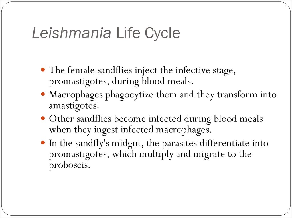
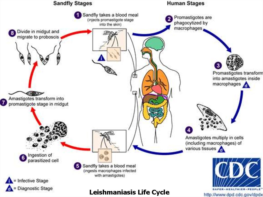
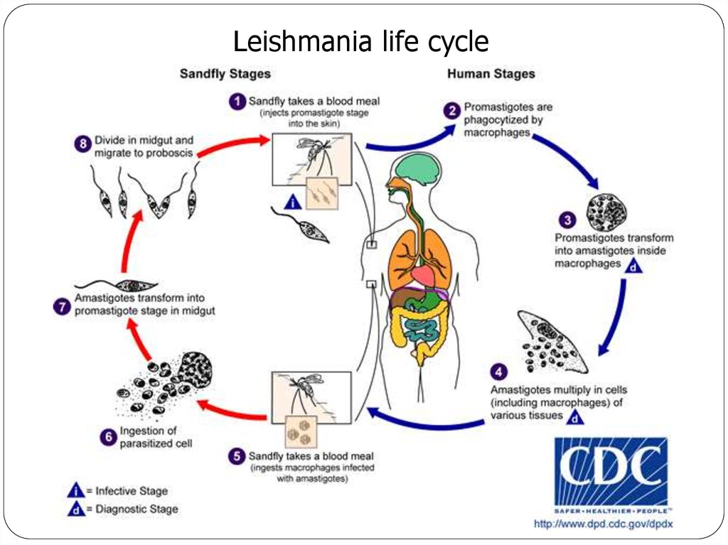
Leishmania Life CycleThe female sandflies inject the infective stage,
promastigotes, during blood meals.
Macrophages phagocytize them and they transform into
amastigotes.
Other sandflies become infected during blood meals
when they ingest infected macrophages.
In the sandfly's midgut, the parasites differentiate into
promastigotes, which multiply and migrate to the
proboscis.
20.
Leishmaniasis Life Cycle21.
Leishmania donovani(Promastigote)
Single flagellum found in sand flies
22.
LeishmaniasisMacrophage
rupturing
Amastogotes
Amastogotes with
nucleus and
kinetoplast
23.
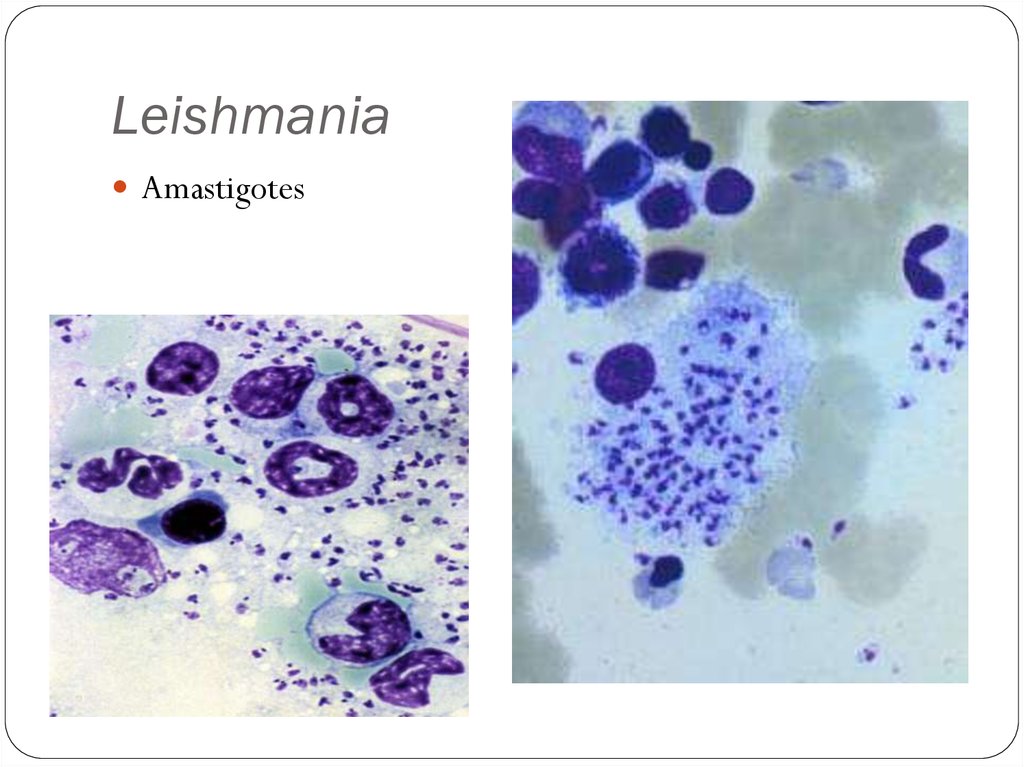
LeishmaniaAmastigotes
24.
SandflyThis looks like a
mosquito, except its
body is hairy and the
wings are feathery.
25.
LeishmaniasisGeographic Distribution:
More than 90 percent of the world's cases of visceral
leishmaniasis are in India, Bangladesh, Nepal, Sudan, and
Brazil.
Leishmaniasis is also found in Mexico, Central America,
and South America, southern Europe, Asia, the Middle
East, and Africa.
26.
LeishmaniasisThere are three forms of Leishmaniasis:
Cutaneous
Mucocutaneus
Visceral
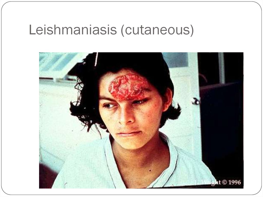
27.
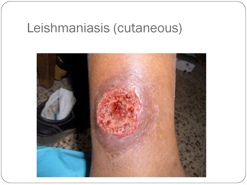
Cutaneous LeishmaniasisThe disease is only at the site of the bite.
This form is seen in Texas, Mexico, Asia, and the Middle East (our
Iraq troops are coming down with this form).
It manifests as a large, wet sore with raised edges. It looks like a
volcano with weepy serum coming out of the center.
The wound is not contagious, just the sandfly bite.
Dogs can get this disease, too.
28.
Leishmaniasis (cutaneous)29.
Leishmaniasis (cutaneous)30.
Leishmaniasis (cutaneous)31.
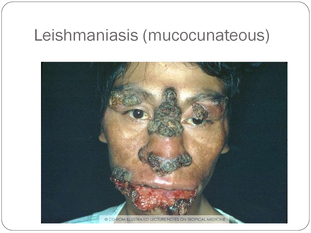
Leishmaniasis (mucocunateous)This is when the disease located in the mucous membranes of
the nose and mouth.
The most gruesome photos are of this form.
32.
Leishmaniasis (mucocunateous)33.
Leishmaniasis (visceral)This is the most serious form. It occurs especially in
immunocompromised people, especially HIV patients.
The amastagotes reproduce inside macrophages.
Only T-cells can kill infected macrophages, but HIV is a disease
that infects T-cells.
This form is known as Kala Azar.
34.
Kala AzarHepatosplenomegaly
35.
Kala Azar (duodenum)36.
Определите тип лейшманиозаА
Б
Visceral leishmaniosis
New World skin and mucous leishmaniosis
Г
В
Old World skin leishmaniosis
New World skin leishmaniosis
(also damages cartilage)
37.
Leishmania life cycle38.
TERMSMastigote = flagella
Promastigote: has single flagella
Amastigote: has no flagella
Kinetoplast: round mass of circular DNA
39.
TrypanosomiasisAfrican Trypanosomiasis
(African Sleeping Sickness)
American Trypanosomiasis
(Chaga’s Disease)
40.
“African Sleeping Sickness”Disease: African Tryptanosomiasis
Causal Agents:
Trypanosoma brucei gambiense
Trypanosoma brucei rhodesiense
41.
42.
Geographic DistributionT. b. gambiense is found in foci in large areas of West and
Central Africa.
Humans are the main reservoir for Trypanosoma brucei gambiense,
but this species can also be found in animals.
T. b. rhodesiense is found in East and Southeast Africa.
Wild game animals are the main reservoir of T. b. rhodesiense.
43.
TrypanosomiasisTrypanosomiasis has a biological vector, the tsetse
(pronounced “set-see”) fly.
Wild animals may also be a reservoir (Zooinotic is when a
disease is transmitted to animals as well as humans.)
44.
TrypanosomiasisThe tsetse fly bites a human and injects the
trypanomastigotes into the skin.
This causes a chanchre (pronounced “shanker”), which is an
ulcer on the skin.
Then it enters the lymphatic system.
45.
TrypanosomiasisIt is characterized by Winterbottom’s Sign: swelling of the
cervical lymph nodes in the head and neck area.
CNS symptoms include a shuffling gait (like a stroke victim),
slurred speech, and malaise (needing to sleep longer and
longer each day).
They are also restless at night.
46.
TrypanosomiasisCNS symptoms
Shuffling gait
Slurred speech
Malaise (sleeping all day)
Treatment
Melarsoprol: which has dangerous side-effects like chemostherapy. This
drug requires administration with a substance called ethylene glycol,
which will break down regular plastic tubing, so the drug must be
administered with special plastic iv tubing.
47.
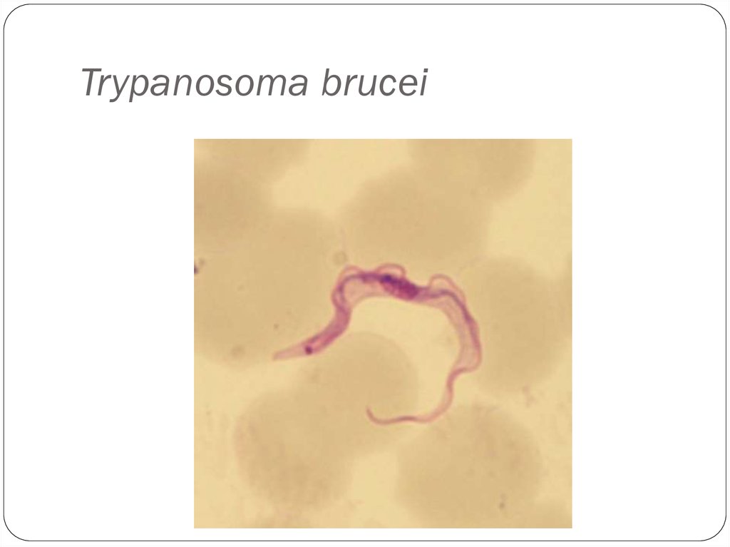
Trypanosoma bruceiTrypomastigote stages are the only form found
in patients.
Posterior kinetoplast
Centrally located nucleus
Undulating membrane
Anterior flagellum
48.
Trypanosoma brucei49.
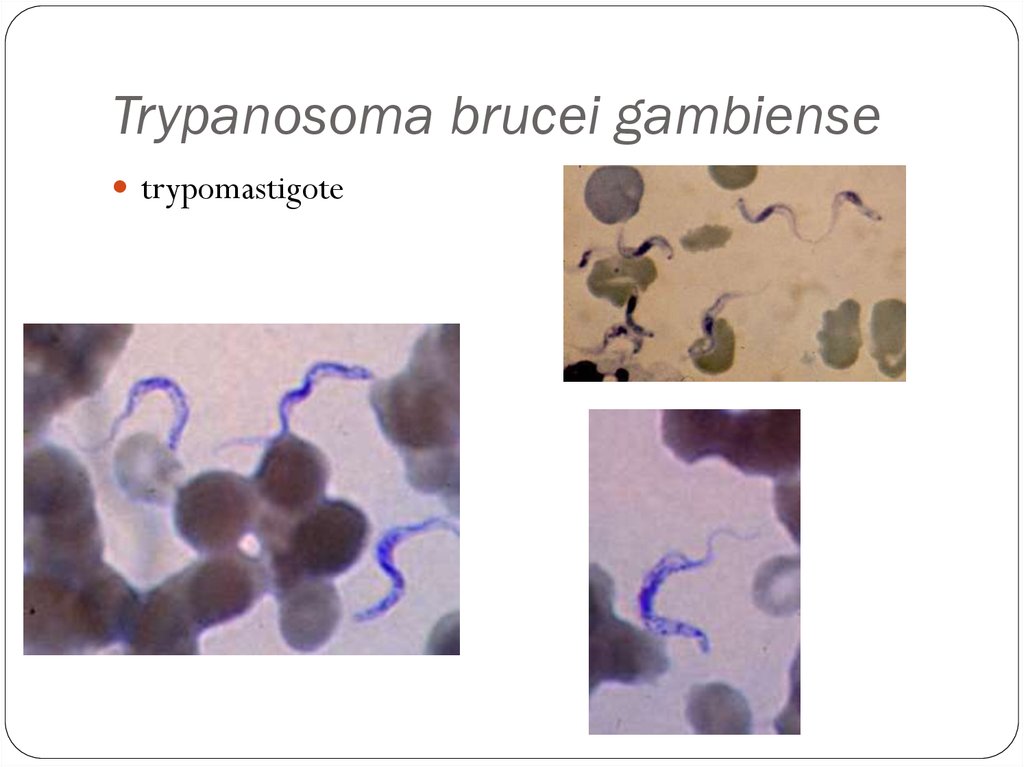
Trypanosoma brucei gambiensetrypomastigote
50.
Trypanosoma brucei rhodesiense51.
Tsetse Fly52.
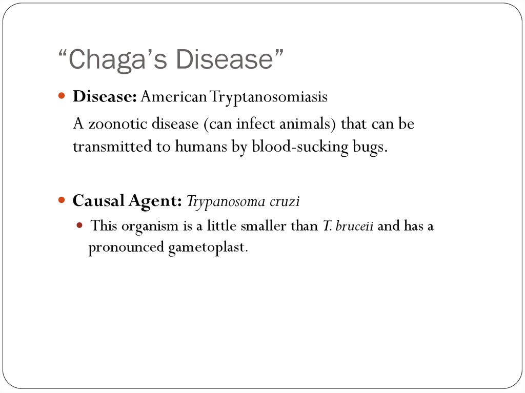
“Chaga’s Disease”Disease: American Tryptanosomiasis
A zoonotic disease (can infect animals) that can be
transmitted to humans by blood-sucking bugs.
Causal Agent: Trypanosoma cruzi
This organism is a little smaller than T. bruceii and has a
pronounced gametoplast.
53.
“Chaga’s Disease”This disease is NOT found in Africa.
This disease is also zoonotic; it can infect animals as well as
humans.
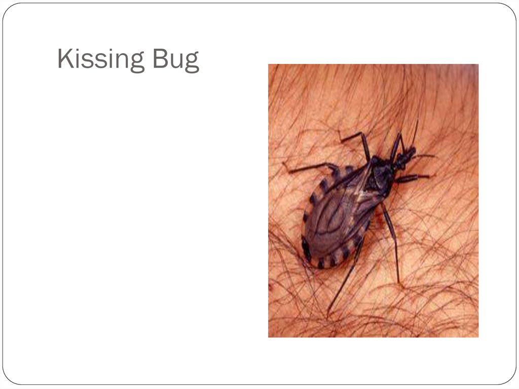
The vector is a large bug called the “Kissing Bug”.
It is found in warm regions and crowded areas, especially in the
cracks of adobe huts.
It comes out at night and crawls on a human while they sleep.
54.
“Chaga’s Disease”It prefers the lips because the blood supply is close to the surface.
It sucks the blood there, but they don’t transmit the organism this
way.
When they suck the blood, they also defecate, and the organism is
in the feces.
When the human wakes up to scratch the itch, feces get into the
tiny wound.
This is a fecal blood route.
55.
“Chaga’s Disease”Symptoms include fever, anorexia, swollen lymph nodes,
hepatosplenomegally (enlarged liver and spleen), and myocarditis
(inflammation of the heart), which usually causes death.
They also have megacolon (large colon) and megaesophagus (large
esophagus).
56.
57.
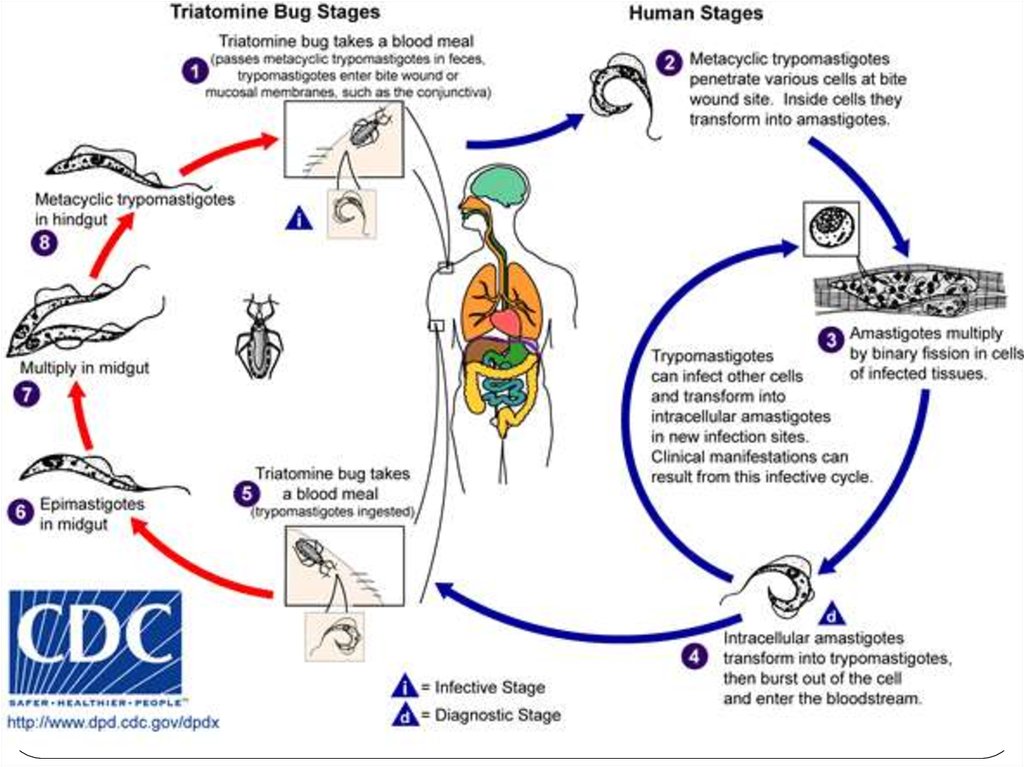
Trypanosoma cruziInsect vector is the “kissing” bug. It takes a blood meal
and releases trypomastigotes in its feces near the site of
the bite wound.
Trypomastigotes enter the host through the wound or
through intact mucosal membranes, such as the
conjunctiva.
Trypanosoma cruzi can also be transmitted through blood
transfusions, organ transplantation, transplacentally, and
in laboratory accidents.
58.
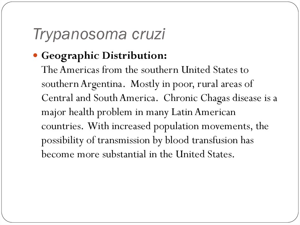
Trypanosoma cruziGeographic Distribution:
The Americas from the southern United States to
southern Argentina. Mostly in poor, rural areas of
Central and South America. Chronic Chagas disease is a
major health problem in many Latin American
countries. With increased population movements, the
possibility of transmission by blood transfusion has
become more substantial in the United States.
59.
Trypanosoma cruzi60.
Trypanosoma cruzi61.
Trypanosoma cruzilarge kinetoplast
62.
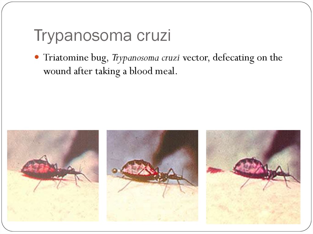
Trypanosoma cruziTriatomine bug, Trypanosoma cruzi vector, defecating on the
wound after taking a blood meal.
63.
Kissing Bug64.
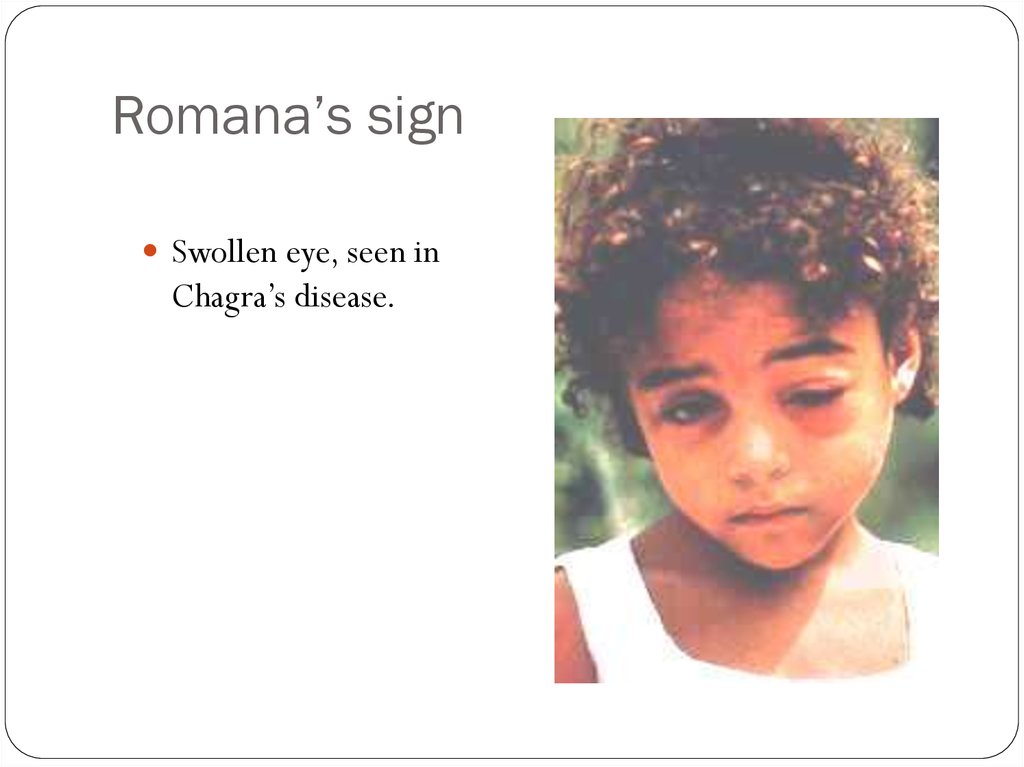
Romana’s signSwollen eye, seen in
Chagra’s disease.