.")

 biology
Similar presentations:


Мм. головы
1. Мышцы головы (musculi capitis).
Мимические мышцы (mm. faciales)Располагаются под кожей головы и
не покрыты фасцией. Большинство из
них начинаются на костях черепа и
прикрепляются к коже головы (т.е.
двигают кожу головы). Выделяют:
Жевательные мышцы
Осуществляют
движение
в
височнонижнечелюстном суставе, обеспечивая жевание,
глотание, зевание, речь. Начинаются на костях
черепа, а прикрепляются к нижней челюсти (т.е.
двигают н. ч.). Выделяют :
1. Мышцы свода черепа
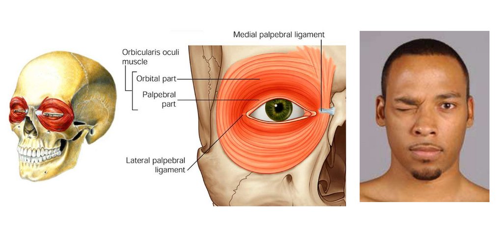
2. Мышцы, окружающие глаз
3. Мышцы, окружающие нос
4. Мышцы, окружающие рот
1. Жевательная м. (m. masseter)
2. Височная м. (m. temporalis)
3. Медиальная крыловидная м. (m. pterygoideus medialis)
4. Латеральная крыловидная м(m. pterygoideus lateralis)
2.
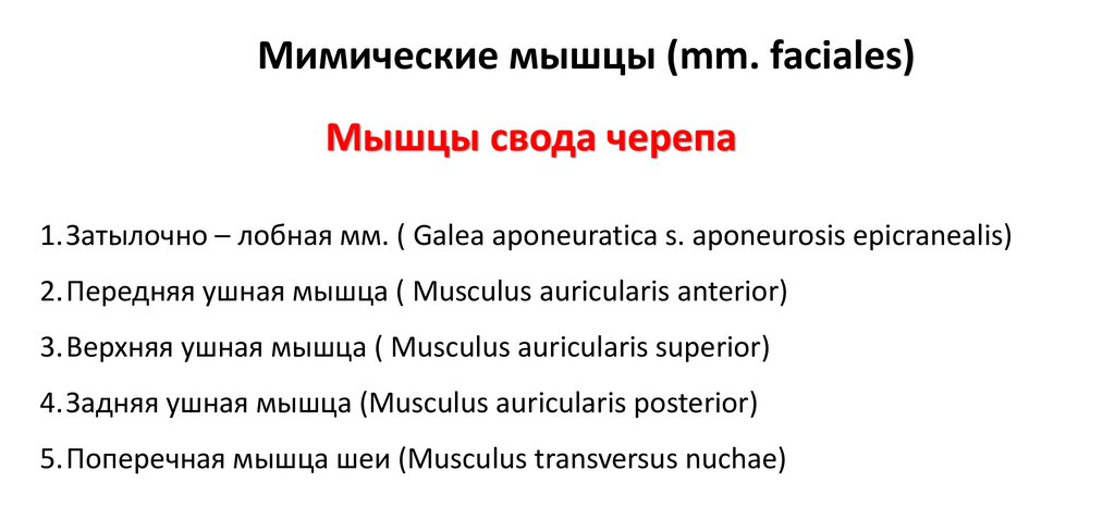
Мимические мышцы (mm. faciales)Мышцы свода черепа
1.Затылочно – лобная мм. ( Galea aponeuratica s. aponeurosis epicranealis)
2.Передняя ушная мышца ( Musculus auricularis anterior)
3.Верхняя ушная мышца ( Musculus auricularis superior)
4.Задняя ушная мышца (Musculus auricularis posterior)
5.Поперечная мышца шеи (Musculus transversus nuchae)
3.
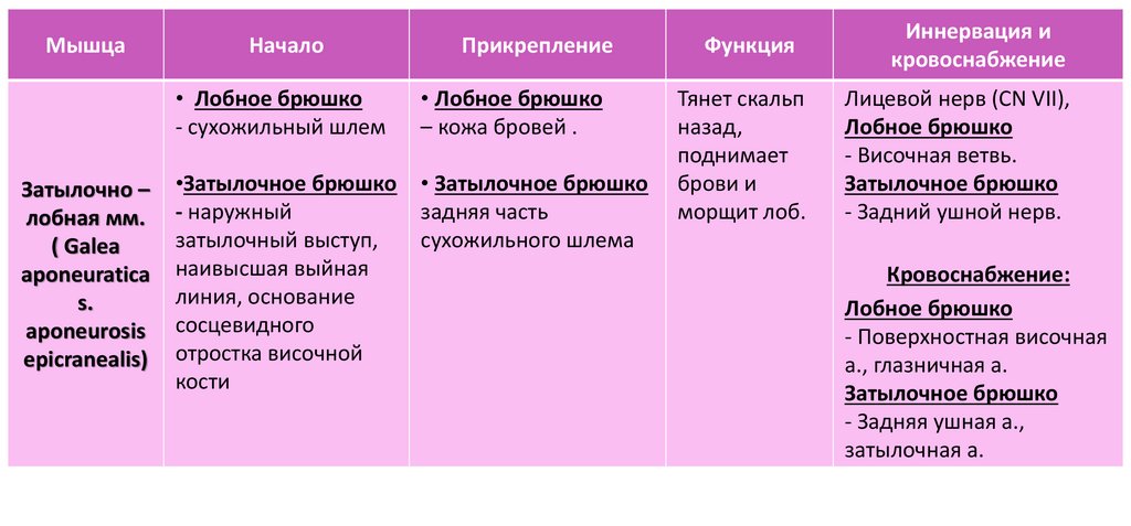
МышцаЗатылочно –
лобная мм.
( Galea
aponeuratica
s.
aponeurosis
epicranealis)
Прикрепление
Функция
Иннервация и
кровоснабжение
• Лобное брюшко
- сухожильный шлем
• Лобное брюшко
– кожа бровей .
•Затылочное брюшко
- наружный
затылочный выступ,
наивысшая выйная
линия, основание
сосцевидного
отростка височной
кости
• Затылочное брюшко
задняя часть
сухожильного шлема
Тянет скальп
назад,
поднимает
брови и
морщит лоб.
Лицевой нерв (CN VII),
Лобное брюшко
- Височная ветвь.
Затылочное брюшко
- Задний ушной нерв.
Начало
Кровоснабжение:
Лобное брюшко
- Поверхностная височная
а., глазничная а.
Затылочное брюшко
- Задняя ушная а.,
затылочная а.
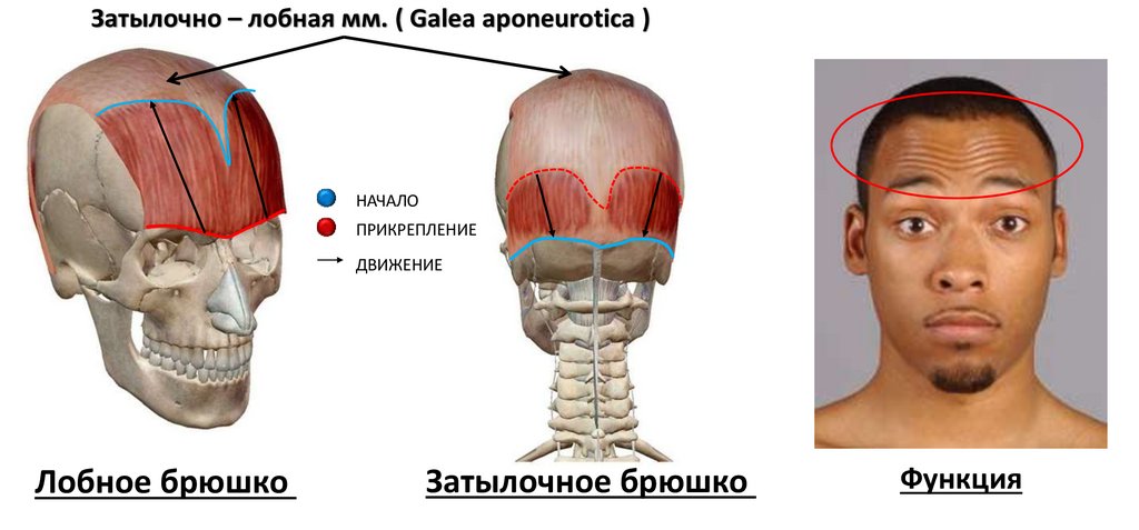
4.
Затылочно – лобная мм. ( Galea aponeurotica )НАЧАЛО
ПРИКРЕПЛЕНИЕ
ДВИЖЕНИЕ
Лобное брюшко
Затылочное брюшко
Функция
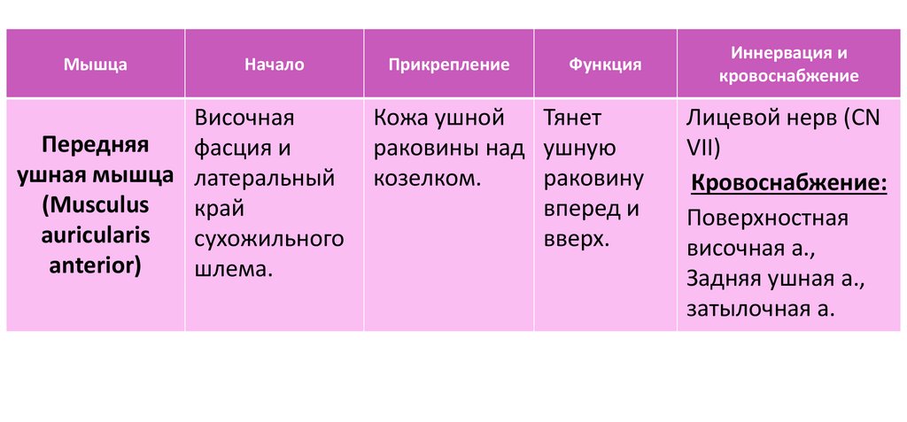
5.
МышцаНачало
Височная
Передняя
фасция и
ушная мышца латеральный
(Musculus
край
auricularis
сухожильного
anterior)
шлема.
Прикрепление
Функция
Кожа ушной
Тянет
раковины над ушную
козелком.
раковину
вперед и
вверх.
Иннервация и
кровоснабжение
Лицевой нерв (CN
VII)
Кровоснабжение:
Поверхностная
височная а.,
Задняя ушная а.,
затылочная а.
6.
МышцаНачало
Прикрепление
Функция
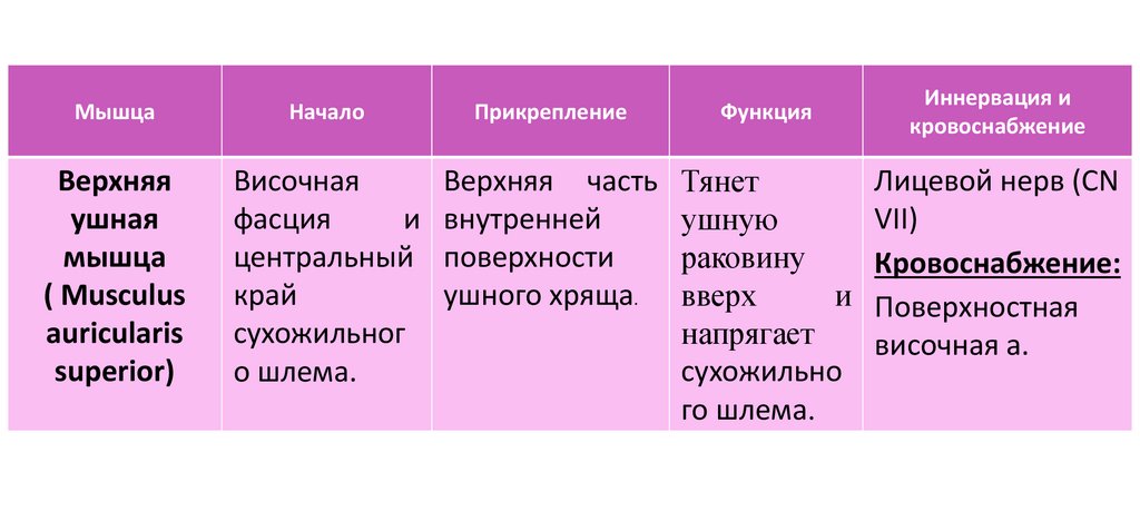
Верхняя
ушная
мышца
( Musculus
auricularis
superior)
Височная
фасция
и
центральный
край
сухожильног
о шлема.
Верхняя часть
внутренней
поверхности
ушного хряща.
Тянет
ушную
раковину
вверх
и
напрягает
сухожильно
го шлема.
Иннервация и
кровоснабжение
Лицевой нерв (CN
VII)
Кровоснабжение:
Поверхностная
височная а.
7.
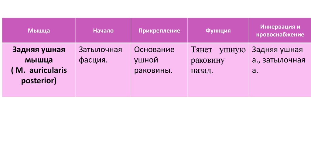
МышцаНачало
Задняя ушная
мышца
( M. auricularis
posterior)
Затылочная
фасция.
Прикрепление
Основание
ушной
раковины.
Функция
Иннервация и
кровоснабжение
Тянет ушную Задняя ушная
а., затылочная
раковину
а.
назад.
8.
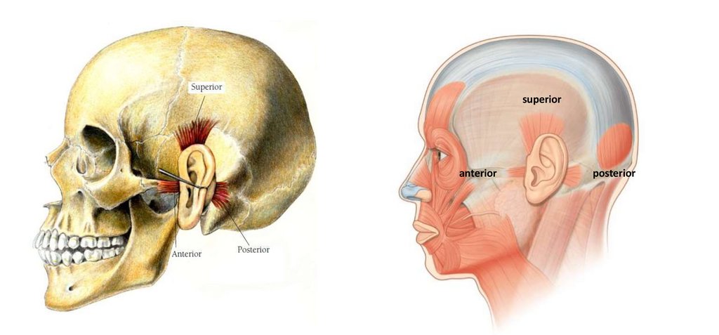
superioranterior
posterior
9.
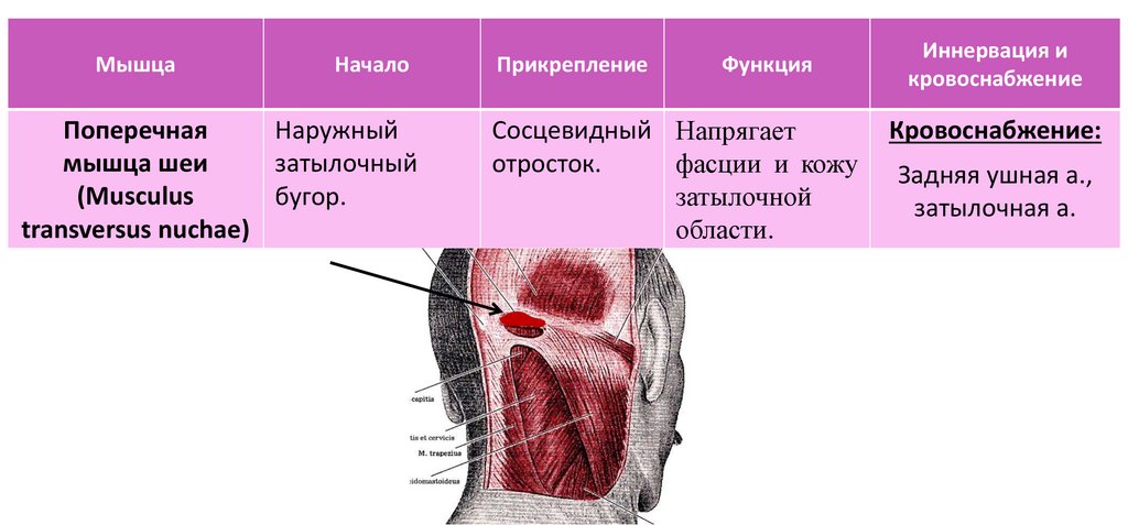
МышцаНачало
Поперечная
Наружный
мышца шеи
затылочный
(Musculus
бугор.
transversus nuchae)
Прикрепление
Функция
Сосцевидный Напрягает
отросток.
фасции и кожу
затылочной
области.
Иннервация и
кровоснабжение
Кровоснабжение:
Задняя ушная а.,
затылочная а.
10.
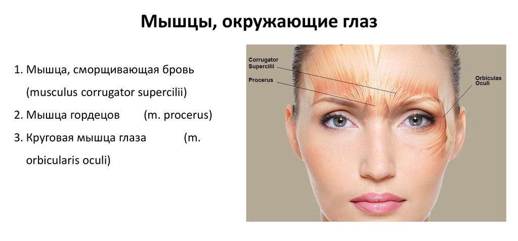
Мышцы, окружающие глаз1. Мышца, сморщивающая бровь
(musculus corrugator supercilii)
2. Мышца гордецов
(m. procerus)
3. Круговая мышца глаза
orbicularis oculi)
(m.
11.
МышцаНачало
Прикрепление
Функция
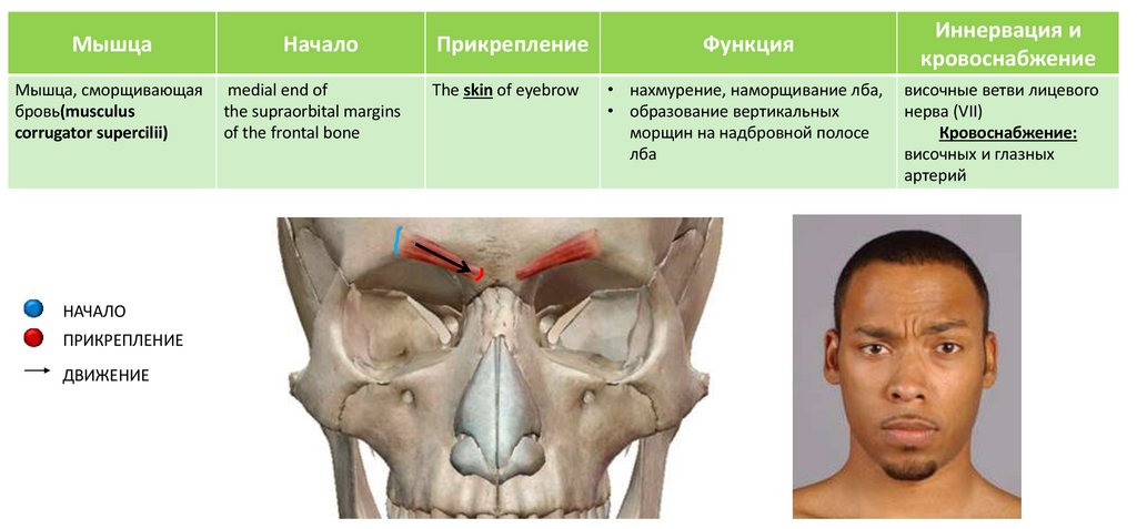
Мышца, сморщивающая
бровь(musculus
corrugator supercilii)
medial end of
the supraorbital margins
of the frontal bone
The skin of eyebrow
• нахмурение, наморщивание лба,
• образование вертикальных
морщин на надбровной полосе
лба
НАЧАЛО
ПРИКРЕПЛЕНИЕ
ДВИЖЕНИЕ
Иннервация и
кровоснабжение
височные ветви лицевого
нерва (VII)
Кровоснабжение:
височных и глазных
артерий
12.
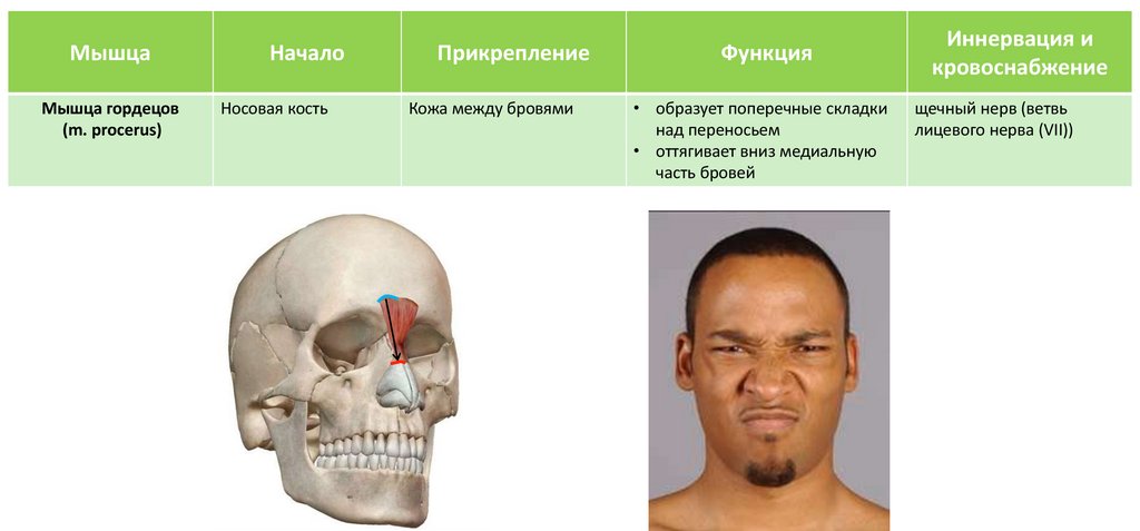
МышцаМышца гордецов
(m. procerus)
Начало
Носовая кость
Прикрепление
Кожа между бровями
Функция
• образует поперечные складки
над переносьем
• оттягивает вниз медиальную
часть бровей
Иннервация и
кровоснабжение
щечный нерв (ветвь
лицевого нерва (VII))
13.
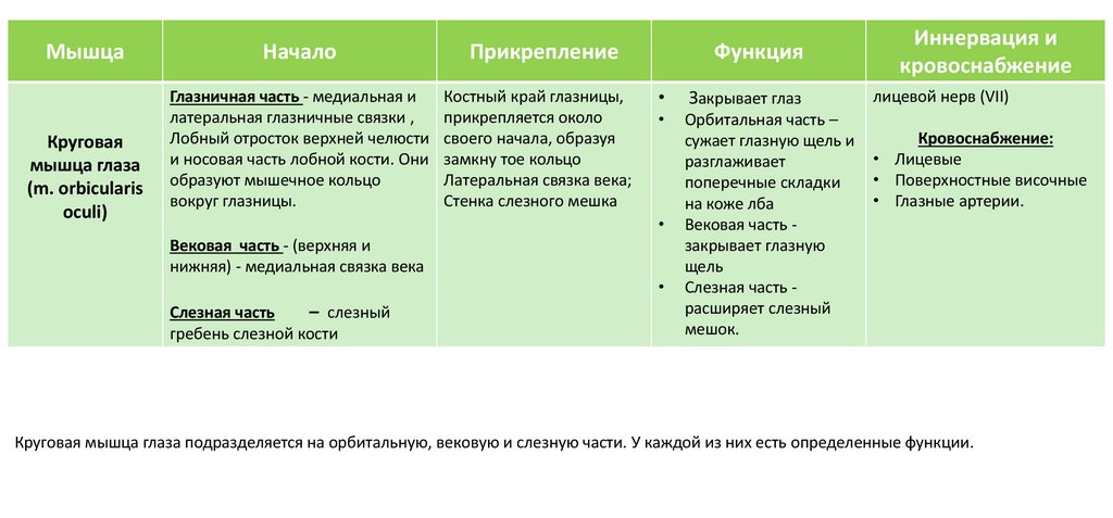
МышцаКруговая
мышца глаза
(m. orbicularis
oculi)
Начало
Прикрепление
Глазничная часть - медиальная и
латеральная глазничные связки ,
Лобный отросток верхней челюсти
и носовая часть лобной кости. Они
образуют мышечное кольцо
вокруг глазницы.
Костный край глазницы,
прикрепляется около
своего начала, образуя
замкну тое кольцо
Латеральная связка века;
Стенка слезного мешка
Функция
Закрывает глаз
Орбитальная часть –
сужает глазную щель и
разглаживает
поперечные складки
на коже лба
Вековая часть закрывает глазную
щель
Слезная часть расширяет слезный
мешок.
Вековая часть - (верхняя и
нижняя) - медиальная связка века
Слезная часть
– слезный
гребень слезной кости
Иннервация и
кровоснабжение
лицевой нерв (VII)
Кровоснабжение:
• Лицевые
• Поверхностные височные
• Глазные артерии.
Круговая мышца глаза подразделяется на орбитальную, вековую и слезную части. У каждой из них есть определенные функции.
14.
15.
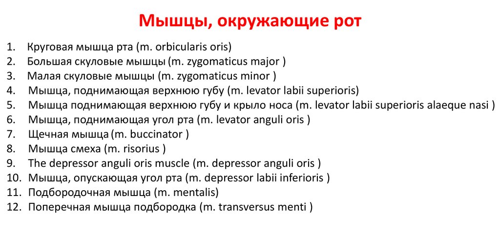
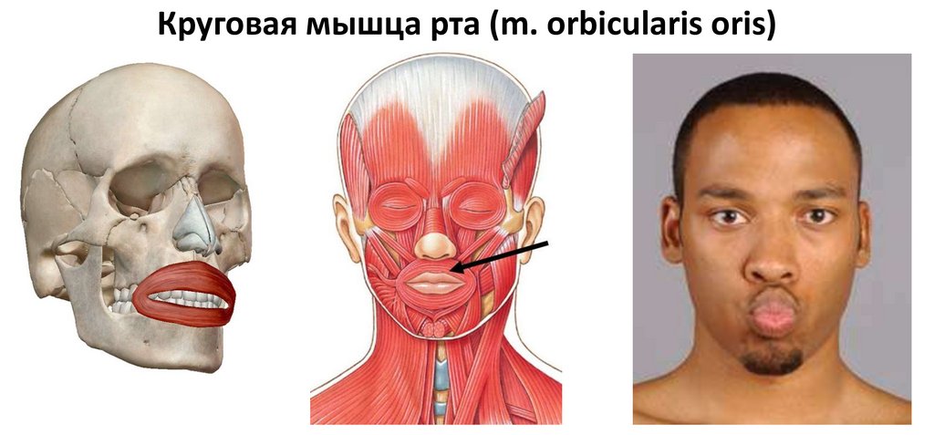
Мышцы, окружающие рот1. Круговая мышца рта (m. orbicularis oris)
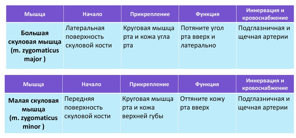
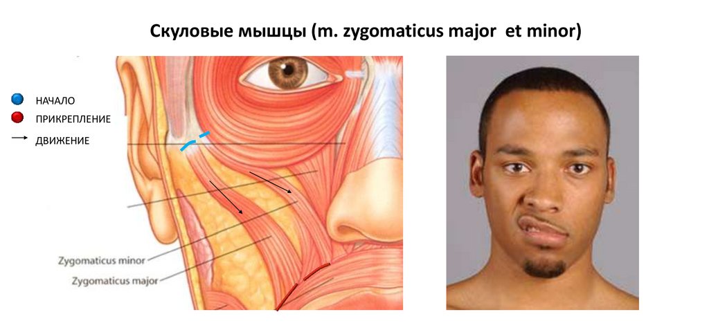
2. Большая скуловые мышцы (m. zygomaticus major )
3. Малая скуловые мышцы (m. zygomaticus minor )
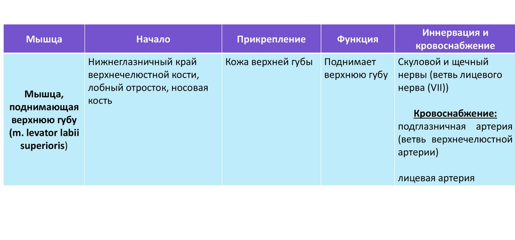
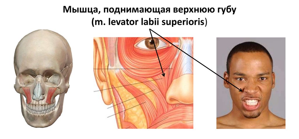
4. Мышца, поднимающая верхнюю губу (m. levator labii superioris)
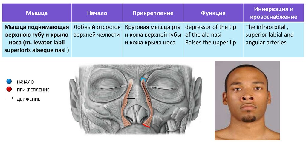
5. Мышца поднимающая верхнюю губу и крыло носа (m. levator labii superioris alaeque nasi )
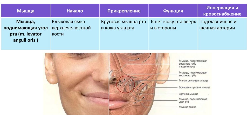
6. Мышца, поднимающая угол рта (m. levator anguli oris )
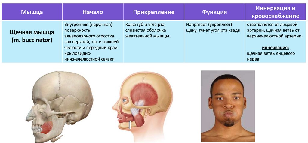
7. Щечная мышца (m. buccinator )
8. Мышца смеха (m. risorius )
9. The depressor anguli oris muscle (m. depressor anguli oris )
10. Мышца, опускающая угол рта (m. depressor labii inferioris )
11. Подбородочная мышца (m. mentalis)
12. Поперечная мышца подбородка (m. transversus menti )
16.
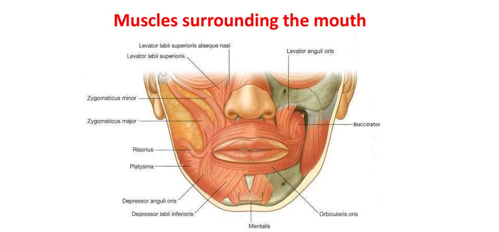
Muscles surrounding the mouth17.
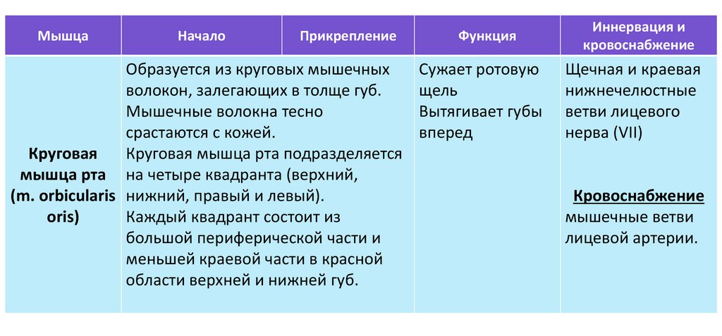
МышцаНачало
Прикрепление
Образуется из круговых мышечных
волокон, залегающих в толще губ.
Мышечные волокна тесно
срастаются с кожей.
Круговая
Круговая мышца рта подразделяется
мышца рта на четыре квадранта (верхний,
(m. orbicularis нижний, правый и левый).
oris)
Каждый квадрант состоит из
большой периферической части и
меньшей краевой части в красной
области верхней и нижней губ.
Функция
Иннервация и
кровоснабжение
Сужает ротовую
щель
Вытягивает губы
вперед
Щечная и краевая
нижнечелюстные
ветви лицевого
нерва (VII)
Кровоснабжение
мышечные ветви
лицевой артерии.
18.
Круговая мышца рта (m. orbicularis oris)19.
МышцаНачало
Латеральная
Большая
поверхность
скуловая мышца
скуловой кости
(m. zygomaticus
major )
Мышца
Начало
Малая скуловая Передняя
поверхность
мышца
(m. zygomaticus скуловой кости
minor )
Прикрепление
Функция
Круговая мышца Потяните угол
рта и кожа угла
рта вверх и
рта
латерально
Прикрепление
Функция
Круговая мышца Оттяните кожу
рта и кожа
рта вверх
верхней губы
Иннервация и
кровоснабжение
Подглазничная и
щечная артерии
Иннервация и
кровоснабжение
Подглазничная и
щечная артерии
20.
Скуловые мышцы (m. zygomaticus major et minor)НАЧАЛО
ПРИКРЕПЛЕНИЕ
ДВИЖЕНИЕ
21.
МышцаМышца,
поднимающая
верхнюю губу
(m. levator labii
superioris)
Начало
Прикрепление
Нижнеглазничный край
верхнечелюстной кости,
лобный отросток, носовая
кость
Кожа верхней губы
Иннервация и
Функция
кровоснабжение
Поднимает
Скуловой и щечный
верхнюю губу нервы (ветвь лицевого
нерва (VII))
Кровоснабжение:
подглазничная артерия
(ветвь верхнечелюстной
артерии)
лицевая артерия
22.
Мышца, поднимающая верхнюю губу(m. levator labii superioris)
23.
МышцаНачало
Прикрепление
Функция
Мышца поднимающая Лобный отросток Круговая мышца рта depressor of the tip
верхнюю губу и крыло верхней челюсти и кожа верхней губы of the ala nasi
носа (m. levator labii
и кожа крыла носа
Raises the upper lip
superioris alaeque nasi )
НАЧАЛО
ПРИКРЕПЛЕНИЕ
ДВИЖЕНИЕ
Иннервация и
кровоснабжение
The infraorbital ,
superior labial and
angular arteries
24.
МышцаНачало
Мышца,
Клыковая ямка
поднимающая угол верхнечелюстной
рта (m. levator
кости
anguli oris )
Иннервация и
кровоснабжение
Круговая мышца рта Тянет кожу рта вверх Подглазничная и
и кожа угла рта
и в стороны.
щечная артерии
Прикрепление
Функция
25.
МышцаЩечная мышца
(m. buccinator)
Начало
Внутренняя (наружная)
поверхность
альвеолярного отростка
как верхней, так и нижней
челюсти и передний край
крыловиднонижнечелюстной связки
Прикрепление
Кожа губ и угла рта,
слизистая оболочка
жевательной мышцы.
Функция
Напрягает (укрепляет)
щеку, тянет угол рта кзади
Иннервация и
кровоснабжение
ответвляется от лицевой
артерии, щечная ветвь от
верхнечелюстной артерии.
иннервация:
щечная ветвь лицевого
нерва
26.
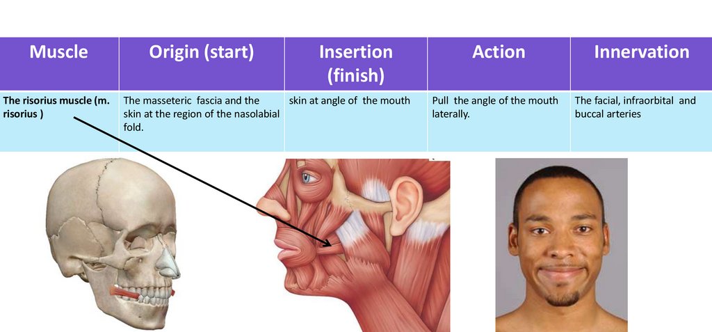
MuscleThe risorius muscle (m.
risorius )
Origin (start)
Insertion
(finish)
The masseteric fascia and the
skin at angle of the mouth
skin at the region of the nasolabial
fold.
Action
Innervation
Pull the angle of the mouth
laterally.
The facial, infraorbital and
buccal arteries
27.
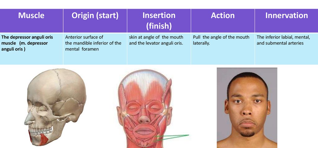
MuscleThe depressor anguli oris
muscle (m. depressor
anguli oris )
Origin (start)
Insertion
(finish)
Action
Innervation
Anterior surface of
the mandible inferior of the
mental foramen
skin at angle of the mouth
and the levator anguli oris.
Pull the angle of the mouth
laterally.
The inferior labial, mental,
and submental arteries
28.
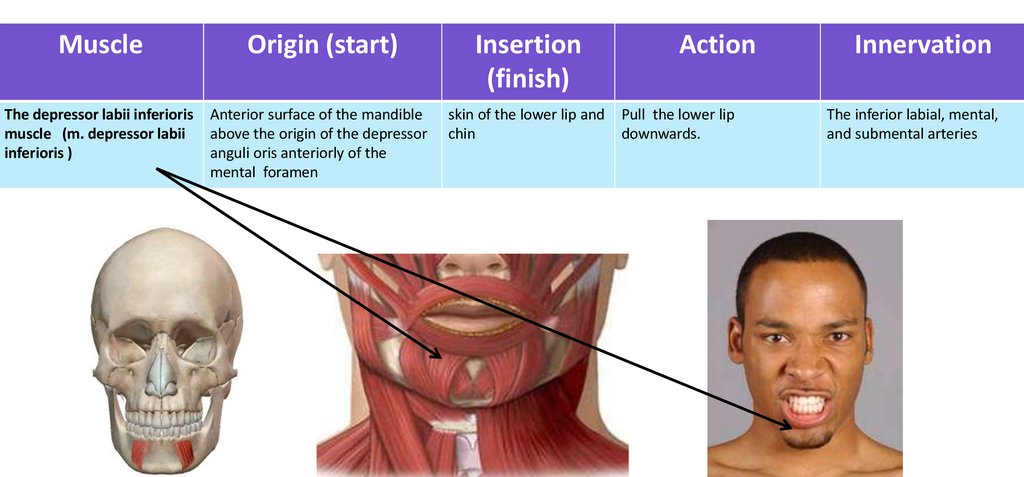
MuscleOrigin (start)
Insertion
(finish)
The depressor labii inferioris
muscle (m. depressor labii
inferioris )
Anterior surface of the mandible
above the origin of the depressor
anguli oris anteriorly of the
mental foramen
skin of the lower lip and
chin
Action
Pull the lower lip
downwards.
Innervation
The inferior labial, mental,
and submental arteries
29.
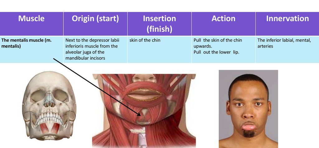
MuscleThe mentalis muscle (m.
mentalis)
Origin (start)
Next to the depressor labii
inferioris muscle from the
alveolar juga of the
mandibular incisors
Insertion
(finish)
skin of the chin
Action
Pull the skin of the chin
upwards.
Pull out the lower lip.
Innervation
The inferior labial, mental,
arteries
30.
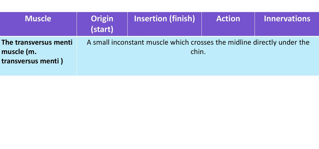
MuscleThe transversus menti
muscle (m.
transversus menti )
Origin
(start)
Insertion (finish)
Action
Innervations
A small inconstant muscle which crosses the midline directly under the
chin.
31.
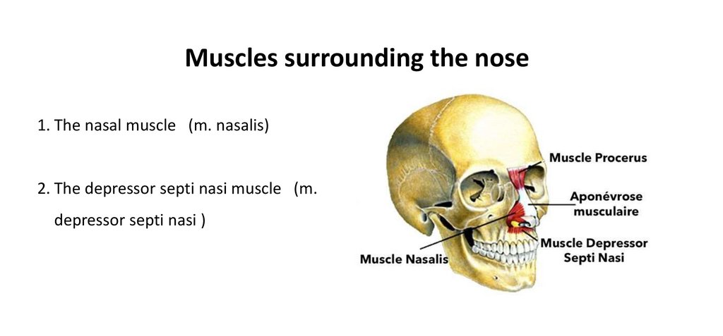
Muscles surrounding the nose1. The nasal muscle (m. nasalis)
2. The depressor septi nasi muscle (m.
depressor septi nasi )
32.
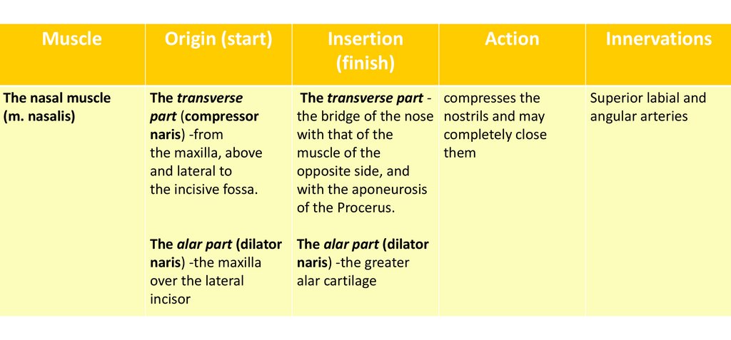
MuscleOrigin (start)
The nasal muscle
(m. nasalis)
The transverse
part (compressor
naris) -from
the maxilla, above
and lateral to
the incisive fossa.
Insertion
(finish)
Action
Innervations
The transverse part - compresses the
the bridge of the nose nostrils and may
with that of the
completely close
muscle of the
them
opposite side, and
with the aponeurosis
of the Procerus.
Superior labial and
angular arteries
The alar part (dilator The alar part (dilator
naris) -the maxilla
naris) -the greater
over the lateral
alar cartilage
incisor
33.
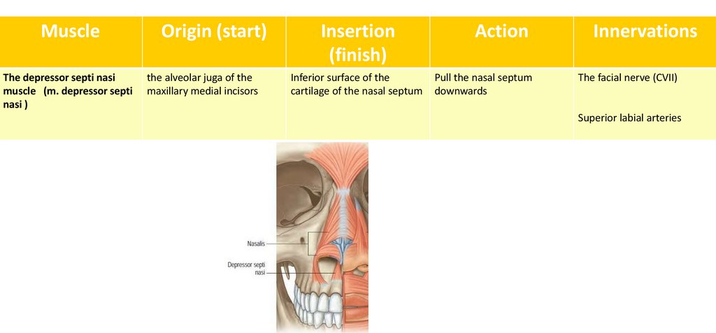
MuscleThe depressor septi nasi
muscle (m. depressor septi
nasi )
Origin (start)
the alveolar juga of the
maxillary medial incisors
Insertion
(finish)
Inferior surface of the
cartilage of the nasal septum
Action
Pull the nasal septum
downwards
Innervations
The facial nerve (CVII)
Superior labial arteries
34.
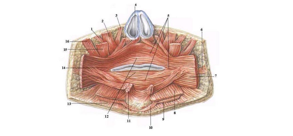
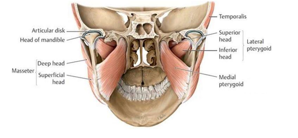
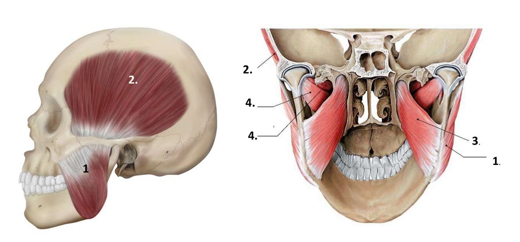
35. The muscles of mastication
1. The masseter muscle (m. masseter)2. The temporal muscle (m. temporalis)
3. The medial pterygoid muscle (m. pterygoideus medialis)
4. The lateral pterygoid muscle (m. pterygoideus lateralis)
36.
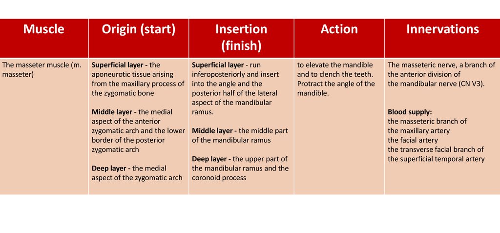
MuscleOrigin (start)
The masseter muscle (m.
masseter)
Superficial layer - the
aponeurotic tissue arising
from the maxillary process of
the zygomatic bone
Insertion
(finish)
Superficial layer - run
inferoposteriorly and insert
into the angle and the
posterior half of the lateral
aspect of the mandibular
ramus.
Middle layer - the medial
aspect of the anterior
zygomatic arch and the lower Middle layer - the middle part
border of the posterior
of the mandibular ramus
zygomatic arch
Deep layer - the upper part of
Deep layer - the medial
the mandibular ramus and the
aspect of the zygomatic arch coronoid process
Action
Innervations
to elevate the mandible
and to clench the teeth.
Protract the angle of the
mandible.
The masseteric nerve, a branch of
the anterior division of
the mandibular nerve (CN V3).
Blood supply:
the masseteric branch of
the maxillary artery
the facial artery
the transverse facial branch of
the superficial temporal artery
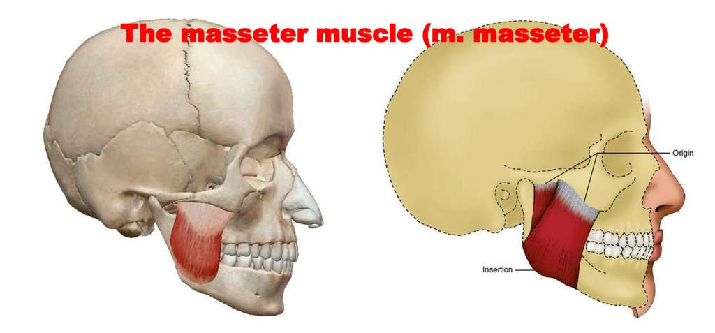
37.
The masseter muscle (m. masseter)38.
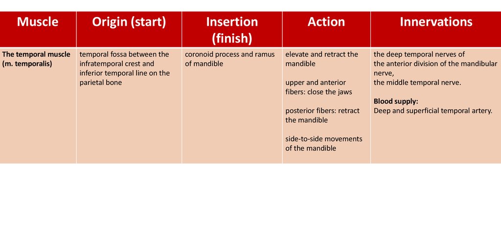
MuscleOrigin (start)
Insertion
(finish)
Action
Innervations
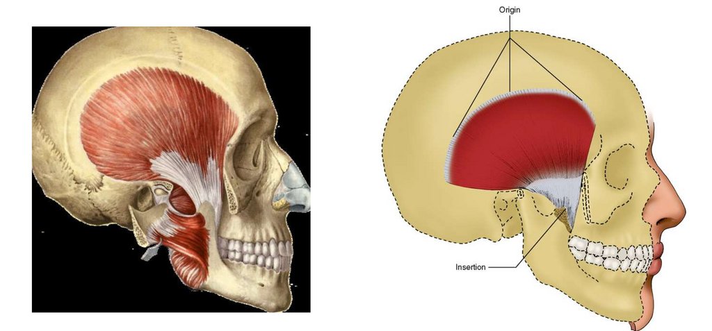
The temporal muscle
(m. temporalis)
temporal fossa between the
infratemporal crest and
inferior temporal line on the
parietal bone
coronoid process and ramus
of mandible
elevate and retract the
mandible
the deep temporal nerves of
the anterior division of the mandibular
nerve,
the middle temporal nerve.
upper and anterior
fibers: close the jaws
posterior fibers: retract
the mandible
side-to-side movements
of the mandible
Blood supply:
Deep and superficial temporal artery.
39.
40.
MuscleOrigin (start)
Insertion
(finish)
Action
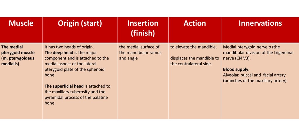
The medial
pterygoid muscle
(m. pterygoideus
medialis)
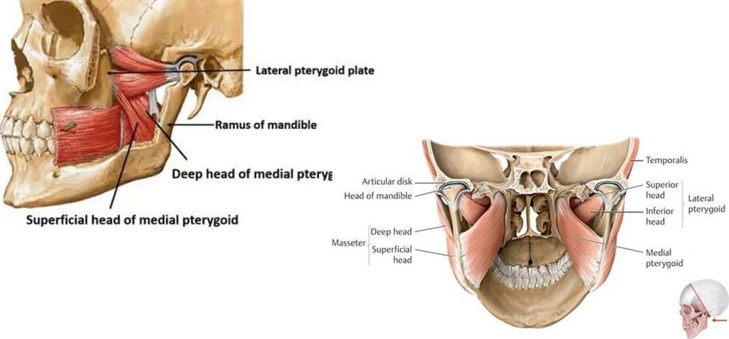
It has two heads of origin.
The deep head is the major
component and is attached to the
medial aspect of the lateral
pterygoid plate of the sphenoid
bone.
the medial surface of
the mandibular ramus
and angle
to elevate the mandible.
The superficial head is attached to
the maxillary tuberosity and the
pyramidal process of the palatine
bone.
Innervations
Medial pterygoid nerve o (the
mandibular division of the trigeminal
displaces the mandible to nerve (CN V3).
the contralateral side.
Blood supply:
Alveolar, buccal and facial artery
(branches of the maxillary artery).
41.
42.
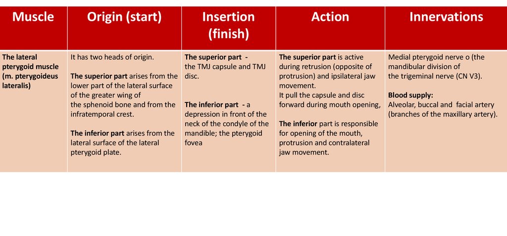
MuscleThe lateral
pterygoid muscle
(m. pterygoideus
lateralis)
Origin (start)
It has two heads of origin.
Insertion
(finish)
The superior part the TMJ capsule and TMJ
The superior part arises from the disc.
lower part of the lateral surface
of the greater wing of
the sphenoid bone and from the The inferior part - a
infratemporal crest.
depression in front of the
neck of the condyle of the
The inferior part arises from the mandible; the pterygoid
lateral surface of the lateral
fovea
pterygoid plate.
Action
The superior part is active
during retrusion (opposite of
protrusion) and ipsilateral jaw
movement.
It pull the capsule and disc
forward during mouth opening,
The inferior part is responsible
for opening of the mouth,
protrusion and contralateral
jaw movement.
Innervations
Medial pterygoid nerve o (the
mandibular division of
the trigeminal nerve (CN V3).
Blood supply:
Alveolar, buccal and facial artery
(branches of the maxillary artery).
43.
44.
2.2.
4.
4.
1
3.
1.