


 medicine
Similar presentations:


Basic and additional methods of examination of a dental patient with dental caries
1.
Basic and additionalmethods of examination
of a dental patient with
dental caries. Caries of
enamel, dentine, cement.
Clinic, diagnosis.
Sorokoumova Alisa Andreevna
2.
Purpose of clinicalexamination of the
patient:
• setting the right diagnosis
• successful treatment of the
patient and prevention of the
disease
3.
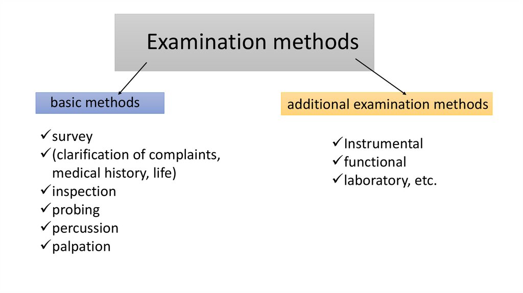
Examination methodsbasic methods
survey
(clarification of complaints,
medical history, life)
inspection
probing
percussion
palpation
additional examination methods
Instrumental
functional
laboratory, etc.
4.
complaints made by patients with diseases ofhard dental tissues:
• pain, often short-term and associated with
external stimuli;
• the presence of cavities on the surface of
the teeth;
• aesthetic defect associated with
irregularities in form and/or tooth color;
• mobility and/or change in tooth position;
• bad breath
• in some cases food retention
5.
anamnesis of the disease• the time of appearance of certain
subjective sensations or visible
changes (appearance of cavities,
chips of hard tissue,
pigmentation, etc.),
• possible reason for their
appearance, dynamics,
• the nature of the interventions
performed and their
effectiveness.
6.
life history• place of birth (to exclude areas where
fluorosis is endemic),
• presence of similar dental lesions in
relatives,
• past and concomitant diseases (especially
affecting the condition of hard dental tissues),
• allergy history(!!!)(really important)
• presence of bad habits,
• working conditions and occupational
hazards.
7.
external examination• general appearance of the patient (constitution, proportionality, height, weight, etc.)
• maxillofacial area (coloring of visible skin and mucous membranes,
• change in facial configuration, condition
• red border of the lips and corners of the mouth, presence of bad breath)
• condition of the lymphatic system of the maxillofacial area (size, soreness, mobility
occipital, parotid, chin, submandibular, superficial cervical, paratracheal)
8.
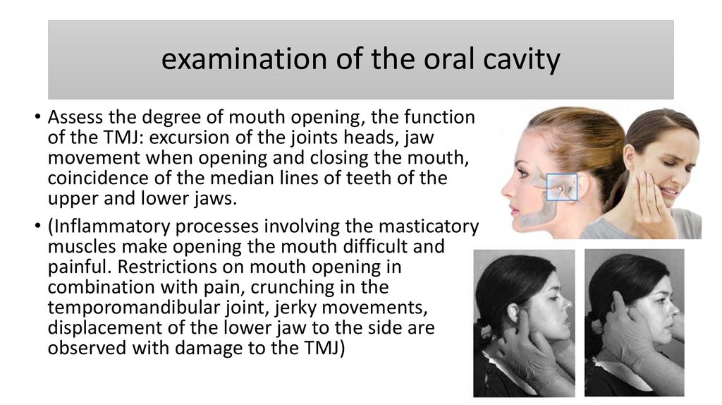
examination of the oral cavity• Assess the degree of mouth opening, the function
of the TMJ: excursion of the joints heads, jaw
movement when opening and closing the mouth,
coincidence of the median lines of teeth of the
upper and lower jaws.
• (Inflammatory processes involving the masticatory
muscles make opening the mouth difficult and
painful. Restrictions on mouth opening in
combination with pain, crunching in the
temporomandibular joint, jerky movements,
displacement of the lower jaw to the side are
observed with damage to the TMJ)
9.
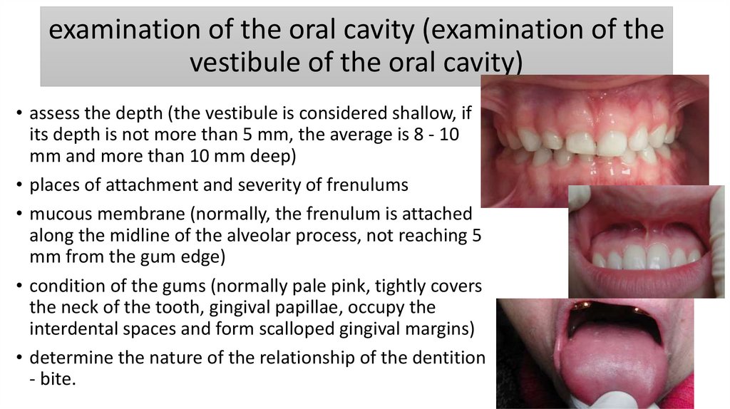
examination of the oral cavity (examination of thevestibule of the oral cavity)
• assess the depth (the vestibule is considered shallow, if
its depth is not more than 5 mm, the average is 8 - 10
mm and more than 10 mm deep)
• places of attachment and severity of frenulums
• mucous membrane (normally, the frenulum is attached
along the midline of the alveolar process, not reaching 5
mm from the gum edge)
• condition of the gums (normally pale pink, tightly covers
the neck of the tooth, gingival papillae, occupy the
interdental spaces and form scalloped gingival margins)
• determine the nature of the relationship of the dentition
- bite.
10.
dental examination• Shape of teeth (rectangular, oval, triangular,
etc.)
• Teeth size (microdentia, macrodentia)
• Teeth color (VITA scale)
• The examination is combined with probing of
fissures, carious cavities and other defects.
• Probing reveals: depth, spread defect of hard
dental tissues, painful areas, consistency, walls
of the defect, etc.
11.
changes in tooth color are observed when:• Carious process
• Non-carious lesions of teeth
• Pulp necrosis
• Under the influence of external factors: smoking
(dark brown color), metal fillings (dark gray
color), root canal treatment (red-brown color
with resorcinol) formaldehyde method)
12.
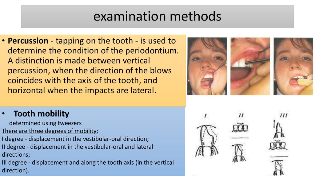
examination methods• Percussion - tapping on the tooth - is used to
determine the condition of the periodontium.
A distinction is made between vertical
percussion, when the direction of the blows
coincides with the axis of the tooth, and
horizontal when the impacts are lateral.
• Tooth mobility
determined using tweezers
There are three degrees of mobility:
I degree - displacement in the vestibular-oral direction;
II degree - displacement in the vestibular-oral and lateral
directions;
III degree - displacement and along the tooth axis (in the vertical
direction).
13.
additional examination methodselectroodontic diagnostics
• Used to study the condition pulp and
periodontium by determining electrical
excitability when exposed to electric
current. When researching the minimum,
threshold force is determined irritation of
nerve receptors in the dental pulp.
• Raising or lowering the threshold sensitivity
indicates various pathological and
physiological processes occurring in the
hard tissues of the tooth.
14.
additional examination methodselectroodontic diagnostics
• The degree of electrical excitability of the pulp
depends on its functional and morphological
state
• Intact teeth respond to a current of 2-6 μA, a
decrease in EDI values to 20-40 μA indicates
the presence of an inflammatory process in the
pulp, up to 100 μA and above indicates pulp
death.
15.
vital stain test• Used to visualize foci of demineralization of
hard dental tissues. During demineralization,
the permeability of hard tissues increases and
the dye is absorbed.
• A 2% solution of methylene blue, a 0.1%
solution of methylene red, a 0.5% solution of
fuchsin, as well as caries detectors are used.
Coloring is carried out for the purpose of:
• Patient motivation;
• Differential diagnosis of caries and non-carious lesions;
• Early diagnosis of caries
• Determining the effectiveness of preventive and therapeutic
• events.
16.
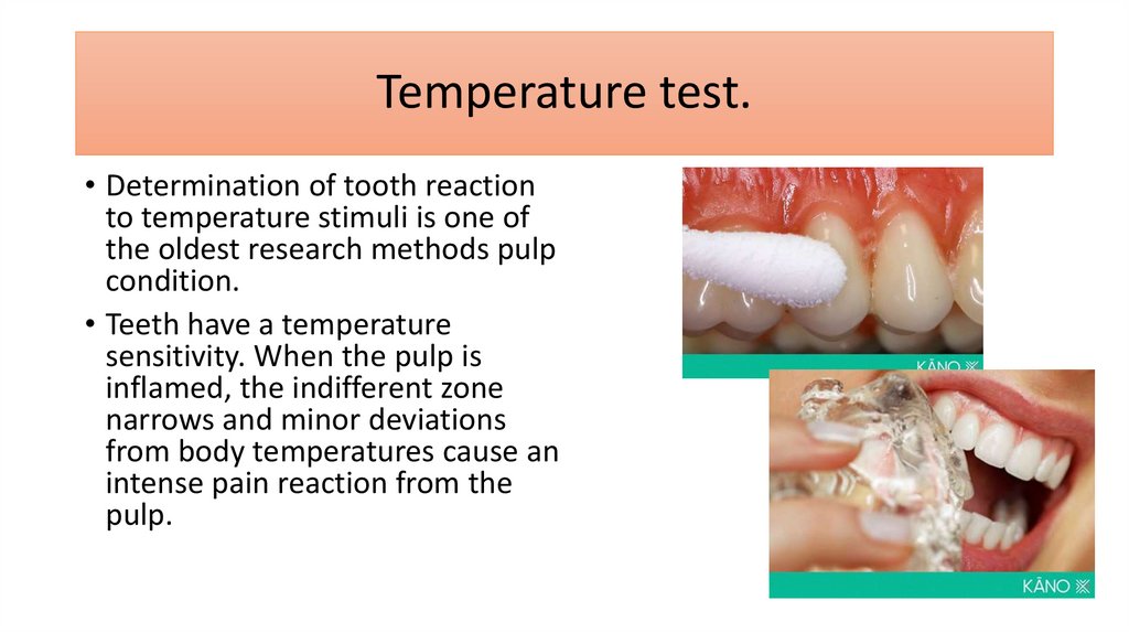
Temperature test.• Determination of tooth reaction
to temperature stimuli is one of
the oldest research methods pulp
condition.
• Teeth have a temperature
sensitivity. When the pulp is
inflamed, the indifferent zone
narrows and minor deviations
from body temperatures cause an
intense pain reaction from the
pulp.
17.
Temperature test.• Heat test.
Use hot water or heated gutta-percha. Guttapercha heated over a flame until it becomes soft
and shiny, but should not be allowed to so that
it smokes (temperature is about 65.5 C). Heated
gutta-percha is placed on the middle third of the
vestibular surface of the crown.
• Cooling test.
To carry out this. It is best to use cold dough
water, dry ice, chlorethyl applied to cotton wool
on a stick.
18.
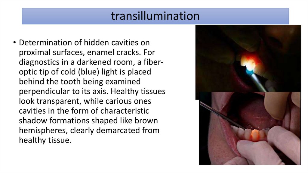
transillumination• Determination of hidden cavities on
proximal surfaces, enamel cracks. For
diagnostics in a darkened room, a fiberoptic tip of cold (blue) light is placed
behind the tooth being examined
perpendicular to its axis. Healthy tissues
look transparent, while carious ones
cavities in the form of characteristic
shadow formations shaped like brown
hemispheres, clearly demarcated from
healthy tissue.
19.
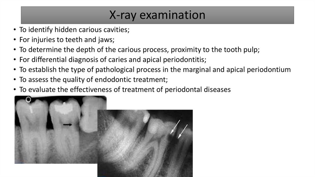
X-ray examination• To identify hidden carious cavities;
• For injuries to teeth and jaws;
• To determine the depth of the carious process, proximity to the tooth pulp;
• For differential diagnosis of caries and apical periodontitis;
• To establish the type of pathological process in the marginal and apical periodontium
• To assess the quality of endodontic treatment;
• To evaluate the effectiveness of treatment of periodontal diseases
20.
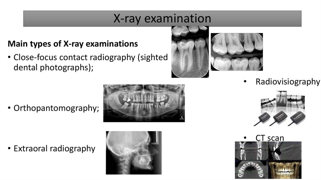
X-ray examinationMain types of X-ray examinations
• Close-focus contact radiography (sighted
dental photographs);
• Radiovisiography
• Orthopantomography;
• Extraoral radiography
• CT scan
21.
The diagnosis of the disease is made on based on:• patient complaints;
• anamnesis data;
• objective examination data;
• results of additional research methods.
22.
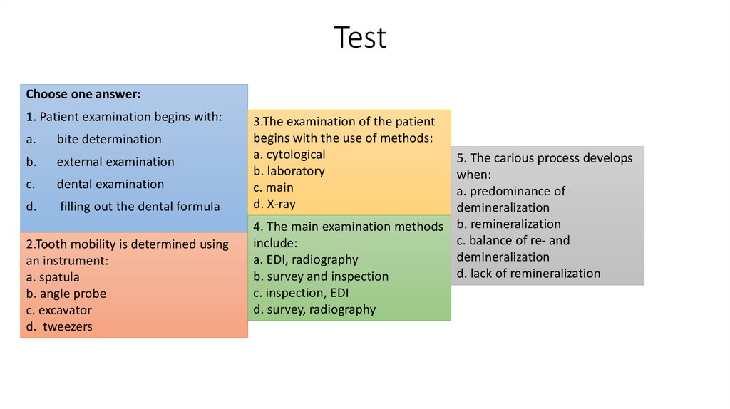
TestChoose one answer:
1. Patient examination begins with:
a.
bite determination
b.
external examination
c.
dental examination
d.
filling out the dental formula
2.Tooth mobility is determined using
an instrument:
a. spatula
b. angle probe
c. excavator
d. tweezers
3.The examination of the patient
begins with the use of methods:
a. cytological
b. laboratory
c. main
d. X-ray
5. The carious process develops
when:
a. predominance of
demineralization
4. The main examination methods b. remineralization
c. balance of re- and
include:
demineralization
a. EDI, radiography
d. lack of remineralization
b. survey and inspection
c. inspection, EDI
d. survey, radiography
23.
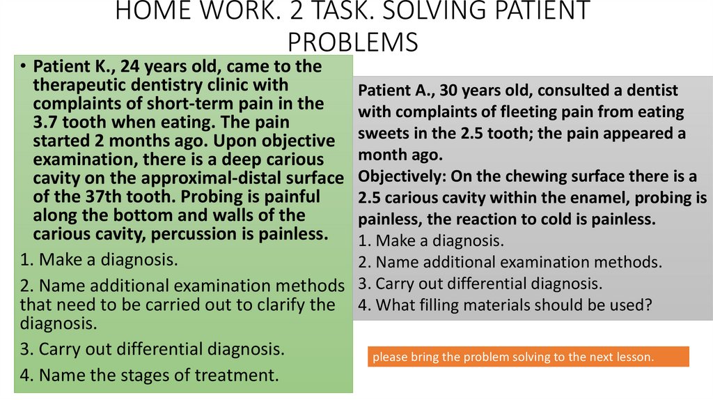
HOME WORK. 2 TASK. SOLVING PATIENTPROBLEMS
• Patient K., 24 years old, came to the
therapeutic dentistry clinic with
Patient A., 30 years old, consulted a dentist
complaints of short-term pain in the
with complaints of fleeting pain from eating
3.7 tooth when eating. The pain
started 2 months ago. Upon objective sweets in the 2.5 tooth; the pain appeared a
month ago.
examination, there is a deep carious
cavity on the approximal-distal surface Objectively: On the chewing surface there is a
of the 37th tooth. Probing is painful
2.5 carious cavity within the enamel, probing is
along the bottom and walls of the
painless, the reaction to cold is painless.
carious cavity, percussion is painless.
1. Make a diagnosis.
1. Make a diagnosis.
2. Name additional examination methods.
2. Name additional examination methods 3. Carry out differential diagnosis.
that need to be carried out to clarify the 4. What filling materials should be used?
diagnosis.
3. Carry out differential diagnosis.
please bring the problem solving to the next lesson.
4. Name the stages of treatment.