


. (A) Control root, supplied with air, with intact cortical cells. (B)")


 chemistry
Similar presentations:


Бетаин (триметилглицин)
1.
Кадаверин (1,5-пентандиамин)Путресцин (1,5-пентандиамин)
Спермин
N,N'-бис(3-аминопропил)бутан-1,4-диамин
Спермидин
N,N'-бис(3-аминопропил)бутан-1,4-диамин
2.
3.
Бетаин(триметилглицин)
4.
Conversion of solar energy intocarbohydrates by a leaf.
5. Shown here are the percentages of light absorbed, reflected, and transmitted, as a function of wavelength. The transmitted and
Optical properties of a bean leaf.Shown here are the percentages
of light absorbed, reflected, and
transmitted, as a function of
wavelength. The transmitted and
reflected green light in the wave
band at 500 to 600 nm gives
leaves their green color. Note that
most of the light above 700 nm is
not absorbed by the leaf.
(From Smith 1986.)
6. Photoprotection by dissipation of excess light energy aided by xanthophyll cycle carotenoids
7.
Xanthophyll cycle8.
9.
Response of frosted orache(Atriplex sabulosa) and
Arizona honeysweet
(Tidestromia oblongifolia) to
heat stress.
Photosynthesis (A) and
respiration (B) were
measured on attached
leaves, and ion leakage (C)
was measured in leaf slices
submerged in water.
(From Bjorkman et al. 1980.)
10.
Организация мембранных микродоменов (рафтов)11.
12.
13.
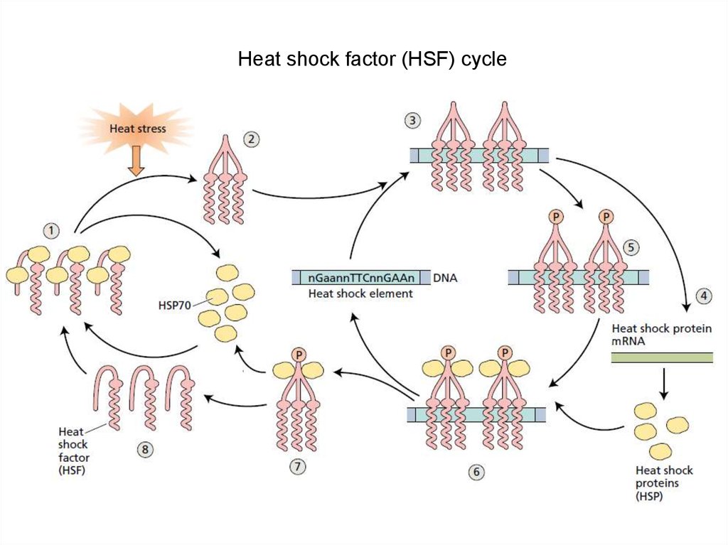
Heat shock factor (HSF) cycle14.
Heat stress Ca-mediated response15.
Low temperature scanning electron microscopy of contro (A) and freezing – stressed tobacco leaves (B-D)
(Ashworth and Pearce, unpubl. res. ). Themicrographs show transverse fractures through the leaves.
Youngpottedplantsweregrowninawarmgreenhouseandweretestedatthetwo-to-fourleafstage.
Theplantsweresprayedwithwaterandcooledat28Chÿ1toÿ208Cthenfreeze®xedinmeltingfreon12.DTAshowedtheleavesfrozebetweenÿ2.08Candÿ3.08C.Detailsofthemicroscopicalmet
hodsareasinPearceandAshworth(1992).A,Controlsampleshowingturgidcellsandabsenceofextracellulariceinal
ltissues(e,epidermis;pm,palisademesophyll;sm,spongymesophyll).Notethatorganellepro®leswerevisiblewhe
rethefractureplanehadcutthroughthecells(starsindicatecrossfracturedcellsinthespongymesophyll;arrowheadsindicateorganellesintwoexamplecells).Theepidermalcells(e)
werealsocrossfractured.BandC,Samplefrozentoÿ208Cshowingextensiveextracellularice(i).Cisanenlargementoftheareaboxe
dinB.Inthisexampleicerami®edextensivelythroughthegasspacesbutdidnotfullyoccludethem.Thewhitearrow(
B)indicatesthecollapsedepidermis.Atlowmagni®cation(B)theiceappearedsuper®ciallysimilartoturgidcells.H
owever,whenenlarged(C)thecrossfractureofthesestructuresshowedthemtocontainnoorganellepro®lesandinsteadthefracturedsurfacehadsteps(ar
rowheads)typicaloffracturedice.Thecellsweremostlyhiddenbytheice.However,intheareaenlargedinCacollapse
dcell(star)waseasilyidenti®edbytheorganellesitcontained:thearrowindicatestheimpressonthecellwalloforgan
ellesinacell.D,Samplefrozentoÿ208C.Icewasremovedfromthespecimenbysublimationinthemicroscope,thusre
vealingthecollapsed,dehydratedcells.Themesophyllcells(pm,palisademesophyll;sm,spongymesophyll)andep
idermis(whitearrows)werecollapsed.Theoutersurfaceofcellwallsshowedanimpressoftheorganelleswithinthec
ells(examplesindicatedbyarrowheads).Starsindicatewherethefractureplanehascutthroughthecells,againreveal
ingorganelles.Allgold-coated.2kV.Bars‹10mm(AandC)or100mm(BandD).
16.
Low temperature scanning electron microscopy of control (A) and freezing –stressed tobacco leaves (B-D) (Ashworth and Pearce, unpubl. res.).
17.
18.
19.
Membrane transport proteins mediating sodium, potassium, and calciumtransport during salinity stress
20. Roots of maize. Scanning electron micrographs (150×). (A) Control root, supplied with air, with intact cortical cells. (B)
Oxygen-deficient root.Note the prominent gas-filled spaces (gs) in the cortex (cx), formed by degeneration of cells. The stele
(all cells interior to the endodermis, En) and the epidermis (Ep) remain intact. X, xylem. (Courtesy of J.
L. Basq and M. C. Drew.)
21.
22.
Metabolic pathways that are active during hypoxia in plantsNarsai et al. 2011
23.
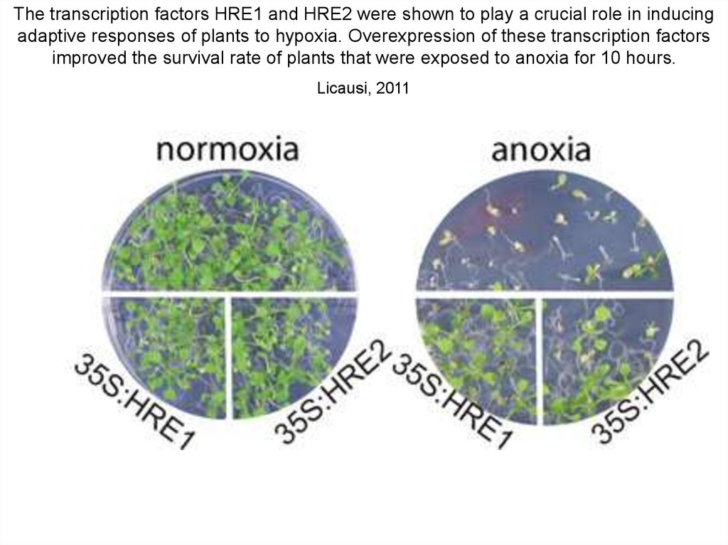
The transcription factors HRE1 and HRE2 were shown to play a crucial role in inducingadaptive responses of plants to hypoxia. Overexpression of these transcription factors
improved the survival rate of plants that were exposed to anoxia for 10 hours.
Licausi, 2011
24. Oxygen sensing in plants is mediated by an N-end rule pathway for protein destabilization
The transcription factor RAP2.12 is constitutively expressed under aerobic conditions. RAP2.12 protein is always present, bound toACBP to prevent RAP2.12 from moving into the nucleus under aerobic conditions and to protect it against proteasomal degradation
in air. Upon hypoxia, RAP2.12 moves into the nucleus, where it activates anaerobic-gene expression. Upon re-oxygenation,
RAP2.12 is rapidly degraded via the N-end rule pathway and proteasome-mediated proteolysis to downregulate the hypoxic
response.