

")

")
")


")


")

")


? Next section– III. Dermis & Hypodermis")


 Glands")
.")


")


? Next section— V. Skin Disorders")


 biology
Similar presentations:


")

The integumentary system
1. Chapter 6
The IntegumentarySystem
6-1
2. Ch. 6 Study Guide
1. Critically read Chapter 6––
–
–
pp. 187-194 before “Skin Color” section
Skip Section 6.2 (Hair and Nails)
Critically read sections 6.3 (Cutaneous glands) and 6.4
(Skin Disorders) pp. 202-207 before “Burns” section
2. Comprehend Terminology (those in bold in the
textbook) within the reading scope above
3. Study-- Figure questions, Think About It questions,
and Before You Go On (section-ending) questions
(within the reading scope above)
4. Do end-of-chapter questions--s
–
–
–
Testing Your Recall— 1-4, 7-17, 20
True or False– All of them (1-10)
Testing Your Comprehension-- 1, 4, 5
6-2
3. § Quotable Quotes (Skin)
Some guys say beauty is only skin
deep. But when you walk into a party,
you don't see somebody's brain. The
initial contact has to be the sniffing.
(James Caan)
Beauty may be skin deep, but ugly
goes clear to the bone. (Redd Foxx)
6-3
4. I. Introduction
6-45. § Overview (1)
• Dermatology– scientific study andmedical treatment of this system
• Largest organ (skin) – covers about 2.0
meter square; 15% of the body weight
• Epidermis
– stratified squamous epithelium
• Dermis
– connective tissue layer
• Hypodermis (NOT part of the skin)– often
what tissue predominates here?
6-5
6. § Overview (2)
• Thickness variable, based on thicknessof Epidermis, two categories-• Thick skin– .5 mm thick (epidermis)
– Locations?
– stratum corneum layer increased
• Sweat glands-- present
• No hair follicles or sebaceous glands
• Thin skin (.1 mm)– The rest of the body
– Has hair follicles, oil glands, and sweat
glands
@Fig. 6.1
6-6
7.
6-78. § Functions of the Skin
• Resistance to trauma/infection– Why? (Fig. 5.28)
– acid mantle (pH 4-6)– acidic film (protection)
• Barrier: to water, UV light, some chemicals;
transdermal patches . . can pass
• Vitamin D synthesis (first step)
• Sensory receptors– what? where?
• Thermoreceptors– in dermis: nerve endings
to the brain, back to blood vessels (Fig. x)
• Nonverbal communication— move the skin
6-8
etc. (Fig. 6.2)
9.
6-910.
Thermoregulationvasoconstri
ction
vasodilation
Less
Heat
loss
Heat
loss
In hot
environment
In cold
environment
6-10
11. Social functions-- Figure 6.2
Skeletal muscles attach to dermal collagenfibers and produce expressions as a smile, a
wrinkle of forehead, and lifting of an eyebrow
6-11
12. II. Epidermis
6-1213. § Cells of the Epidermis (1)
Five types of cells-1. Keratinocytes – most of the skin cells;Named b/c keratin synthesis
2. Dendritic (Langerhans) cells
– MACROPHAGES guard against pathogens
– Locations– the epidermis and epithelia of
oral cavity, esophagus, and vagina
Fig. 6.3 and X
6-13
14.
The Epidermis— Fig. 6.26-14
15.
6-1516. § Cells of the Epidermis (2)
Location of the following types of cells—
stratum ___________
3. Stem cells
– undifferentiated cells for keratinocytes
4. Melanocytes
– synthesize ________that shield UV rays
– “sunny side” phenomenon (Fig. x)
5. Tactile (Merkel) cells (for touch)
– receptor cells associated with nerve fibers
– They are Meissner corpuscles
6-16
17.
KeratinocytesMelanocyte
6-17
18.
§ Layers of the Epidermis—Next five slides (1-5)
from deep to superficial and from
youngest to oldest keratinocytes
6-18
19. 1. Stratum Basale (deepest layer)
• Single layer cells on basementmembrane (Fig. 6.3)
• Cell types in this layer (A review)
– Stem cells and keratinocytes
• undergo mitosis to replace epidermis
– Melanocytes
• distribute melanin through cell processes
• melanin picked up by kerotinocytes
– Merkel cells are touch receptors
• form Merkel disc
6-19
20. Figure 6.2a
6-2021. 2. Stratum Spinosum– above stratum basale
• Several layers of keratinocytes (flattened asthey cease dividing toward apical side; Why)
– appear spiny due to shrinkage
of keratinocytes (histological preparation)
– What are these spiny structures?
– Thickest stratum in most skin except in ______
• Contains dendritic (Langerhans) cells
– macrophages from bone marrow
that migrate to the epidermis
– help protect body against pathogens by
“presenting” them to the immune system
6-21
22. 3. Stratum Granulosum
3 to 5 layers flat keratinocytes: three
developments occur to them-A. Contain keratohyalin granules (dark-stained)
–
Granules release a substance bonding with
cytoskeleton and convert them to keratin
B. Granules release a glycolipid by exocytosis
to waterproof the skin
–
–
called epidermal water barrier
Other structures contribute to this— TJs, proteins
C. Programmed cell death (apoptosis)—dander
& dandruff
6-22
23. 4. Stratum Lucidum— superficial to the stratum granulosum
• Thin translucent zone seen only in thickskin
• Keratinocytes are densely packed with
eleidin, a precursor to keratin
– Eleidin does not stain well (pale appearance)
• In addition, cells (keratinocytes) here
have no nucleus or organelles
– Appearance– Pale and featureless
Fig. x
6-23
24.
6-2425. 5. Stratum Corneum
• Up to 30 layers of dead, scaly,keratinized cells
– surface cells flake off (exfoliate)
– Especially in thick skin--palms, soles
and corresponding fingers/toes
6-25
26. § Life History of Keratinocytes
• Produced by stem cells in stratumbasale
• New cells push others toward surface
– cells grow flat and fill with vesicles (lipids)
• Cells filled with keratin
– forms epidermal water barrier
• Cells die and exfoliate (relating to dust
mites, “house dust allergy”--Fig. 6.4)
6-26
27.
Fig. 6.4 The House Dust Mite, DermatophagoidesThey are about 0.5 mm
in length
Feed on _______,
edible flakes of keratin
Esp. in pillows,
mattresses, and
upholstery
We actually allergy to
the feces of these mites
6-27
6-27
28. Questions (muddiest points)? Next section– III. Dermis & Hypodermis
Questions (muddiestpoints)?
Next section–
III. Dermis & Hypodermis
6-28
29. § Dermis- a C.T. layer
• Thickness = 0.2 to 4.0 mm• Composition
– Collagen (mainly), elastic and reticular fibers,
– Cells– fibroblast etc. --Blood supply (yes/no)
– Sweat glands, sebaceous glands, nerve endings
• Dermal papillae – fingerlike extensions of
the dermis into the epidermis
• Layers (fig. 6.5) in dermis:
– papillary layer, thin and rich in capillaries,
areolar tissue
– reticular layer, deeper part, Dense irregular C.T.;
striae— stretch marks (tearing of collagens) 6-29
30. Fig. 6.5 layers of the dermis
Epidermalridges
Fig. 6.5 layers of the dermis
Areolar Tissue
Dense irregular CT
Dermal
papillae
6-30
6-30
31. § Hypodermis
1. Other names-Subcutaneous tissue;superficial fascia
2. Mostly adipose tissue;
Uniformly distributed?; 8%
thicker in women
3. Functions
– energy reservoir
– thermal insulation
4. Hypodermic injections
(to subcutaneous tissue)
– highly vascular; absorb
drugs easily
6-31
32. Questions? Next section— IV. Cutaneous Glands
6-3233. Table 6.2— summary of cutaneous glands 1. Sweat glands 2. Oil glands 3. Ceruminous glands 4. Mammary glands
Table 6.2— summaryof cutaneous glands
1.
2.
3.
4.
Sweat glands
Oil glands
Ceruminous glands
Mammary glands
6-33
34.
§ CutaneousGlands
6-34
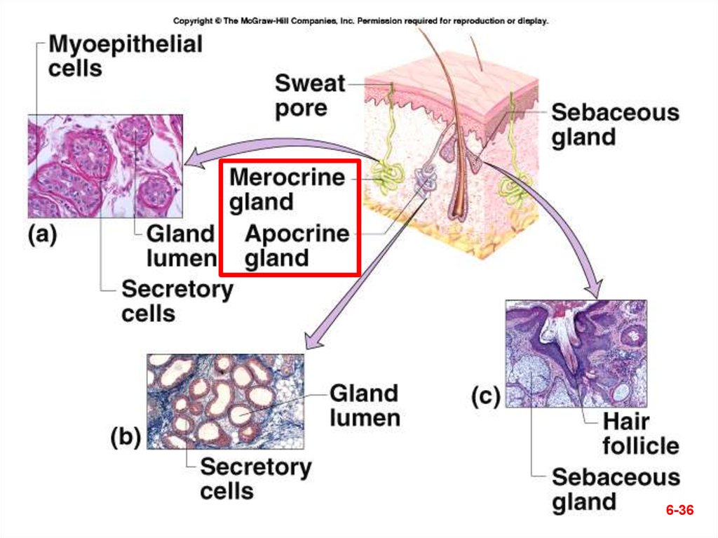
35. 1. Two kinds of Sweat Glands
Filtrate of plasma and some waste products
– insensible perspiration; @ 500 ml a day
– sweating with visible wetness is diaphoresis
A. Merocrine (eccrine) glands is simple tubular
gland; what in the sweat?
B. Apocrine glands (larger lumen) produce
sweat containing fatty acids; are scent
glands—produce pheromones
– Locations-- near hair follicles and respond to
stress and sexual stimulation
– bromhidrosis is disagreeable body odor produced
by bacterial action on fatty acids; poor hygiene
Fig. 6.11
6-35
36.
6-3637. 2. Sebaceous (Oil) Glands
• Oily secretion called sebum thatcontains broken-down cells
– Due to mitosis replacement at the base of
the gland
– Sebum keeps the skin/hair from becoming
dry
– lanolin in skin creams is sheep sebum
• Flask-shaped glands with duct that opens
into hair follicle
Fig. 6.11c
6-37
38. ID specific cutaneous glands (A & B).
ID specificcutaneous
glands (A & B).
A.
B.
Which specific kind?
6-38
39. 3. Ceruminous Glands
A. Found only in external ear canalB. Their secretion combines with sebum
to produce earwax (called cerumen)
– Waterproofs the auditory canal
– Keeps eardrum flexible
– Bitterness repel mites and other pests
– Has a bactericidal effect
Fig. X
6-39
40. Ceruminous glands—inappropriate interventions
Ceruminous glands—inappropriate interventions
6-40
41. Cotton-tipped applicator (a no-no)
Cottontippedapplicator
(a no-no)
6-41
42. ᵡ Ear Candling!?
6-4243. 4. Mammary Glands
1. Breasts of both sexes rarely containmammary glands
– secondary sexual characteristic of females
2. Mammary glands (within female breast)
– produce milk--during lactation and pregnancy
• Mammary ridges or milk lines
– Mammals-- 2 rows of mammary glands
– Primates-- kept only anteriormost glands
• Additional nipples (polythelia)
– may develop along milk line
Fig. x
6-43
44. Mammary Glands
AreolaNipple
6-44
45. Check Point Questions
1. (True/False) The three layers of theskin are the epidermis, dermis, and
hypodermis.
2. How do merocrine and apocrine sweat
glands differ in structure and
function?
6-45
46. Questions (muddiest points)? Next section— V. Skin Disorders
6-4647. § Skin Cancer
1. Cause– the ultraviolet rays of the sun– There is no such thing as a healthy suntan
– Controversial on suncreens (Read Insight 6.4)
2. Types– named for the epidermal cells they
originate and the appearance of their
lesions (zones of tissue injury):
A. Basal cell carcinoma
B. Squamous cell carcinoma
C. Malignant melanoma
6-47
48. A. Basal cell carcinoma
1. Most common type and the leastdangerous one
2. Origination- by cells of the stratum
basale
Fig. 6.12a
6-48
49. Fig. 6.12a
A. Basal cell carcinomaFig. 6.12a
6-49
50. B. Squamous cell carcinoma
1. Chance of recovery is good withearly detection and surgical
removal. But it can be lethal when
metastasize
2. Origination- from the keratinocytes
of the stratum spinosum (the layer
right above the basale)
Fig. 6.12b
6-50
51.
B. Squamous cell carcinoma6-51
52. C. Malignant melanoma
1. Most deadly skin cancer but accountsfor only 5% of all cases
2. Origination- from the melanocytes of
preexisting mole.
3. Distinguish a mole from this cancer
(ABCD rule):
–
–
–
–
Asymmetry
Border irregularity
Color (mixture of brown, black, tan etc.)
Diameter (greater than 6 mm)
Fig. 6.12c
6-52
53.
C. Malignant melanoma; which ofthe ABCD rules can you identify
6-53
54. Video watching
• Preventing melanoma (1 min 30 sec),when available and time allows
6-54