


 medicine
Similar presentations:


")


")


Obstructive jaundice and cholangitis
1.
Obstructive jaundice andcholangitis
Professor Eduard I. Galperin
2.
Anatomy of the bile ductsAnatomy and flow of bile. Sphincter of Oddi regulates bile flow. Liver secretes bile
constantly, closed sphincter of Oddi ensures filling of gallbladder, open sphincter
is followed by bile drainage from the gallbladder towards duodenum.
Bile duct obstruction causes biliary hypertension.
2
Reliability of biliary system – bile duct lumen of 1 mm is enough for daily bile
Bile Duct Diseases - Harvard Health
passage.
3.
Anatomy of hepatic lobule1
2
3
5
4
6
1 – bile capillary
2 – layer of hepatocytes
3 – space of Disse
4 – sinusoid
5 – portal vein branch
6 – hepatic artery branch
Substrates from sinusoid normally pass into the hepatocytes. After metabolic transformation,
some metabolites (bile, bile acids, etc.) pass into the bile capillary. A part of metabolized
substrates return to sinusoid and pass into systemic circulation.
2a
4.
Cholestasis3
Definition: any impairment of secretion and release of bile from the hepatocyte
to the major duodenal papilla is called cholestasis. Thus, this concept includes
both biochemical and mechanical disorders.
Cholestasis is a universal liver reaction against any type of lesion (ischemic,
toxic, obstructive, metabolic, autoimmune).
Types of cholestasis: extrahepatic (biliary hypertension, obstructive jaundice),
intrahepatic, combined.
5.
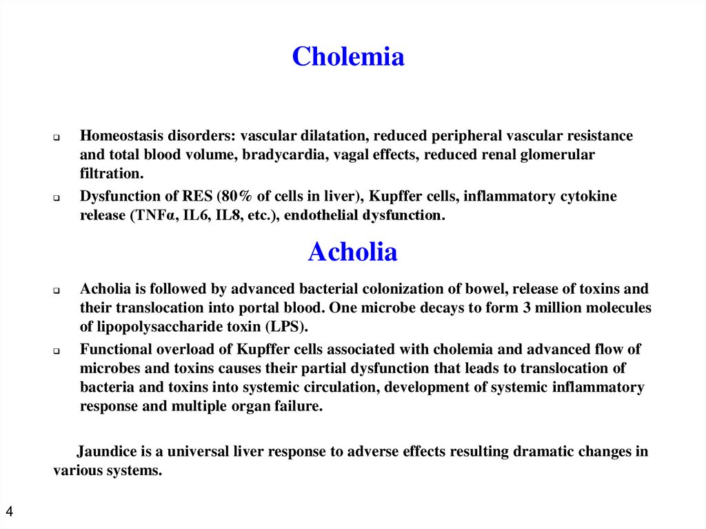
CholemiaHomeostasis disorders: vascular dilatation, reduced peripheral vascular resistance
and total blood volume, bradycardia, vagal effects, reduced renal glomerular
filtration.
Dysfunction of RES (80% of cells in liver), Kupffer cells, inflammatory cytokine
release (TNFα, IL6, IL8, etc.), endothelial dysfunction.
Acholia
Acholia is followed by advanced bacterial colonization of bowel, release of toxins and
their translocation into portal blood. One microbe decays to form 3 million molecules
of lipopolysaccharide toxin (LPS).
Functional overload of Kupffer cells associated with cholemia and advanced flow of
microbes and toxins causes their partial dysfunction that leads to translocation of
bacteria and toxins into systemic circulation, development of systemic inflammatory
response and multiple organ failure.
Jaundice is a universal liver response to adverse effects resulting dramatic changes in
various systems.
4
6.
Painful and painless obstructive jaundiceVarious rates of biliary hypertension development (fast, sudden or slow,
gradual) determine occurrence of painful or painless obstructive jaundice.
Sudden biliary hypertension is followed by acute pain in the right upper
abdominal quadrant. Slow progression of biliary hypertension determines
painless jaundice. Fast development of biliary hypertension is mainly
observed in cholangiolithiasis, a slow one – in patients with bile duct
tumors.
4
7.
PAINFUL OBSTRUCTIVE JAUNDICE5
8.
Cholangiolithiasis1.
2.
3.
6
Gallstone migration from the
gallbladder.
Obstruction of common hepatic
duct and common bile duct.
Obstruction of major duodenal
papilla.
9.
Stenosis of major duodenal papillaCauses: cholangitis, pancreatitis, instrumental injury, gallstone passage, parapapillary
diverticulum, functional disorders of the sphincter of Oddi.
7
Morphological changes: fibrosclerotic disorders.
Symptoms: pain, intermittent jaundice, urine and stool discoloration.
Diagnosis: endoscopic examination of major duodenal papilla, ERCP, PTC, manometry.
Treatment: endoscopic or surgical intervention for severe stenosis.
10.
Choledocholithiasis. Ultrasoundа – ultrasound: common bile
duct enlargement and
calculus (arrow).
б – ultrasound: calculus
inside the common bile duct
(arrow).
в – endoscopic ultrasound: a
small calculus is visualized
(arrow).
Dilatation of the bile ducts, doubling of the tubular structure
Calculi in the bile ducts
Endoscopic ultrasound – small calculi
Sensitivity of ultrasound – 28-50%, endoscopic ultrasound– 98%.
8
11.
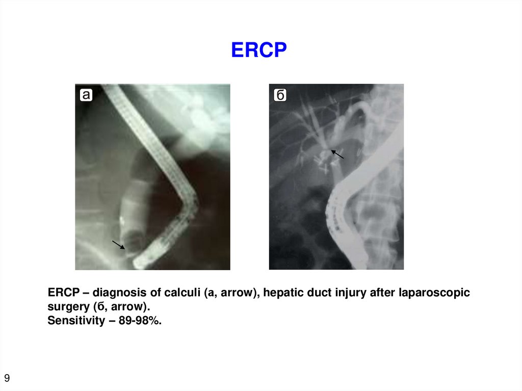
ERCPERCP – diagnosis of calculi (а, arrow), hepatic duct injury after laparoscopic
surgery (б, arrow).
Sensitivity – 89-98%.
9
12.
PTCMirizzi syndrome. Hepatic duct calculi.
Sensitivity - 90-100%.
10
13.
MRCP. МR-cholangiographyImaging of gallbladder, bile ducts and calculi (arrows) without contrast
enhancement.
Sensitivity 85–88%.
11
14.
ERCP vs. MRCPCalculi
Calculi
ERCP
MRCP
A relatively invasive method requires
contrast agent injection into the bile
ducts, biliary hypertension and
irradiation.
Non-invasive method without the need for direct
contrast enhancement of the bile ducts.
Less clear image, but no irradiation.
Images were obtained from the same patient. Calculi were removed via
endoscopic approach.
12
15.
Mirizzi syndromeType I - narrowing of
common hepatic duct
caused by calculus-induced
compression of Hartmann's
pouch
13
Type II - fistula between the
gallbladder and hepatic duct.
Hypertension of lobar hepatic ducts
16.
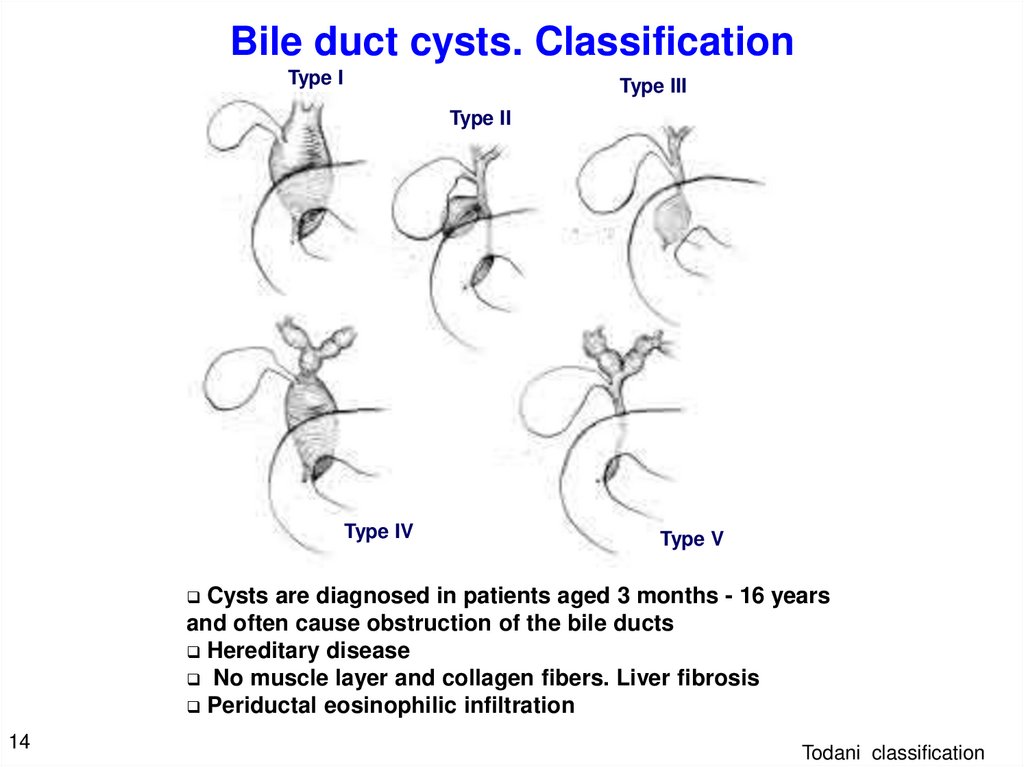
Bile duct cysts. ClassificationType I
Type III
Type II
Type IV
Type V
Cysts are diagnosed in patients aged 3 months - 16 years
and often cause obstruction of the bile ducts
Hereditary disease
No muscle layer and collagen fibers. Liver fibrosis
Periductal eosinophilic infiltration
14
Todani classification
17.
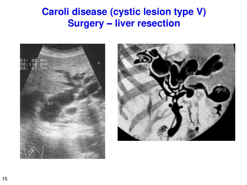
Caroli disease (cystic lesion type V)Surgery – liver resection
15
18.
Primary sclerosing cholangitisIdiopathic gallbladder obliteration, autoimmune disease, frequent
combination with ulcerative colitis, Crohn's disease. Higher-thannormal levels of AST and ALP.
Chronic course
Cause is unclear
Multiple strictures and dilatations of
intrahepatic bile ducts
Outcome: liver cirrhosis,
cholangiocarcinoma is rarer
Diagnosis: direct contrast enhanced
methods, MRCP
Radical approach – liver
transplantation
16
19.
HaemobiliaDamage to the liver or
intrahepatic bile ducts, local liver
necrosis. Haemobilia is a
secondary sign of the underlying
disease.
Right upper quadrant abdominal
pain, melena, transient jaundice.
Endoscopic examination of major
duodenal papilla, ERCP,
angiography, ultrasound, CT, MRI
Mortality rate 32–50%.
17
20.
Parasitic invasionOpisthorchiasis (Ob, Volga basin, East Asia)
Echinococcosis, alveococcosis, ascariasis,
Fascioliasis– Fasciola gigantica
Schistosomiasis – tropical helminth
Parasites penetrate into the bile ducts from the
duodenum
18
21.
Symptoms and diagnosis of painful obstructivejaundice
19
Acute onset
Scleral icterus
Pain attack
Dark urine, stool
discoloration
Common previous cholelithiasis
Ultrasound, ERCP, PTC, MRCP, CT,
endoscopic ultrasound.
Laboratory survey.
22.
ACUTE CHOLANGITIS20
23.
Acute cholangitis (AC)AC is an infectious inflammation of the bile ducts. Most often,
AC develops on the background of obstructive jaundice and
biliary hypertension. AC is associated with penetration of
microbes (Escherichia coli, Klebsiella, Proteus) from the
duodenum into the bile ducts.
Metabolic products of microbial cells cause acute biliary
hypertension.
21
23
24.
Cholangiovenous reflux1
2
3
1 – bile capillary
2 – layer of hepatocytes
3 – space of Disse
4 – sinusoid
5 – portal vein branch
6 – hepatic artery branch
5
4
6
Corrosion casting. Scanning electron microscopy
Ductal pressure, mm H2O
200
Penetration of corrosive particles (1,7 µm)*
Intact bile ducts
200–500
Particles achieve sinusoids
500–800
Particles achieve central veins
* Particle of 1,7 µm – is a size of microbe
Secretory pressure. Microbial metabolite pressure. L., Pellegrini C.A., Way L.W. Am. J.
Surg., 1988; 155: 23–28.
22
25.
Symptoms of acute cholangitis• Chills, fever
• Leukocytosis
• Infection
• Symptoms associated with biliary hypertension and obstructive
jaundice
• Recurrent systemic inflammatory response with organ dysfunction
Charcot's triad:
fever, rigors, jaundice
23
Reynolds’ pentad :
Charcot's triad +
altered mental status, hypotension
26.
Causes of short-term SIRS and symptoms of sepsisTwo factors:
1.
Large purulent surface of gallbladder, direct biliovenous reflux - sinusoidal
endotoxemia
2.
Kupffer cell failure caused by cholemia - systemic endotoxemia, increased release
of pro-inflammatory cytokines – systemic inflammatory response, sepsis.
The difference between cholangitis and other purulent diseases is
high sinusoidal endotoxemia combined with Kupffer cell failure.
24
27.
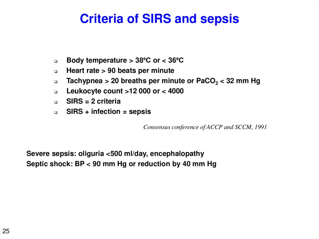
Criteria of SIRS and sepsisBody temperature > 38ºC or < 36ºC
Heart rate > 90 beats per minute
Tachypnea > 20 breaths per minute or PaCO2 < 32 mm Hg
Leukocyte count >12 000 or < 4000
SIRS = 2 criteria
SIRS + infection = sepsis
Consensus conference of ACCP and SCCM, 1991
Severe sepsis: oliguria <500 ml/day, encephalopathy
Septic shock: BP < 90 mm Hg or reduction by 40 mm Hg
25
28.
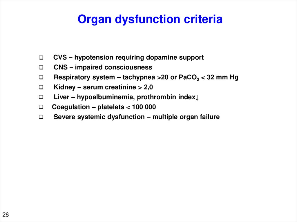
Organ dysfunction criteria26
CVS – hypotension requiring dopamine support
CNS – impaired consciousness
Respiratory system – tachypnea >20 or PaСO2 < 32 mm Hg
Kidney – serum creatinine > 2,0
Liver – hypoalbuminemia, prothrombin index↓
Coagulation – platelets < 100 000
Severe systemic dysfunction – multiple organ failure
29.
Renal failure in acute cholangitisKidney is a main organ secreting bile components – cholemic nephropathy.
Cholemia and endotoxemia cause renin and aldosterone release, increased level of atrial
natriuretic peptide, vasodilation, reducing total blood volume, renal and glomerular blood
flow, arterial and venous thrombosis.
Renal failure is more common in patients with jaundice and cholangitis compared to those
without jaundice.
The difference of cholangitis from other purulent diseases is high incidence of
renal failure
27
30.
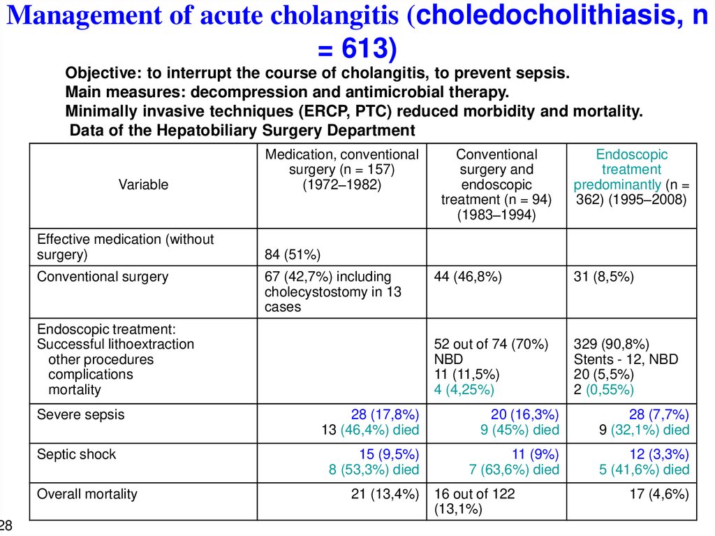
Management of acute cholangitis (choledocholithiasis, n= 613)
28
Objective: to interrupt the course of cholangitis, to prevent sepsis.
Main measures: decompression and antimicrobial therapy.
Minimally invasive techniques (ERCP, PTC) reduced morbidity and mortality.
Data of the Hepatobiliary Surgery Department
Variable
Effective medication (without
surgery)
Conventional surgery
Medication, conventional
surgery (n = 157)
(1972–1982)
Conventional
surgery and
endoscopic
treatment (n = 94)
(1983–1994)
Endoscopic
treatment
predominantly (n =
362) (1995–2008)
44 (46,8%)
31 (8,5%)
52 out of 74 (70%)
NBD
11 (11,5%)
4 (4,25%)
329 (90,8%)
Stents - 12, NBD
20 (5,5%)
2 (0,55%)
84 (51%)
67 (42,7%) including
cholecystostomy in 13
cases
Endoscopic treatment:
Successful lithoextraction
other procedures
complications
mortality
Severe sepsis
28 (17,8%)
13 (46,4%) died
20 (16,3%)
9 (45%) died
28 (7,7%)
9 (32,1%) died
Septic shock
15 (9,5%)
8 (53,3%) died
11 (9%)
7 (63,6%) died
12 (3,3%)
5 (41,6%) died
Overall mortality
21 (13,4%)
16 out of 122
(13,1%)
17 (4,6%)
31.
Treatment of pyogenic liver abscesses in acutecholangitis (n = 19)
The main requirement is biliary decompression + puncture or
drainage of the abscess.
Mean duration of acute cholangitis – 8 days. Solitary – 7, multiple – 2, miliary – 10. Symptoms
are significantly determined by severity of cholangitis.
Percutaneous puncture (abscess volume – 15–120 ml):
≤80 ml – efficacy 80%, >80 ml – efficacy 33%
Percutaneous drainage (abscess volume 30–160 ml).
Volume < 120 ml – efficiency is 15 times higher than for volume > 120 ml.
Duration of drainage – 10–55 days.
Surgery: drainage, liver lobe resection – 3.
Miliary abscesses – antimicrobial therapy without surgery – 10, 4 patients died.
Severe sepsis – 5, 2 patients died.
Septic shock – 7, 4 patients died.
Risk factors: miliary abscesses, septic shock, inadequate drainage, high
creatinine.
29
32.
Stages of acute cholangitis and Tokyo Guidelines(2007)
Stage of acute cholangitis
Criterion
mild
(I)
moderate
(II)
severe
(III)
Organ dysfunction
no
no
yes
Response to therapy*
yes
no
no
*Overall and antimicrobial therapy
30
Emergency biliary decompression is required for severe acute cholangitis
(stage III)
In mild stages, it is important to determine response to therapy. Emergency
biliary decompression is required if there is no response (moderate, stage II).
Antibiotics enter the bile only after medical or surgical decompression. An
importance of decompression in emergency surgery.
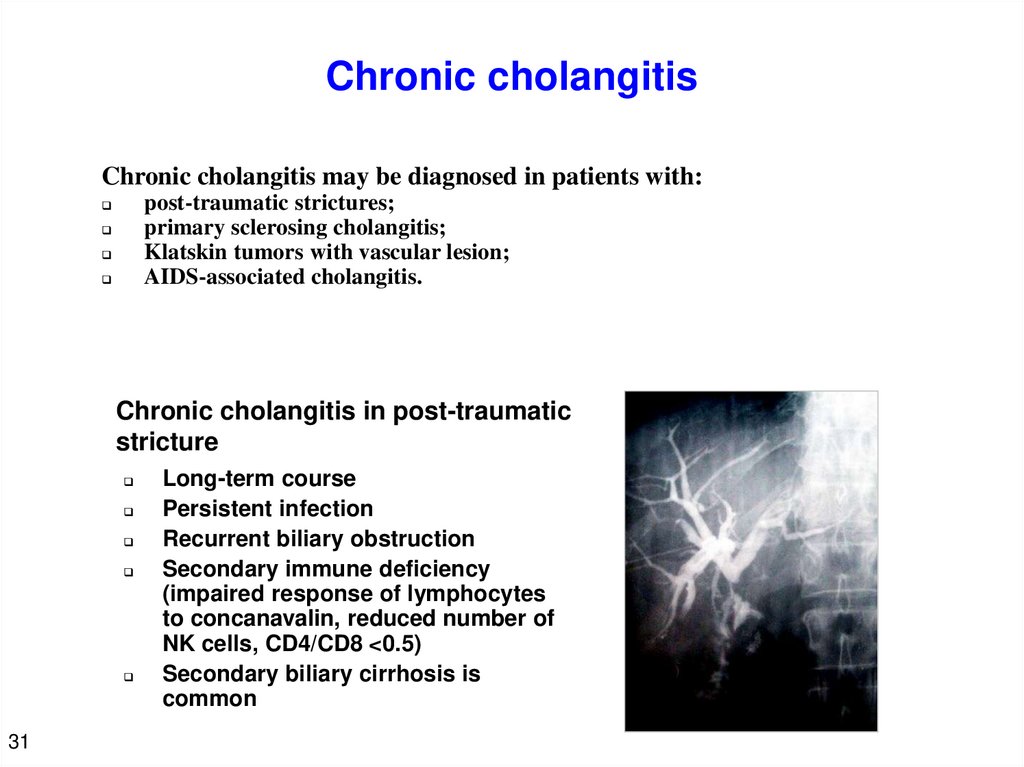
33.
Chronic cholangitisChronic cholangitis may be diagnosed in patients with:
post-traumatic strictures;
primary sclerosing cholangitis;
Klatskin tumors with vascular lesion;
AIDS-associated cholangitis.
Chronic cholangitis in post-traumatic
stricture
31
Long-term course
Persistent infection
Recurrent biliary obstruction
Secondary immune deficiency
(impaired response of lymphocytes
to concanavalin, reduced number of
NK cells, CD4/CD8 <0.5)
Secondary biliary cirrhosis is
common
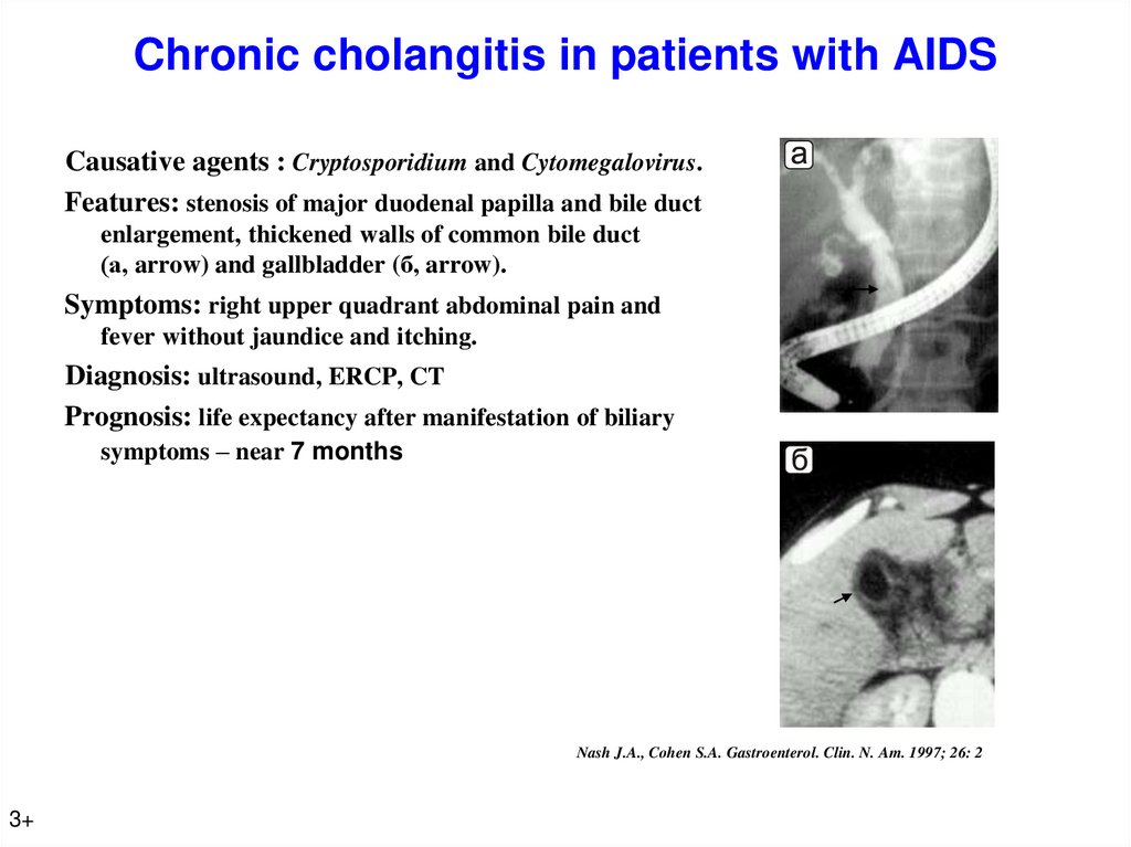
34.
Chronic cholangitis in patients with AIDSCausative agents : Cryptosporidium and Cytomegalovirus.
Features: stenosis of major duodenal papilla and bile duct
enlargement, thickened walls of common bile duct
(а, arrow) and gallbladder (б, arrow).
Symptoms: right upper quadrant abdominal pain and
fever without jaundice and itching.
Diagnosis: ultrasound, ERCP, CT
Prognosis: life expectancy after manifestation of biliary
symptoms – near 7 months
Nash J.A., Cohen S.A. Gastroenterol. Clin. N. Am. 1997; 26: 2
3+
35.
Conclusion34
Acute cholangitis is characterized by purulent process proceeding on the background of
cholemia and acholia caused by obstructive jaundice. This determines the features of
course in comparison with other purulent diseases.
Features of acute cholangitis:
Large purulent surface of gallbladder. Cholangiovenous reflux.
Advanced portal endotoxemia combined with cholemia-induced Kupffer cell
failure. Systemic endotoxemia. Short-term SIRS and organ failure.
High incidence of renal failure.
Endoscopic methods ensure simultaneous diagnosis and treatment of acute cholangitis,
early decompression of gallbladder in critically ill patients, and lithoextraction in 90%
of patients. Endoscopic treatment reduces the risk of severe sepsis and septic shock,
results low mortality and is currently preferable treatment strategy.
A distinctive feature of chronic cholangitis is recurrent gallbladder obstruction and
secondary immune deficiency.
36.
PAINLESS OBSTRUCTIVE JAUNDICE35
37.
Tumors of bile ductsProgressive biliary obstruction. Jaundice is
the first, but not an early symptom. Features
of Klatskin tumor and pancreatic cancer.
36
38.
Symptoms of painless obstructive jaundice37
Icteric sclera and skin
Itching
Dark urine and stool discoloration
No pain as a rule
Signs of tumor growth are sometimes observed: body
mass loss, no appetite, weakness
39.
Cancer of hepatic and common bile ductsCommon hepatic duct
(Klatskin tumor)
56%
• Obstructive
jaundice following a
small tumor
• No metastases for
a long time (nodular
and papillary forms
as a rule)
• Proximal growth
38
Common bile duct
44%
• Early local invasion
• Early lymphogenous
metastasizing
40.
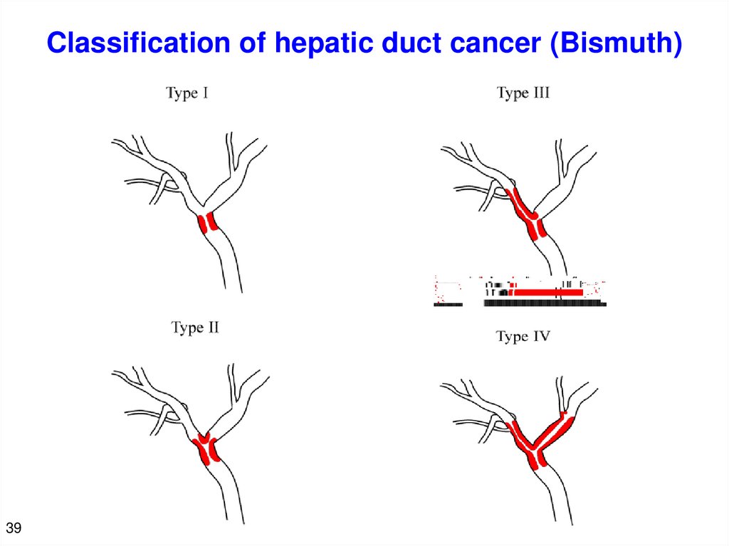
Classification of hepatic duct cancer (Bismuth)39
41.
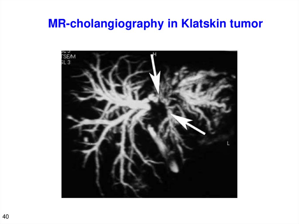
MR-cholangiography in Klatskin tumor40
42.
Pancreatic head cancer. MRCPCBD
PD
41
43.
Differential diagnosis of obstructive andparenchymatous jaundice
42
Patients with a painless obstructive jaundice do not notice anything for many
days because severe symptoms are absent. Patients often admit to infectious
disease departments with a diagnosis of hepatitis and undergo examination for
a long time. Differential diagnosis is simple - DETECTION OF ENLARGED
common bile duct during ultrasound or other surveys.
44.
Functional and morphological features of liver inpainless obstructive jaundice
Increased levels of direct and indirect bilirubin and alkaline phosphatase. A slight
increase in AST, ALT, LDH in some patients. No other biochemical disorders.
Reduced prothrombin, platelets, rare signs of mild encephalopathy in long-standing
obstructive jaundice (over 3-4 weeks).
Hepatocyte proliferation is replaced by their wrinkling and degeneration after 2-3
days (foci, fields). After 4 - 5 weeks, hepatocyte dimensions are reduced by 30-40%,
their volume decreases from 97% to 40%.
Bile duct proliferation from the first day. After 4–5 weeks, their volume is increased
from 2% to 40%,volume of stroma - from 1% to 20%.
Phlebitis of portal vein branches, neutrophilic infiltration, increased permeability of
intercellular junctions.
Wu P.C. et al. – J. Pathol. 1981,133: 61–74.
43
45.
Metabolic disorders in painless obstructive jaundiceReduced ATP and local blood flow velocity are associated with
long-standing obstructive jaundice and progressive
hyperbilirubinemia (p <0.05)
HGF release - regeneration regulator (ATP interval 0.53-0.6 μmol/g
of tissue) – the 9th day
TNF-α release - apoptosis factor (decrease in ATP below 0.365
μmol/g of tissue) - after the 12th day
44
46.
Conclusion on disorders arising in painlessobstructive jaundice
45
Painless obstructive jaundice causes severe functional and morphological
disorders in liver associated with biliary hypertension, cholestasis,
cholemia and acholia.
These disturbances result extrahepatic disorders, microbial colonization of
gastrointestinal tract, portal endotoxemia, RES dysfunction, especially
Kupffer cells, systemic toxemia followed by organ dysfunction.
Intra- and extrahepatic disorders together with persistent obstructive
jaundice and progressive hyperbilirubinemia acquire a critical nature
(fragile stability state), and additional effect ("second impact") can result
SIRS and multiple organ failure.
47.
PREOPERATIVE DECOMPRESSION46
48.
Preoperative decompression of the bile ductsSecondary obstructive jaundice is often more dangerous for the patient's life than
the underlying disease that caused this syndrome. Therefore, minimally invasive
correction of obstructive jaundice is essential in some patients. These patients
undergo a two-stage surgery: stage 1 – bile duct decompression, correction of
hyperbilirubinemia, stage 2 – total resection of tumor.
Methods of preoperative biliary decompression:
Percutaneous cholangiostomy.
Endoscopic nasobiliary drainage or stenting.
Cholecystostomy: open, ultrasound-assisted laparoscopic
percutaneous.
Open choledocho- or hepaticostomy.
A fairly complete restoration of liver and other systems requires prolonged
decompression of the bile ducts (3-4 weeks). Therefore, various stents are
often used for this purpose. For short-term decompression, drainage tubes
are used. The last ones may be later replaced with stents.
47
49.
Can bile duct decompression per se impair liver function?ATP restoration after decompression in 6- and 12-day obstructive jaundice
µmol/g of tissue
0,9
0,6
0,3
Days after decompression
0
0
3
Норма
6
6 дн желтуха
12
18
12 дн желтуха
In a 12-day obstructive jaundice, decrease of ATP, progressive energy deficit
of liver tissue and hyperbilirubinemia have been observed for 3 days. This
the so-called post-decompression syndrome is probably associated with fast
decompression of the bile ducts (similar to fast drainage of abdominal
effusion).
48
50.
Comparison of various methods of bile ductdecompression (n = 205)
NBD-85, PTC-37, cholecystostomy-63, CBD decompression -20
NBD was followed by gradual
decrease in biliary pressure
throughout the entire period of
decompression. An acceptable
pressure was achieved by the 7th
day. Other methods were
characterized by faster decrease in
biliary pressure. Decompression
rate may be adjusted by raising or
lowering the outer end of drainage
tube.
NBD – В = 4,6; PTC – В = 10; cholecystostomy– В = 9
49
51.
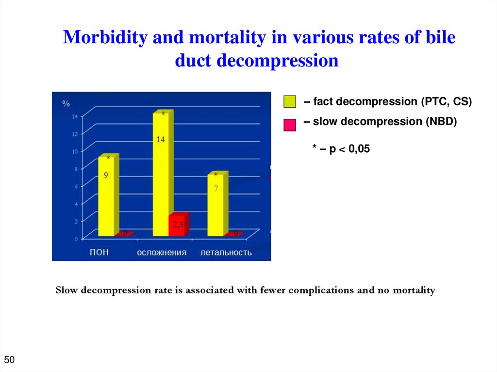
Morbidity and mortality in various rates of bileduct decompression
– fact decompression (PTC, CS)
– slow decompression (NBD)
* – p < 0,05
Slow decompression rate is associated with fewer complications and no mortality
50
52.
Positive and negative aspects of preoperative biliarydecompression
Advantages
Improved function of liver and
other organs and systems
Improved immune status
Improved liver microcirculation
Restored energy potential of liver
tissue
Drawbacks
Enhanced microbial contamination of
the bile ducts (18-97%)
Chronic inflammation in bile ducts and
gallbladder
Stenting can cause some adverse
events: acute pancreatitis, cholangitis,
bleeding, stent obstruction, etc.
Opinions on stenting are still controversial. However, there are absolute
indications for preoperative biliary decompression: cholangitis, neoadjuvant
chemotherapy, inoperable tumor, risk of radical surgery.
Additional factors in favor of preoperative biliary decompression: serum
bilirubin > 200 mmol / L and Klatskin tumor.
51
53.
Features of preoperative biliary decompression inKlatskin tumor
52
1.
Radical surgery for Klatskin tumor implies extended liver resection.
Future liver remnant (preferably at least 30%) must be functionally
adequate that is facilitated by preoperative decompression of the
bile ducts.
2.
Percutaneous transhepatic selective drainage of segmental bile
duct of future liver remnant is of particular importance. This
procedure together with increased portal blood flow facilitate fast
regeneration and enlargement of liver remnant.
54.
Conclusion on bile duct decompressionOpinions on preoperative biliary decompression are still controversial. Absolute
indications for decompression are acute cholangitis, neoadjuvant chemotherapy,
inoperable tumor and high risk of radical surgery.
Factors in favor of decompression are serum bilirubin > 200 mmol / L and
scheduled total resection of Klatskin tumor.
Preoperative biliary decompression should be slow in patients with long-standing
obstructive jaundice and serum bilirubin > 200 mmol/L. NBD with standard
drainage tube (length 180 cm and diameter 2 mm), as well as its height in relation
to common bile duct ensure gradual decompression.
Fast biliary decompression deteriorates liver disorders within 2–3 days in
patients with high serum bilirubin and should not be used in these cases.
53