


 industry
Similar presentations:


Inclusion in emeralds
1. Включения в изумрудах
2. Natural
Minute fluid inclusions parallel to the basal plane are the cause of asterismin this emerald from Madagascar. Thin-film interference causes
rainbow colors visible at high magnification. Field of view 1.8 mm.
3.
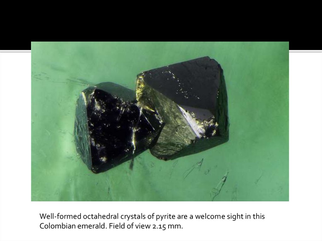
Well-formed octahedral crystals of pyrite are a welcome sight in thisColombian emerald. Field of view 2.15 mm.
4.
Jagged three-phase inclusions consisting of salt solution, a gas bubble,and a salt crystal are commonly seen in Colombian emeralds. Field of
view 0.91 mm.
5.
The etched prism face of an emerald from Alexander County, NorthCarolina, is shown in false, high-contrast color using differential interference
contrast microscopy. Field of view 0.61 mm.
6.
Gota de aceite results from rapid columnar growth, causing a roiledappearance in some Colombian emeralds. Field of view 2.60 mm.
7.
Brassy pyrite crystals are often seen in Colombian emeralds. Field of view 5.25mm.
8.
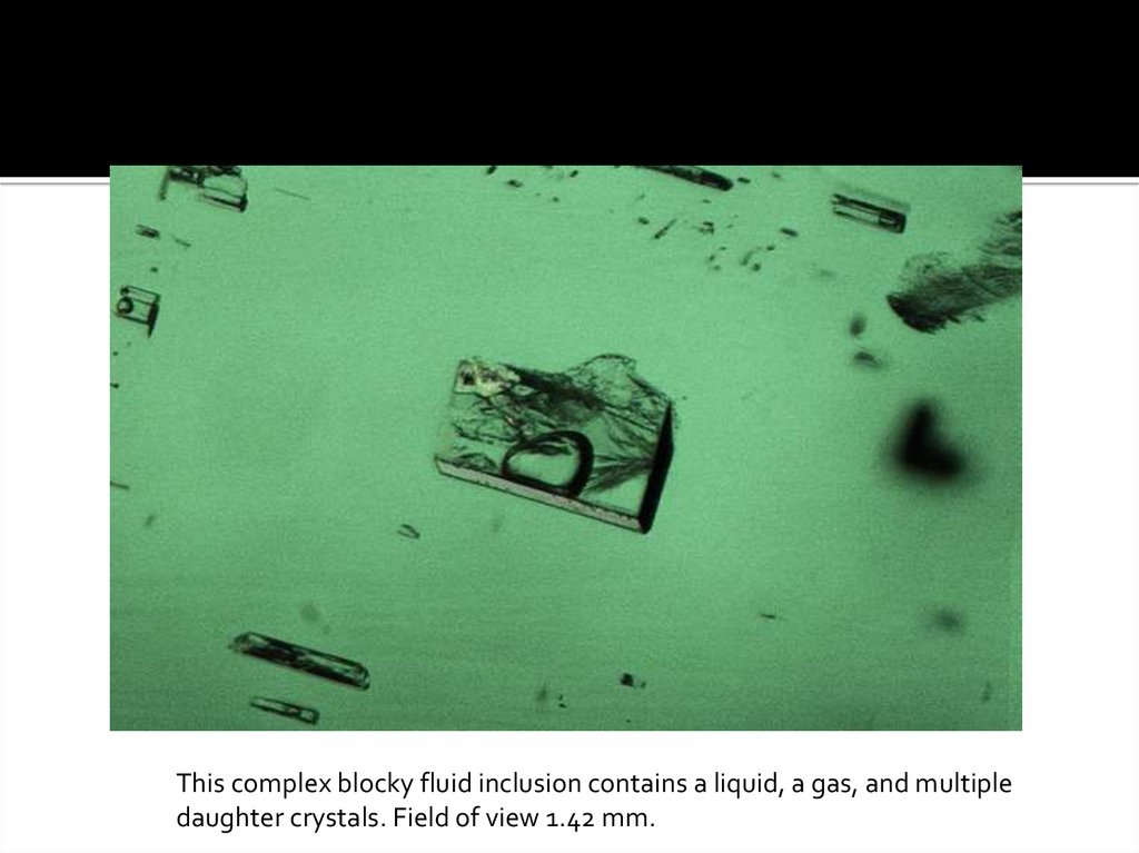
This complex blocky fluid inclusion contains a liquid, a gas, and multipledaughter crystals. Field of view 1.42 mm.
9.
Biotite crystals, shown here in polarized light, are common in emeraldsfrom schist-hosted deposits. Field of view 2.15 mm.
10.
This emerald from North Carolina contains vibrant orangy brown rutileinclusions. Field of view 6.25 mm.
11.
Amphibole crystals are occasionally seen in emeralds from Zambia.Field of view 1.72 mm.
12.
Skeletal platy crystals of ilmenite are scattered throughout this Zambianemerald. Field of view 1.91 mm.
13.
This Colombian emerald shows prominent angular color zoning reminiscentof a mountain range. Field of view 14.52 mm.
14.
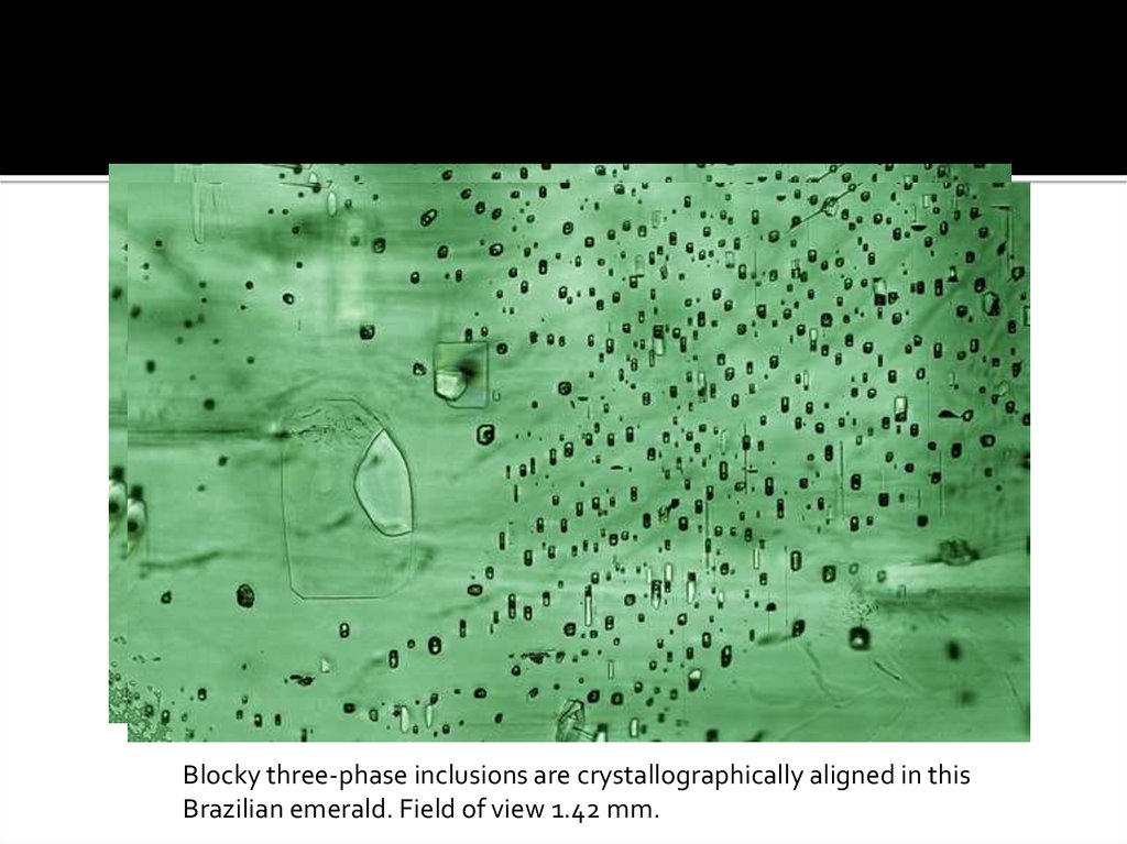
Blocky three-phase inclusions are crystallographically aligned in thisBrazilian emerald. Field of view 1.42 mm.
15.
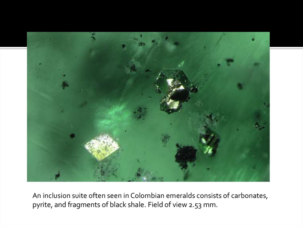
An inclusion suite often seen in Colombian emeralds consists of carbonates,pyrite, and fragments of black shale. Field of view 2.53 mm.
16.
Several rhombohedral magnesite crystals are present in this emeraldfrom Santa Terezinha de Goiás, Brazil. Field of view 8.68 mm.
17.
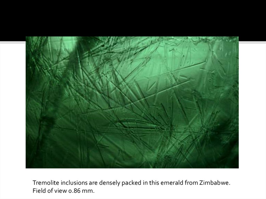
Tremolite inclusions are densely packed in this emerald from Zimbabwe.Field of view 0.86 mm.
18.
A very rare inclusion of parisite is diagnostic of Colombian origin.Field of view 0.82 mm.
19.
This Colombian emerald contains a carbonate crystal that is includedwith a pyrite crystal. Field of view 2.34 mm.
20.
A rare inclusion of purple fluorite is seen in high contrast to its greenemerald host. Field of view 2.34 mm.
21.
Russian emeralds often host brightly colored reflective thin-film fluidinclusions. These are oriented perpendicular to the optic axis. Field of
view 2.15 mm.
22. Treated
Clarity-enhancing resin showing a flow structure is present in the fractureof a natural emerald. Field of view 3.75 mm.
23.
A partially filled fracture shows vibrant interference colors in the unfilledareas and a dendritic pattern where the oil filler has wicked into
the fracture, reducing its visibility. Field of view 6.40 mm.
24.
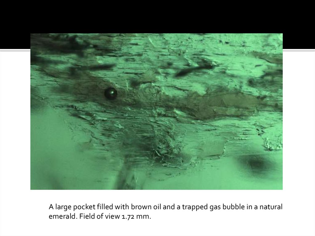
A large pocket filled with brown oil and a trapped gas bubble in a naturalemerald. Field of view 1.72 mm.
25.
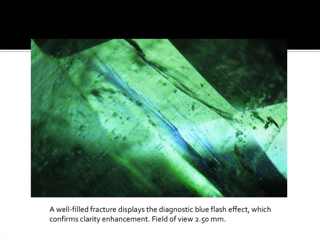
A well-filled fracture displays the diagnostic blue flash effect, whichconfirms clarity enhancement. Field of view 2.50 mm.
26.
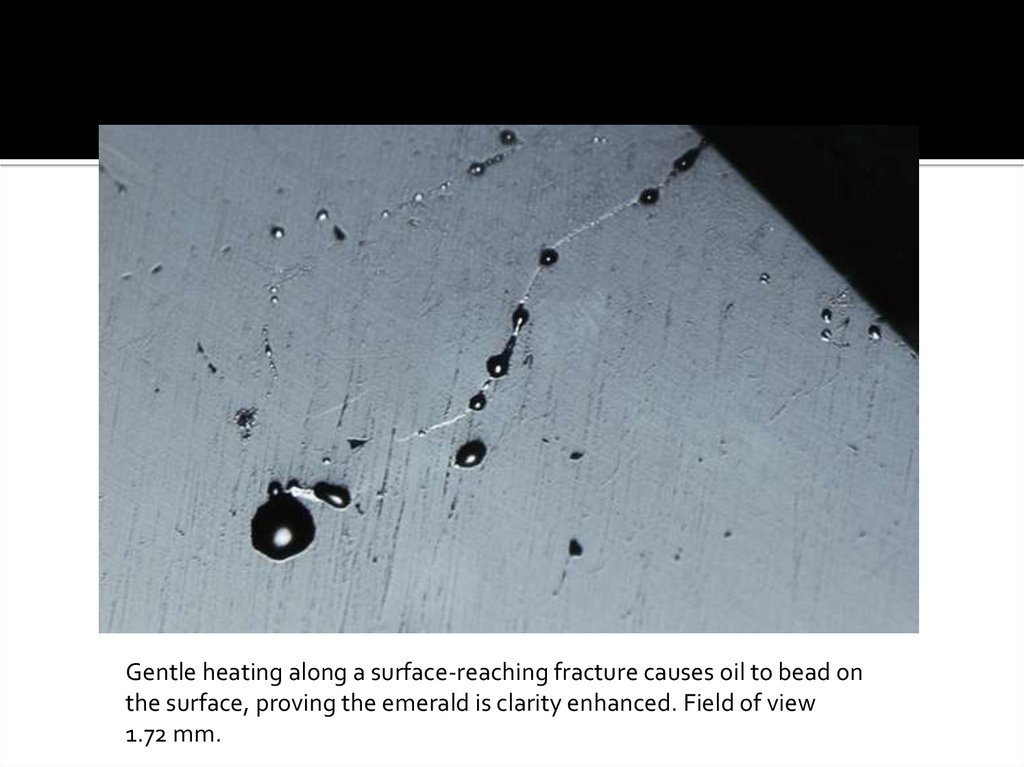
Gentle heating along a surface-reaching fracture causes oil to bead onthe surface, proving the emerald is clarity enhanced. Field of view
1.72 mm.
27. Synthetic
Nail-head spicules, as seen in this Regency hydrothermal syntheticemerald, are indicative of synthetic origin. Field of view 1.42 mm.
28.
Healed whitish feather-like inclusions of flux residue are present inthis Gilson synthetic emerald. Field of view 4.75 mm.
29.
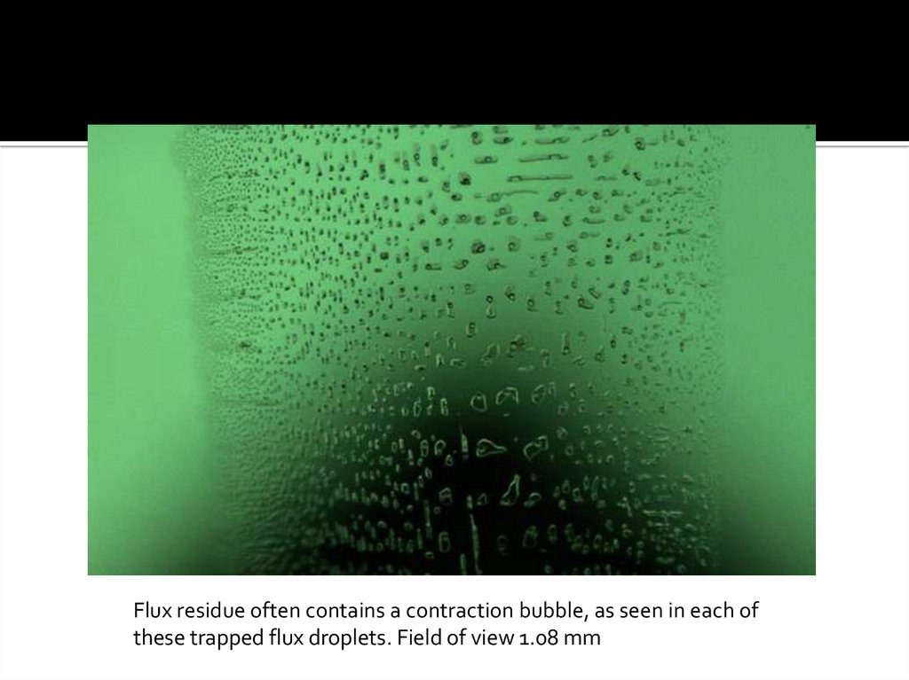
Flux residue often contains a contraction bubble, as seen in each ofthese trapped flux droplets. Field of view 1.08 mm
30.
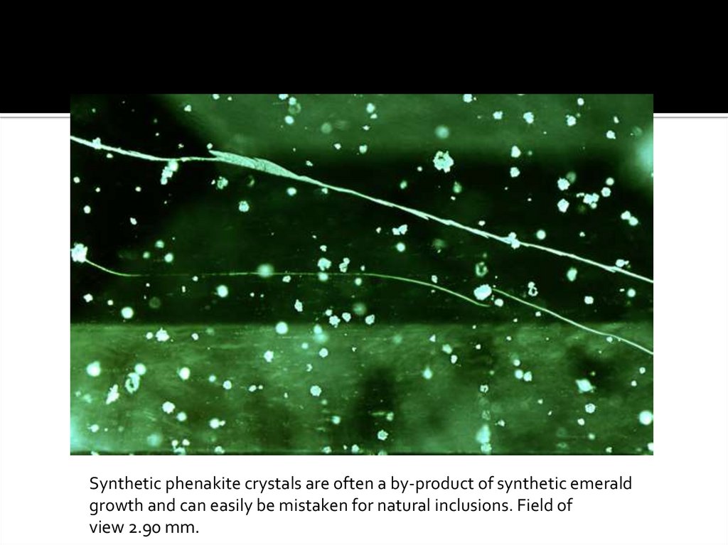
Synthetic phenakite crystals are often a by-product of synthetic emeraldgrowth and can easily be mistaken for natural inclusions. Field of
view 2.90 mm.
31.
This roiled, chevron-like growth is characteristic of hydrothermallygrown synthetic emeralds. Field of view 1.72 mm.
32. MICRO-FEATURES OF EMERALD
This chart contains a selection of photomicrographs of natural, synthetic, andtreated emeralds. It is by no means comprehensive. The images show the visual
appearance of numerous features a gemologist might observe when viewing
emeralds with a microscope.
Published in conjunction with Nathan D. Renfro, John I. Koivula, Jonathan Muyal,
Shane F. McClure, Kevin Schumacher, and James E. Shigley (2016), “Inclusions in
Natural, Synthetic, and Treated Emerald,” Gems & Gemology, Vol. 52, No. 4, pp.
402–403. Photomicrographs by Nathan D. Renfro, John I. Koivula, and Jonathan
Muyal.
© 2016 Gemological Institute of America